Note: Descriptions are shown in the official language in which they were submitted.
WO 2023/079498 PCT/IB2022/060621
1
SYSTEMS FOR DELIVERING DEVICES FOR REGULATING BLOOD
PRESSURE ACROSS AN ATRIAL SEPTUM
CROSS-REFERENCE TO RELATED APPLICATION
[0001] This application claims priority to U.S. Provisional Patent
Appl. No. 63/263,535,
filed November 4, 2021, the entire contents of which are incorporated herein
by reference.
FIELD OF USE
[0002] This application generally relates to devices and methods
for delivering implantable
devices to the atrial septum, particularly in subjects with heart pathologies
such as pulmonary
arterial hypertension (PAH), congestive heart failure (CHF) or myocardial
infarction (MI).
BACKGROUND
[0003] Pulmonary arterial hypertension (PAH) occurs when the
pressure within the blood
vessels and lungs becomes too high. PAH may be caused by obstruction in the
arteries in the
lung such as the development of scar tissue in the blood vessels of the lungs,
but in many cases,
the cause is unknown. Under normal conditions, the pressure within the right
side of the heart
and the blood vessels of the lungs is lower than the rest of the body which
maximizes
oxygenation of the blood in the lungs. With PAH, the heart must work harder
under greater
pressure to pump blood through the arteries in the lungs, weakening the heart
muscles over time.
As a result, the heart may be unable to sufficiently pump blood to the lungs
to be oxygenated to
keep the body functioning normally.
[0004] Heart failure is the physiological state in which cardiac
output is insufficient to meet
the needs of the body or to do so only at a higher filling pressure. There are
many underlying
causes of HF, including myocardial infarction, coronary artery disease,
valvular disease,
hypertension, and myocarditis. Chronic heart failure is associated with
neurohormonal
activation and alterations in autonomic control. Although these compensatory
neurohormonal
mechanisms provide valuable support for the heart under normal physiological
circumstances,
they also play a fundamental role in the development and subsequent
progression of HF.
CA 03237108 2024- 5-2
WO 2023/079498
PCT/1B2022/060621
2
[0005] For example, one of the body's main compensatory mechanisms
for reduced blood
flow in HF is to increase the amount of salt and water retained by the
kidneys. Retaining salt and
water, instead of excreting it via urine, increases the volume of blood in the
bloodstream and
helps to maintain blood pressure. However, the larger volumes of blood also
cause the heart
muscle, particularly the ventricles, to become enlarged. As the heart chambers
become enlarged,
the wall thickness decreases and the heart's contractions weaken, causing a
downward spiral in
cardiac function. Another compensatory mechanism is vasoconstriction of the
arterial system,
which raises the blood pressure to help maintain adequate perfusion, thus
increasing the load that
the heart must pump against.
[0006] In low ejection fraction (EF) heart failure, high pressures
in the heart result from the
body's attempt to maintain the high pressures needed for adequate peripheral
perfusion.
However, as the heart weakens as a result of such high pressures, the disorder
becomes
exacerbated. Pressure in the left atrium may exceed 25 mmHg, at which stage
fluids from the
blood flowing through the pulmonary circulatory system transudate or flow out
of the pulmonary
capillaries into the pulmonary interstitial spaces and into the alveoli,
causing lung congestion
and, if untreated, the syndrome of acute pulmonary edema and death.
[0007] Table 1 lists typical ranges of right atrial pressure (RAP),
right ventricular pressure
(RVP), left atrial pressure (LAP), left ventricular pressure (LVP), cardiac
output (CO), and
stroke volume (SV) for a normal heart and for a heart suffering from HF. In a
normal heart
beating at around 70 beats/minute, the stroke volume needed to maintain normal
cardiac output
is about 60 to 100 milliliters. When the preload, after-load, and
contractility of the heart are
normal, the pressures required to achieve normal cardiac output are listed in
Table 1. In a heart
suffering from HF, the hemodynamic parameters change (as shown in Table 1) to
maintain
peripheral perfusion.
Table 1
Parameter Normal Range HF Range
RAP (mmHg) 2-6 6-20
RVSP (mmHg) 15-25 20-80
LAP (mmHg) 6-12 15-50
LVEDP (mmHg) 6-12 15-50
CO (liters/minute) 4-8 2-6
SV (milliliters/beat) 60-100 30-80
CA 03237108 2024- 5-2
WO 2023/079498 PCT/1B2022/060621
3
[0008] HF is generally classified as either systolic heart failure
(SHF) or diastolic heart
failure (DHF). In SHF, the pumping action of the heart is reduced or weakened.
A common
clinical measurement is the ejection fraction, which is the volume of blood
ejected out of the left
ventricle (stroke volume) divided by the maximum volume in the left ventricle
at the end of
diastole or relaxation phase. A normal ejection fraction is greater than 50%.
Systolic heart
failure generally causes a decreased ejection fraction of less than 40%. Such
patients have heart
failure with reduced ejection fraction (HFrEF). A patient with HFrEF may
usually have a larger
left ventricle because of a phenomenon called "cardiac remodeling" that occurs
secondary to the
higher ventricular pressures.
[0009] In DHF, the heart generally contracts normally, with a
normal ejection fraction, but is
stiffer, or less compliant, than a healthy heart would be when relaxing and
filling with blood.
Such patients are said to have heart failure with preserved ejection fraction
(HFpEF). This
stiffness may impede blood from filling the heart and produce backup into the
lungs, which may
result in pulmonary venous hypertension and lung edema. HFpEF is more common
in patients
older than 75 years, especially in women with high blood pressure.
[0010] Both variants of HF have been treated using pharmacological
approaches, which
typically involve the use of vasodilators for reducing the workload of the
heart by reducing
systemic vascular resistance, as well as diuretics, which inhibit fluid
accumulation and edema
formation, and reduce cardiac filling pressure. No pharmacological therapies
have been shown
to improve morbidity or mortality in HFpEF whereas several classes of drugs
have made an
important impact on the management of patients with HFrEF, including renin-
angiotensin
antagonists, beta blockers, and mineralocorticoid antagonists. Nonetheless, in
general, HF
remains a progressive disease and most patients have deteriorating cardiac
function and
symptoms over time. In the U.S., there are over 1 million hospitalizations
annually for acutely
worsening HF and mortality is higher than for most forms of cancer.
[0011] In more severe cases of HFrEF, assist devices such as
mechanical pumps are used to
reduce the load on the heart by performing all or part of the pumping function
normally done by
the heart. Chronic left ventricular assist devices (LVAD), and cardiac
transplantation, often are
used as measures of last resort. However, such assist devices typically are
intended to improve
CA 03237108 2024- 5-2
WO 2023/079498 PCT/1B2022/060621
4
the pumping capacity of the heart, to increase cardiac output to levels
compatible with normal
life, and to sustain the patient until a donor heart for transplantation
becomes available. Such
mechanical devices enable propulsion of significant volumes of blood
(liters/min), but are
limited by a need for a power supply, relatively large pumps, and pose a risk
of hemolysis,
thrombus formation, and infection. Temporary assist devices, intra-aortic
balloons, and pacing
devices have also been used.
[0012] Various devices have been developed using stents to modify
blood pressure and flow
within a given vessel, or between chambers of the heart. Implantable
interatrial shunt devices
have been successfully used in patients with severe symptomatic heart failure.
By diverting or
shunting blood from the left atrium (LA) to the right atrium (RA), the
pressure in the LA is
lowered or prevented from elevating as high as it would otherwise (left atrial
decompression).
Such an accomplishment would be expected to prevent, relieve, or limit the
symptoms, signs,
and syndromes associated with pulmonary congestion. These include severe
shortness of breath,
pulmonary edema, hypoxia, the need for acute hospitalization, mechanical
ventilation, and death.
[0013] Percutaneous implantation of interatrial shunts generally
requires transseptal
catheterization immediately preceding shunt device insertion. The transseptal
catheterization
system is placed from an entrance site in the femoral vein, across the
interatrial septum in the
region of fossa ovalis (FO), which is the central and thinnest region of the
interatrial septum.
This is the same general location where a congenital secundum atrial septal
defect (ASD) would
be located. The FO in adults is typically 15-20 mm in its major axis dimension
and <3 mm in
thickness, but in certain circumstances may be up to 10 mm thick. LA chamber
access may be
achieved using a host of different techniques familiar to those skilled in the
art, including but not
limited to: needle puncture, stylet puncture, screw needle puncture, and radi
frequency ablation.
The passageway between the two atria is dilated to facilitate passage of a
shunt device having a
desired orifice size. Dilation generally is accomplished by advancing a
tapered sheath/dilator
catheter system or inflation of an angioplasty type balloon across the FO. A
limitation of
advancing a typical separate tapered dilator is that after dilating the
septum, the dilator must be
removed from the sheath before any device to be delivered can be loaded into
the sheath and
advanced for deployment.
CA 03237108 2024- 5-2
WO 2023/079498 PCT/1B2022/060621
[0014] Moreover, devices such as those described in U.S. 5,312,341
to Turi, have been
theorized for transseptal catheterization. Specifically, these devices have a
retaining means such
as an inflatable balloon that is inflated within the left atrium of the
patient to prevent inadvertent
retraction of the distal tip of the sheath from the left atrium during
subsequent portions of the
catheterization procedure.
[0015] In view of the foregoing, it would be desirable to provide
devices for delivering
implantable devices to the atrial septum of the heart to reduce left atrial
pressure, while reducing
the number of delivery tools required.
[0016] It would further be desirable to provide devices and methods
for controlled
positioning and delivery of atrial shunt devices.
SUMMARY
[0017] The present disclosure overcomes the drawbacks of previously
known systems and
methods by providing systems and methods for delivering a shunt to an atrial
septum of a
patient. For example, the apparatus may include a sheath having a proximal
region, a distal
region, and a sheath lumen extending therethrough, the sheath lumen sized and
shaped to receive
the shunt in a collapsed delivery state, and a balloon catheter configured to
be moveably
disposed within the sheath lumen. The balloon catheter may include a balloon
configured to
transition between a deflated collapsed state and an inflated expanded state
adjacent to the distal
region to form a continuous, step-free transition between the balloon and the
distal region of the
sheath. The apparatus further may include a handle having one or more
actuators configured to
be actuated to deploy the shunt at the atrial septum.
[0018] In addition, the apparatus may include a pusher slidably
disposed within the sheath
lumen. The pusher may be operatively coupled to a pusher actuator of the one
or more actuators
of the handle, such that the pusher actuator may be configured to be actuated
to move the pusher
within the sheath lumen. For example, the pusher actuator may be configured to
be actuated to
move the pusher distally relative to the sheath, such that a distal end of the
pusher engages with a
proximal portion of the shunt in the collapsed delivery state to thereby move
the shunt distally
CA 03237108 2024- 5-2
WO 2023/079498 PCT/1B2022/060621
6
relative to the sheath until a distal portion of the shunt is exposed beyond
the distal region of the
sheath and transitions from the collapsed delivery state to an expanded
deployed state.
[0019] The apparatus further may include a release knot slidahly
disposed within the sheath
lumen, the release knot configured to be releasably engaged with the shunt,
e.g., at a proximal
portion of the shunt, via a hitch knot, e.g., a painters hitch knot or a Quick
Tie and Release
(QTaR) hitch knot. For example, a first end of the release knot may be
operatively coupled to a
release actuator of the one or more actuators of the handle and a second end
of the release knot
may be operatively coupled to a retrieval actuator of the one or more
actuators of the handle,
such that actuation of the release actuator causes the hitch knot to
disassemble and disengage the
release knot from the shunt, and actuation of the retrieval actuator causes
retraction of the shunt
proximally within the sheath lumen via the hitch knot for halfway retrieval of
the shunt.
Alternatively, a first end of the release knot may be operatively coupled to a
release actuator of
the one or more actuators of the handle and a second end of the release knot
may be operatively
coupled to a distal portion of the pusher, such that actuation of the release
actuator causes the
hitch knot to disassemble and disengage the release knot from the shunt, and
actuation of the
pusher actuator causes retraction of the shunt proximally within the sheath
lumen via the hitch
knot for halfway retrieval of the shunt.
[0020] Moreover, the balloon may be configured to be deflated to
permit deployment of the
shunt through the distal region of the sheath. The balloon catheter may
include a fluid lumen
configured to fluidically couple the balloon and a fluid source. In addition,
the balloon catheter
may be operatively coupled to a balloon catheter actuator of the one or more
actuators of the
handle, such that actuation of the balloon catheter actuator causes the
balloon catheter to move
relative to the sheath_
[0021] In accordance with another aspect of the present disclosure,
a method for delivering a
shunt to an atrial septum of a patient is provided. The method may include
inflating a balloon
adjacent to a distal region of a sheath to form a continuous, step-free
transition between the
balloon and the distal region of the sheath, the balloon disposed on a distal
portion of a balloon
catheter slidably disposed within a lumen of the sheath; delivering the
inflated balloon and the
sheath through an opening of the atrial septum, such that the inflated balloon
and the sheath
CA 03237108 2024- 5-2
WO 2023/079498 PCT/1B2022/060621
7
dilates the opening of the atrial septum; deflating the balloon; advancing the
shunt distally within
the lumen of the sheath in a collapsed delivery state until a distal portion
of the shunt is exposed
beyond the distal region of the sheath and transitions to an expanded deployed
state within a first
atrium; and retracting the sheath proximally relative to the atrial septum
until a proximal portion
of the shunt is exposed beyond the distal region of the sheath and transitions
to the expanded
deployed state within a second atrium, such that the shunt is deployed at the
atrial septum.
[0022] The shunt may releasably engaged with a release knot, such
that pulling a first end of
the release knot causes the release knot to disengage from the shunt, and
pulling a second end of
the release knot causes retraction of the shunt proximally within the lumen of
the sheath for
halfway retrieval of the shunt. For example, the release knot may be
releasably engaged with the
shunt via a painters hitch knot or a Quick Tie and Release (QTaR) hitch knot.
[0023] Accordingly, prior to retracting the sheath proximally
relative to the atrial septum
until the proximal portion of the shunt is exposed beyond the distal region of
the sheath, the
method further may include pulling the first end of the release knot to
disengage the release knot
from the shunt. Moreover, prior to retracting the sheath proximally relative
to the atrial septum
until the proximal portion of the shunt is exposed beyond the distal region of
the sheath, the
method further may include pulling the second end of the release knot to
transition the distal
portion of the shunt to the collapsed delivery state within the lumen of the
sheath for halfway
retrieval of the shunt. In addition, prior to retracting the sheath proximally
relative to the atrial
septum until the proximal portion of the shunt is exposed beyond the distal
region of the sheath,
the method further may include retracting the sheath and the shunt proximally
relative to the
atrial septum until the distal portion of the shunt contacts the atrial septum
from within the first
atrium.
[0024] Advancing the shunt distally within the lumen of the sheath
may include advancing a
pusher distally within the lumen of the sheath, such that a distal end of the
pusher engages with
the proximal portion of the shunt in the collapsed delivery state within the
lumen of the sheath.
Accordingly, prior to retracting the sheath proximally relative to the atrial
septum until the
proximal portion of the shunt is exposed beyond the distal region of the
sheath, the method
further may include retracting the shunt proximally within the lumen of the
sheath to transition
CA 03237108 2024- 5-2
WO 2023/079498 PCT/1B2022/060621
8
the distal portion of the shunt to the collapsed delivery state within the
lumen of the sheath for
halfway retrieval of the shunt. In some embodiments, prior to retracting the
shunt proximally
within the lumen of the sheath, the method may include retracting the pusher
distally relative to
the sheath. The method further may include retracting the balloon catheter and
the deflated
balloon proximally within the lumen of the sheath to a position proximal to
the shunt in the
collapsed delivery state prior to advancing the shunt within the lumen of the
sheath. In addition,
the method may include removing the sheath and the balloon catheter from the
patient.
[0025] In accordance with another aspect of the present disclosure,
another apparatus for
delivering a shunt to an atrial septum of a patient is provided. The apparatus
may include a
sheath configured to be advanced through a hole in the atrial septum, the
sheath having a
proximal region, a distal region, and a sheath lumen extending therethrough,
the sheath lumen
sized and shaped to receive the shunt in a collapsed delivery state, and a
dilator moveably
disposed within the sheath lumen. The dilator may include an expandable
portion configured to
transition between a first state and a second state where the expandable
portion engages with the
distal region of the sheath. Accordingly, the dilator and the sheath may be
configured to dilate
the hole in the atrial septum as tissue surrounding the hole is smoothly
guided over the distal
portion of the dilator and the sheath as the apparatus is advanced through
hole in the atrial
septum. The dilator may include a guidewire lumen sized and shaped to receive
a guidewire.
[0026] In accordance with one aspect of the present disclosure, the
dilator further may
include a dilation catheter moveably disposed within the sheath lumen, and a
cone-shaped tip
coupled to a distal end of the dilation catheter, and to a distal portion of
the expandable portion
of the dilator. For example, in the first state, the proximal portion of the
expandable portion may
be contracted radially inward toward the dilation catheter, and, in the second
state, a proximal
portion of the expandable portion may be removeably engaged with the distal
region of the
sheath to form a continuous, step-free transition between the sheath and the
expandable portion
of the dilator. The dilation catheter may be configured to be moved distally
relative to the sheath
to cause the expandable portion of the dilator to transition from the second
state to the first state.
Moreover, the expandable portion of the dilator may be biased toward the first
state.
CA 03237108 2024- 5-2
WO 2023/079498
PCT/1B2022/060621
9
[0027] In the second state, the distal portion of the expandable
portion of the dilator may
engage with an outer surface of the distal region of the sheath. Accordingly,
the apparatus may
have a continuous, step-free transition between the sheath and the expandable
portion of the
dilator when the expandable portion is in the second state. The guidewire
lumen may extend
through the cone-shaped tip and the dilation catheter. In addition, the sheath
may he configured
to be moved proximally relative to the dilator when the expandable portion of
the dilator is in the
first state to deploy the shunt at the atrial septum. The apparatus further
may include a hollow
catheter, e.g., a PEEK tube, moveably disposed within the sheath lumen, the
hollow catheter
sized and shaped to receive the dilation catheter. For example, in the first
state, the proximal
portion of the expandable portion of the dilator may be disposed within a
distal region of the
hollow catheter.
[0028] In accordance with another aspect of the present disclosure,
the distal region of the
sheath may be configured to transition between a contracted state and an
expanded state, and the
expandable portion of the dilator may include a proximal portion coupled to an
outer tube
moveably disposed within the sheath lumen, and a cone-shaped distal portion
coupled to an inner
tube moveably disposed within the outer tube such that the cone-shaped distal
portion is
moveable relative to the proximal portion between the second state where the
distal region of the
sheath is sandwiched between the proximal portion and the cone-shaped distal
portion in the
contracted state, and the first state where the distal region of the sheath
disengages with the
proximal portion and the cone-shaped distal portion to transition to the
expanded state.
Accordingly, the apparatus may have a continuous, step-free transition between
the cone-shaped
distal portion and the distal region of the sheath when the distal legion of
the sheath is in the
contracted state.
[0029] In the contracted state, the distal region of the sheath may
have a plurality of
longitudinal slits disposed circumferentially along the distal region, and
extending from a distal
end of the distal region toward the proximal region of the sheath. Moreover,
in the expanded
state, the distal region of the sheath may be expanded along the plurality of
longitudinal slits
such that each of the plurality of longitudinal slits comprises a V-shape. The
distal region of the
sheath may be biased toward the expanded state. Moreover, the distal region of
the sheath may
include an elastic material encapsulated with a biocompatible material. For
example, the elastic
CA 03237108 2024- 5-2
WO 2023/079498
PCT/1B2022/060621
material may be superelastic Nitinol, and the biocompatible material may be a
polyether block
amide. The guidewire lumen may extend through the cone-shaped distal portion
and the inner
tube.
[0030] In accordance with another aspect of the present disclosure,
the expandable portion of
the dilator may include an expandable braided tip coupled to an inner tube
moveably disposed
within the sheath lumen, the expandable braided tip configured to transition
between the first
state and the second state. Moreover, a distal portion of the shunt may be
configured to
transition between the collapsed delivery state where the distal portion of
the shunt forms a
continuous, step-free transition between the distal portion of the shunt and
the expandable
braided tip when the expandable braided tip is in the second state, and an
expanded deployed
state. A proximal end of the expandable braided tip may be coupled to an outer
tube and a distal
end of the expandable braided tip may be coupled to the inner tube, and the
inner tube may be
moveably disposed within a lumen of the outer tube, such that the proximal end
of the
expandable braided tip is moveable relative to the distal end of the
expandable braided tip to
transition the expandable braided tip between the first state and the second
state. In addition, the
distal portion of the shunt may be configured to transition from the collapsed
delivery state to the
expanded deployed state upon application of heat. Accordingly, the sheath
further may include a
fluid lumen configured to deliver heated liquid to the distal portion of the
shunt. The apparatus
may have a continuous, step-free transition between the distal portion of the
shunt and the distal
region of the sheath when the distal portion of the shunt is in the collapsed
delivery state. The
guidewire lumen may extend through the inner tube.
[0031] In accordance with another aspect of the present disclosure,
the distal region of the
sheath may be configured to transition between a contracted state and an
expanded state, and the
expandable portion of the dilator may include a balloon coupled to balloon
catheter configured to
be moveably disposed within the sheath lumen. The balloon may be configured to
be inflated
from the first state to the second state to transition the distal region from
the contracted state to
the expanded state. For example, in the contracted state, the distal region of
the sheath may
define an opening and may have a plurality of longitudinal slits disposed
circumferentially along
the distal region, and extending from the opening toward the proximal region
of the sheath, and
in the expanded state, the distal region of the sheath may be expanded along
the plurality of
CA 03237108 2024- 5-2
WO 2023/079498
PCT/1B2022/060621
11
longitudinal slits such that each of the plurality of longitudinal slits
comprises a V-shape. A
distal tip of the balloon may be configured to extend through the opening to
form a continuous,
step-free transition between the balloon and the distal region of the sheath.
In addition, the
plurality of longitudinal slits may define a plurality of fingers of the
distal region, and a distal
end of each of the plurality of fingers may have a round shape_ Moreover, the
distal region of
the sheath may include a shape-memory material configured to cause the distal
region to return
to the contracted state upon exposure to heat.
[0032] In accordance with another aspect of the present disclosure,
the expandable portion of
the dilator may include a balloon coupled to a balloon catheter configured to
be moveably
disposed within the sheath lumen. The balloon may be configured to transition
between the first
state and the second state adjacent to the distal region to form a continuous,
step-free transition
between the balloon and the distal region of the sheath. The apparatus further
may include a
pusher slidably disposed within the sheath lumen. The pusher may have a pusher
lumen sized
and shaped to slidably receive the balloon catheter therethrough, and a distal
end configured to
engage with a proximal portion of the shunt in the collapsed delivery state to
thereby move the
shunt distally relative to the sheath. The apparatus further may include a
release knot slidably
disposed within the sheath lumen. For example, the release knot may be
configured to be
releasably engaged with the shunt via a hitch knot, e.g., a painters hitch
knot or a Quick Tie and
Release (QTaR) hitch knot, such that pulling a first end of the release knot
causes the release
knot to disengage from the shunt, and pulling a second end of the release knot
causes retraction
of the shunt proximally within the sheath lumen for halfway retrieval of the
shunt.
[0033] In some embodiments, the release knot may be configured to
be releasably engaged
with a proximal portion of the shunt. Additionally, the first and second ends
of the release knot
may pass through a central passageway of the shunt toward a middle portion of
the shunt, and
loop around an outer surface of the middle portion of the shunt and back
towards the hitch knot,
such that pulling the second end of the release knot causes the shunt to
transition toward the
collapsed delivery state. Alternatively, the release knot may be configured to
be releasably
engaged with a middle portion of the shunt, and the first and second ends of
the release knot may
loop around an outer surface of the middle portion of the shunt, such that
pulling the second end
of the release knot causes the shunt to transition toward the collapsed
delivery state. The balloon
CA 03237108 2024- 5-2
WO 2023/079498
PCT/1B2022/060621
12
may be configured to be deflated to permit deployment of the shunt through the
distal region of
the sheath. Moreover, the balloon catheter may have a fluid lumen configured
to fluidically
couple the balloon and a fluid source.
BRIEF DESCRIPTION OF THE DRAWINGS
[0034] FIGS. IA to ID illustrate an exemplary device for delivering
an interatrial shunt
device to the atrial septum in accordance with the present disclosure.
[0035] FIGS. 2A to 2F illustrate an alternative exemplary device
for delivering an interatrial
shunt device to the atrial septum in accordance with the present disclosure.
[0036] FIGS. 3A to 3G illustrate another alternative exemplary
device for delivering an
interatrial shunt device to the atrial septum in accordance with the present
disclosure.
[0037] FIGS. 4A and 4B illustrate yet another alternative exemplary
device for delivering an
interatrial shunt device to the atrial septum in accordance with the present
disclosure.
[0038] FIG. 5A illustrates yet another alternative exemplary device
for delivering an
interatrial shunt device to the atrial septum in accordance with the present
disclosure_
[0039] FIG. 5B illustrates an exemplary handle for actuating the
delivery device of FIG. 5A,
constructed in accordance with the principles of the present disclosure.
[0040] FIG. 6A illustrates an exemplary knot mechanism of the
delivery device of FIG. 5A.
[0041] FIG. 6B illustrates an alternative exemplary knot mechanism
in accordance with the
principles of the present disclosure.
[0042] FIGS. 7A to 7H illustrate exemplary method steps for
delivering an interatrial shunt
device to the atrial septum using the delivery device of FIGS. 5A and 5B in
accordance with the
present disclosure.
[0043] FIGS. 71 to 7K illustrate exemplary method steps for half-
way retrieval of the
interatrial shunt device using the delivery device of FIGS. 5A and 5B in
accordance with the
present disclosure.
CA 03237108 2024- 5-2
WO 2023/079498
PCT/1B2022/060621
13
[0044] FIGS. 8A to 8G illustrate exemplary method steps for
delivering an interatrial shunt
device to the atrial septum using another exemplary delivery device in
accordance with the
present disclosure.
[0045] FIGS. 8H and 81 illustrate exemplary method steps for half-
way retrieval of the
interatrial shunt device using the delivery device of FIGS. 8A to 8G in
accordance with the
present disclosure.
DETAILED DESCRIPTION
[0046] Embodiments of the present invention are directed to devices
for delivering
implantable devices to a wall of the heart such as the atrial septum, and thus
may be useful in
treating subjects suffering from heart failure, myocardial infarction,
pulmonary hypertension, or
other disorders associated with elevated atrial pressure. For example, the
inventive device may
be designed to deliver an hourglass or "diabolo" shaped shunt device,
preferably formed of a
shape memory metal as described in U.S. Patent No. 9,629,715 to Nitzan, U.S.
Patent No.
10,076,403 to Eigler, and U.S. Patent No. 11,458,287 to Eigler, each assigned
to the assignee of
the present invention, the entire contents of each of which are incorporated
herein by reference.
The delivery devices described herein are configured to lodge the shunt
securely in a hole in a
heart wall such as the atrial septum, preferably the fossa ovalis, to function
as an interatrial
shunt, allowing blood flow between the left atrium and the right atrium.
[0047] Referring now to FIGS. lA to 1D, exemplary delivery device
100 for delivering
interatrial shunt device 10 to the atrial septum is provided. As shown in FIG.
1A, delivery
device 100 includes sheath 110 removeably coupled to dilator 103. Sheath 110
has a lumen
extending from distal region 112 of sheath 110 to the proximal region of the
sheath external to
the patient. The lumen of sheath 110 is sized and shaped to receive shunt 10
in its collapsed
delivery state. As shown in FIG. 1A, distal region 112 of sheath 110 may have
an outer diameter
that is less than the outer diameter of rest of the length of sheath 110
extending from distal region
112 toward the proximal region of sheath 110.
[0048] Dilator 103 includes dilation catheter 102 moveably disposed
within the lumen of
sheath 110, such that dilation catheter 102 may be moved relative to sheath
110, e.g., via
CA 03237108 2024- 5-2
WO 2023/079498
PCT/1B2022/060621
14
actuation of a handle external to the patient that is independently coupled to
sheath 110 and
dilation catheter 102. In addition, dilator 103 includes dilator tip 104
coupled to the distal end of
dilation catheter 102. Dilator tip 104 may be a low durometer soft tip and may
have an
atraumatic cone shape that may be inserted through a puncture of the atrial
septum to enlarge the
puncture without damaging the surrounding tissue. Alternatively, dilator tip
104 may have a
sharp needle tip that may be used to create the puncture within the atrial
septum, such that further
advancement of dilator tip 104 across the atrial septum enlarges the puncture.
[0049] Dilator 103 may have a guidewire lumen 106 sized and shaped
to receive a guidewire
therethrough, such that device 100 may be advanced over a conventional
guidewire across the
atrial septum. Accordingly, guidewire lumen 106 may extend through dilator tip
104 and
dilation catheter 102. Moreover, dilator 103 may include expandable portion
108. Expandable
portion 108 may be coupled at its distal portion to dilator tip 104, and
extend toward sheath 110.
In one embodiment, dilator tip 104 and expandable portion 108 are formed of a
unitary
construction. As shown in FIG. 1D, at least expandable portion 108 of dilator
103 may be
encapsulated with biocompatible material 109, e.g., polyether block amide
(PEBA), such as
PEBAX (made available by Arkema, Colombes, France).
[0050] Expandable portion 108 may be formed of an elastic material,
e.g., superelastic
Nitinol, and may be transitionable between an expanded state and a contracted
state. For
example, expandable portion 108 may be heat-set during manufacturing in the
contracted state,
such that expandable portion 108 is biased toward the contracted state.
Accordingly, prior to
insertion into the patient, expandable portion 108 may be expanded and fit
over distal region 112
of sheath 110. Specifically, as shown in FIG. 1A, in the expanded state, the
proximal portion of
expandable portion 108 may be engaged with the outer surface of distal region
112, to thereby
form a step-free transition between sheath 110 and expandable portion 108 of
dilator 103 when
expandable portion 108 is in the expanded state. Thus, when the proximal
portion of expandable
portion 108 is engaged with the outer surface of distal region 112 of sheath
110, the proximal
portion of expandable portion 108 has the same outer diameter as the portion
of sheath 110
adjacent to distal region 112. The step-free transition between sheath 110 and
expandable
portion 108 results from distal region 112 having an outer diameter that is
less than the outer
diameter of the rest of sheath 110. Although FIG. lA illustrates sheath 110
having a constant
CA 03237108 2024- 5-2
WO 2023/079498
PCT/1B2022/060621
thickness along its longitudinal length, e.g., including distal region 112,
and thus having an inner
diameter varying from sheath 110 to distal region 112, alternatively, sheath
110 and distal region
112 may have a constant inner diameter along its longitudinal length.
[0051] As shown in FIG. 1A, device 100 may include hollow catheter
114, e.g., PEEK tube,
moveably disposed within the lumen of sheath 110. Hollow catheter 114 may be
sized and
shaped to receive dilation catheter 102, and at least a portion of the
proximal portion of
expandable portion 108 therethrough. Accordingly, dilation catheter 102,
hollow catheter 114,
shunt 10 in its collapsed delivery state, and sheath 110 may be concentric.
Dilation catheter 102,
hollow catheter 114, and sheath 110 may each be coupled at their proximal
regions to a handle
for use by a clinician, such that each component may be independently actuated
via the handle.
[0052] As shown in FIG. 1B, upon movement of dilator 103 distally
relative to sheath 110,
expandable portion 108 of dilator 103 will disengage with distal region 112 of
sheath 110 and
return to its contracted state, as distal region 112 will no longer exert a
radially outward force on
the inner surface of the proximal portion of expandable portion 108.
Accordingly, expandable
portion 108 will be contracted radially inward toward dilation catheter 102.
In its contracted
state, the proximal portion of expandable portion 108 may be adjacent to the
opening into hollow
catheter 114. Accordingly, upon retraction of dilation catheter 102 relative
to hollow catheter
114, at least a portion of the proximal portion of expandable portion 108 may
be received by the
distal region of hollow catheter 114, thereby causing expandable portion 108
to contract even
further. Shunt 10 may be deployed by retracting sheath 110 proximally relative
to hollow
catheter 114 and dilator 103, as shown in FIG. 1C, to thereby implant shunt 10
at atrial septum
AS.
[0053] Loading of shunt 10 into delivery device 100, e.g., during
manufacturing or in a
preparatory step, may proceed as follows. First, hollow catheter 114 may be
advanced over
dilation catheter 102 until the distal region of hollow tube 114 is adjacent
to expandable portion
108 of dilator 103. Hollow catheter 114 may be further advanced over at least
a portion of the
proximal portion of expandable portion 108 to receive the proximal portion of
expandable
portion 108 therein, providing rigidity to the combined structure of hollow
catheter 114 and
dilator 103. Hollow catheter 114 and dilator 103 together may be advanced
distally through the
CA 03237108 2024- 5-2
WO 2023/079498
PCT/1B2022/060621
16
lumen of sheath 110 until at least dilator 103 is exposed beyond proximal
region 112 of sheath
110, such that dilation catheter 102, hollow catheter 114, shunt 10 in its
collapsed delivery state,
and sheath 110 are concentric. Alternatively, the combined structure of hollow
catheter 114 and
dilator 103 may be back-loaded through the distal opening of sheath 110 until
dilator 103 is
adjacent to distal region 112.
[0054] Next, dilation catheter 102 may be moved distally relative
to hollow catheter 114,
such that expandable portion 108 is no longer within hollow catheter 114 and
may be expanded
radially outwardly away from dilation catheter 102 and positioned over the
outer surface of distal
region 112 of sheath 110. Upon release of expandable portion 108 over distal
region 112, distal
region 112 will maintain expandable portion 108 in its expanded state, such
that the proximal
portion of expandable portion 108 is fitted onto distal region 112.
Accordingly, sheath 110,
expandable portion 108, and tip 104 form a smooth and continuous dilator
assembly, as shown in
FIG. 1A, suitable for inserting into a blood vessel over a guidewire and
advancing across the
interatrial septum.
[0055] Next, shunt 10 may be collapsed to its collapsed delivery
state within sheath 110, for
example, using tools as described in U.S. Patent No. 9,713,696 to Yacoby or
U.S. Patent App.
Pub. No. 2020/0315599 to Nae, each assigned to the assignee of the present
invention, the entire
contents of each of which are incorporated by reference herein. Delivery
device 100 is then
ready to deliver shunt 10. Delivery of shunt 10 using delivery device 100
described above may
proceed as follows. Guidewire 101 may be advanced to the target location
through a puncture of
the atrial septum, e.g., within the left atrium of the patient. Device 100 may
be advanced over
guidewire 101 via guidewire lumen 106 until dilator tip 104 comes into contact
with the puncture
of the atrial septum. Device 100 may be further advanced such that dilator 103
enlarges the
puncture of the atrial septum as the tissue surrounding the puncture is
smoothly guided over
dilator tip 104, followed by expandable portion 108, and sheath 110. Unlike
other known
delivery systems, a separate dilator is not required to enlarge the puncture
of the atrial septum,
which must be subsequently removed prior to loading shunt into sheath 110.
[0056] Under visualization methods such as fluoroscopy and/or
ultrasound imaging such as
trans-esophageal echo (TEE) or intracardiac echo (ICE), the target position of
device 100 relative
CA 03237108 2024- 5-2
WO 2023/079498
PCT/1B2022/060621
17
to the atrial septum may be verified, e.g., via a radiopaque marker on sheath
110. Next, dilation
catheter 102 may be moved distally relative to sheath 110, thereby causing
expandable portion
108 to disengage from distal region 112 and transition from its expanded state
to its contracted
state toward dilation catheter 102, e.g., by virtue of its superelasticity, as
shown in FIG. 1B.
Dilation catheter 102 may then he moved proximal relative to hollow catheter
114 such that at
least a portion of the proximal portion of expandable portion 108 is received
into hollow catheter
114. While hollow catheter 114, dilator 103, and shunt 10 remain stationary
relative to the atrial
septum, sheath 110 may be retracted proximally to expose the distal portion of
shunt 10 such that
the distal portion of shunt 10 deploys within the left atrium. Shunt 10 may be
maintained
stationary relative to the atrial septum using devices within sheath 110 such
as those described in
W02020202046, the entire contents of which is incorporated by reference
herein. For example,
a device having a plurality of hooks may be used to engage with the proximal
portion of shunt 10
within sheath 110.
[0057] After the distal portion of shunt 10 is deployed within the
left atrium, delivery device
100 may be retracted proximally until the distal portion of shunt 10 contacts
the atrial septal
wall. Then, sheath 110 may be further retracted proximally while shunt 10 is
maintained
stationary relative to the atrial septum until the proximal portion of shunt
10 is exposed from
distal region 112 of sheath 110 and deploys within the right atrium of the
patient as shown in
FIG. 1C. Delivery device 100 may then be removed from the patient, leaving
shunt 10
implanted at the atrial septum.
[0058] Referring now to FIGS. 2A to 2F, exemplary delivery device
200 for delivering
interatrial shunt device 10 to the atrial septum is provided. As shown in
FIGS. 2A and 2D,
delivery device 200 includes sheath 212 removeably coupled to dilator 203.
Dilator 203 includes
proximal portion 208 coupled to outer tube 210, and distal portion 204 coupled
to inner tube 202
moveably disposed within outer tube 210. Dilator 203 may be expandable, such
that proximal
portion 208 may be moved relative to distal portion 204, e.g., via actuation
of a handle external
to the patient that is independently coupled to outer tube 210 and inner tube
202. As shown in
FIG. 2A, the proximal surface of distal portion 204 may have a geometry
corresponding to the
distal surface of proximal portion 208. For example, distal portion 204 may
have an arrowhead
shape.
CA 03237108 2024- 5-2
WO 2023/079498
PCT/1B2022/060621
18
[0059] Distal portion 204 may be a low durometer soft tip and may
have an atraumatic cone
shape that may be inserted through a puncture of the atrial septum to enlarge
the puncture
without damaging the surrounding tissue. Alternatively, distal portion 204 may
have a sharp
needle tip that may be used to create the puncture within the atrial septum,
such that further
advancement of distal portion 204 across the atrial septum enlarges the
puncture. Dilator 203
may have a guidewire lumen 206 sized and shaped to receive a guidewire
therethrough, such that
device 200 may be advanced over a conventional guidewire across the atrial
septum.
Accordingly, guidewire lumen 206 may extend through distal portion 204 and
inner tube 202.
[0060] Sheath 212 has a lumen extending from distal region 214 of
sheath 212 to the
proximal region of the sheath external to the patient. The lumen of sheath 212
is sized and
shaped to receive shunt 10 in its collapsed delivery state. In addition, outer
tube 210 may be
moveably disposed within the lumen of sheath 212, such that dilator 203 may be
moved relative
to sheath 212, e.g., via actuation of the handle that is independently coupled
to outer tube 210,
inner tube 202, and sheath 212.
[0061] Distal region 214 of sheath 212 may be formed of an elastic
material, e.g.,
superelastic Nitinol, and may be transitionable between a contracted state and
an expanded state
where sheath 212 has a generally tubular shape. For example, distal region 214
may be heat-set
during manufacturing in the expanded state, such that distal region 214 is
biased toward the
expanded state, as shown in FIG. 2E. Accordingly, prior to insertion into the
patient, distal
region 214 may be contracted and positioned between distal portion 204 and
proximal portion
208 of dilator 203, as shown in FIG. 2A. For example, distal portion 204 and
proximal portion
208 may initially be decoupled, e.g., spaced apart from each other, thereby
providing a gap
therebetween, and upon contraction of distal region 214 of sheath 212, such
that when the distal
end of distal region 214 is contracted radially inward toward inner tube 202,
distal portion 204
and proximal portion 208 may be moved toward each other to sandwich distal
region 214
therebetween, thereby maintaining distal region 214 in its contracted state.
Moreover, as shown
in FIG. 2B, at least distal region 214 of sheath 212 may be encapsulated with
biocompatible
material 215, e.g., polyether block amide (PEBA), such as PEBAXO (made
available by
Arkema, Colombes, France). As shown in FIG. 2B, a proximal portion of distal
region 214, e.g.,
within the distal portion of sheath 212, may remained unencapsulated. As
further shown in FIG.
CA 03237108 2024- 5-2
WO 2023/079498
PCT/1B2022/060621
19
2B, the inner surface of sheath 212 may be lined with layer 217, e.g.,
polytetrafluoroethylene
(PTFE). Accordingly, at least a portion of sheath 212 and layer 217 may
sandwich the proximal
portion of distal region 214. Alternatively, in some embodiments, sheath 212
and distal region
214 may be formed of a unitary construction.
[0062] As shown in FIG. 2A, in its contracted state, distal region
214 may have a dome
shape, such that the distal end of distal region 214 has a smaller inner
diameter than the inner
diameter of the portion of sheath 212 proximal to distal region 214. The
curvature of distal
region 214 may be selected such that there is a step-free transition between
distal region 214 and
distal portion 204 when distal region 214 is sandwiched between distal portion
204 and proximal
portion 208 of dilator 203, thereby forming a continuous dilator.
[0063] FIG. 2C illustrates an example distal region of the sheath.
As will be understood by a
person having ordinary skill in the art, the axial length of distal region 214
may be longer, as
shown in FIG. 2A. As shown in FIG. 2C, in its contracted state, distal region
214 may have a
plurality of tapered slots, e.g., longitudinal slits 216, disposed
circumferentially along distal
region 214. Each of the plurality of longitudinal slits 216 extend from the
distal end of distal
region 214 toward the proximal region of sheath 212, and have a length
selected such that the
fingers formed therebetween may be crimped to fit snugly around dilator 203
between distal
portion 204 and proximal portion 208 in the contracted state.
[0064] As shown in FIG. 2D, distal portion 204 and proximal portion
208 may be decoupled
by either moving distal portion 204 distally relative to proximal portion 208,
or moving proximal
portion 208 proximally relative to distal portion 204, or both, thereby
releasing/disengaging
distal region 214 of sheath 212. Accordingly, distal region 214 will return to
its natural,
expanded tubular configuration, as shown in FIG. 2E. As shown in FIG. 2E,
distal region 214 of
sheath 212 is expanded along plurality of longitudinal slits 216 such that
each of the longitudinal
slits forms a V-shape. The width of the distal end of each finger between
adjacent longitudinal
slits may depend on the number of slits of distal region 214. For example,
distal region 214 may
have 2, 4, 8, or more longitudinal slits 216, forming an equal number of
fingers such that each
finger has a trapezoidal shape. In one embodiment, the distal ends of the
fingers may be rounded
or smoothed to prevent them from damaging shunt 10 during its delivery, or
injuring tissue
CA 03237108 2024- 5-2
WO 2023/079498
PCT/1B2022/060621
during withdrawal of the sheath from the patient. Shunt 10 may be deployed by
retracting sheath
212 proximally relative to dilator 203, as shown in FIG. 2F, to thereby
implant shunt 10 at atrial
septum AS.
[0065] Delivery of shunt 10 using delivery device 200 described
above may proceed as
follows. First, shunt 10 may be collapsed to its collapsed delivery state
within sheath 212 in a
similar manner as described above with regard to sheath 110. Next, inner tube
202 may be
received through the distal end of outer tube 210, and outer tube 210 may be
advanced over inner
tube 202 until proximal portion 208 is adjacent to distal portion 204. Dilator
203 may then be
advanced through the lumen of sheath 212 until distal portion 204 and proximal
portion 208 are
in proximity of distal region 214 of sheath 212. Distal portion 204 and
proximal portion 208
may be spaced apart enough such that distal region 214 may be contracted to
its contracted state
so that the distal end of distal region 214 is positioned between distal
portion 204 and proximal
portion 208. Distal portion 204 and proximal portion 208 may be moved toward
each other to
sandwich the distal end of distal region 214 therebetween, and locked in
place. Inner tube 202,
outer tube 210, and sheath 212 may each be coupled at their proximal regions
to a handle for use
by a clinician, such that each component may be independently actuated via the
handle.
[0066] Guidewire 101 may be advanced to the target location through
a puncture of the atrial
septum, e.g., within the left atrium of the patient. Device 200 may be
advanced over guidewire
101 via guidewire lumen 206 until distal portion 204 comes into contact with
the puncture of the
atrial septum. Device 200 may be further advanced such that dilator 203
enlarges the puncture
of the atrial septum as the tissue surrounding the puncture is smoothly guided
over distal portion
204, followed by distal region 214 and sheath 212. Unlike other known delivery
systems, a
separate dilator is not required to enlarge the puncture of the atrial septum,
and subsequently
removed.
[0067] Under visualization methods such as fluoroscopy and/or
ultrasound imaging, the
target position of device 200 relative to the atrial septum may be verified,
e.g., via a radiopaque
marker on sheath 212. Next, distal portion 204 and proximal portion 208 may be
moved apart
from each other, to thereby release/disengage distal region 214 such that
distal region 214
expands to its expanded, tubular shape. While dilator 203 and shunt 10 remain
stationary
CA 03237108 2024- 5-2
WO 2023/079498
PCT/1B2022/060621
21
relative to the atrial septum, sheath 212 may be retracted proximally to
expose the distal portion
of shunt 10 such that the distal portion of shunt 10 deploys within the left
atrium. Shunt 10 may
be maintained stationary relative to the atrial septum using devices within
sheath 212 such as
those described in W02020202046, the entire contents of which is incorporated
by reference
herein. For example, a device having a plurality of hooks may be used to
engage with the
proximal portion of shunt 10 within sheath 212.
[0068] After the distal portion of shunt 10 is deployed within the
left atrium, delivery device
200 may be retracted proximally until the distal portion of shunt 10 contacts
the atrial septal
wall. Then, sheath 212 may be further retracted proximally while shunt 10 is
maintained
stationary relative to the atrial septum until the proximal portion of shunt
10 is exposed from
distal region 214 of sheath 212 and deploys within the right atrium of the
patient as shown in
FIG. 2F. Delivery device 200 may then be removed from the patient, leaving
shunt 10 implanted
at the atrial septum.
[0069] Referring now to FIGS. 3A to 3G, exemplary delivery device
300 for delivering
interatrial shunt device 10 to the atrial septum is provided. Delivery device
300 includes sheath
302 and balloon catheter 310. Sheath 302 may have a lumen sized and shaped to
receive balloon
catheter 310 therein. As described in further detail below, balloon catheter
310 has inflatable
balloon 312 disposed at its distal region. The lumen of sheath 302 is further
sized and shaped to
receive shunt 10 in its collapsed delivery state, and includes distal portion
304 having a plurality
of longitudinal slits 306. Distal portion 304 may be formed of a malleable
material, e.g.,
martensitic Nitinol or stainless steel, such that distal portion 304 is in its
contracted state prior to
delivery/deployment of shunt 10 at the atrial septum.
[0070] As shown in FIG. 3A, the distal end of fingers 307 formed by
longitudinal slits 306 of
sheath 302 may have a rounded shape and contact each other in the contracted
state. The
rounded shape of the distal end of fingers 307 define an opening when distal
portion 304 is in its
contracted state. Accordingly, as further shown in FIG. 3A, distal tip 313 of
balloon 312 may be
pointed, such that distal tip 313 protrudes through the opening formed by
fingers 307, and
thereby forming a continuous dilator with distal portion 304. The round shape
of the distal end
of fingers 307 of distal portion 304 assist in protecting balloon 312 and
shunt 10 from damage
CA 03237108 2024- 5- 2
WO 2023/079498
PCT/1B2022/060621
22
during balloon expansion of fingers 307 as well as during deployment of shunt
10, as described
in further detail below. In addition, as shown in FIG. 3A, sheath 302 further
may include
radiopaque marker 308 to assist in verification of delivery device 300 with
respect to the atrial
septum during delivery/deployment of shunt 10.
[0071] As shown in FIG. 3B, balloon catheter 310 has inflatable
balloon 312 disposed at its
distal region, and includes fluid lumen 311 for introducing fluid to balloon
312 to inflate/deflate
balloon 312. Accordingly, as shown in FIG. 3B, balloon 312 may be positioned
within the
lumen of sheath 302 adjacent to longitudinal slits 306 in an inflated state,
such that distal tip 313
of balloon 312 extends beyond the distal end of distal portion 304 when distal
portion 304 is in
its contracted state, thereby forming a continuous dilator. Balloon 312 may be
inflated prior to
delivery of delivery device 300 to the atrial septum, or alternatively,
balloon 312 may be in a
deflated state until delivery device 300 is delivered to the atrial septum,
and then inflated. In the
inflated state, balloon 312 may provide additional support to distal portion
304 during delivery of
delivery device 300, as shown in FIG. 3B.
[0072] Moreover, delivery device 300 may include guidewire lumen
314 extending through
balloon catheter 310, sized and shaped to receive a guidewire therethrough.
Accordingly, device
300 may be advanced over guidewire 101 via guidewire lumen 314 until distal
tip 313 of balloon
312 comes into contact with the puncture of the atrial septum. Device 300 may
be further
advanced such that distal tip 313 and distal portion 304 enlarges the puncture
of the atrial septum
as the tissue surrounding the puncture is smoothly guided over distal tip 313,
followed by distal
portion 304 and sheath 302.
[0073] Under visualization methods such as fluoroscopy and/or
ultrasound imaging, the
target position of device 300 relative to the atrial septum may be verified,
e.g., via radiopaque
marker 308 on sheath 302. Next, balloon catheter 310 may be advanced distally
such that
balloon 312, in its inflated state, pushes against distal portion 304, causing
distal portion 304 to
expand radially outward and transition to an expanded state, as shown in FIG.
3C. Radiopaque
markers 316 on the outer surface of balloon 312 may be used to visualize
balloon 312 under
fluoroscopy relative to sheath 302 to ensure that distal portion 304 is
sufficiently expanded.
CA 03237108 2024- 5-2
WO 2023/079498
PCT/1B2022/060621
23
Balloon 312 may then be deflated, as shown in FIG. 3D, while distal portion
304 remains in its
expanded state.
[0074] Shunt 10 may be maintained stationary relative to the atrial
septum using devices
within sheath 302 such as those described in W02020202046, the entire contents
of which is
incorporated by reference herein. For example, pusher 318 may have a plurality
of hooks that
may be used to engage with the proximal portion of shunt 10 within sheath 302.
Next, pusher
318 slidably disposed within the lumen of sheath 302, may be advanced distally
relative to
sheath 302 to push shunt 10 distally through the lumen of sheath 302 until the
distal portion of
shunt 10 is exposed beyond distal portion 304 such that the distal portion of
shunt 10 deploys
within the left atrium, as shown in FIG. 3E.
[0075] After the distal portion of shunt 10 is deployed within the
left atrium, delivery device
300 may be retracted proximally until the distal portion of shunt 10 contacts
the atrial septal
wall. Then, sheath 302 may be further retracted proximally while shunt 10 is
maintained
stationary relative to the atrial septum until the proximal portion of shunt
10 is exposed from
distal portion 304 of sheath 302 and deploys within the right atrium of the
patient as shown in
FIG. 3F. Next, balloon 312 may be re-advanced through sheath 302 and re-
inflated such that
balloon 312 is adjacent to distal portion 304 to thereby re-form a continuous
device, as shown in
FIG. 3G. Delivery device 300 may then be removed from the patient, leaving
shunt 10
implanted at the atrial septum.
[0076] Alternatively, in some embodiments, distal portion 304 may
be formed of a shape-
memory material, e.g., martensitic Nitinol, with an austenitic finish (AF)
temperature above
body temperature. Thus, distal portion 304 may be heat set to its contracted
state for delivery of
delivery device 300. Balloon 312 may then be advanced distally relative to
sheath 302 to expand
distal portion 304 to its expanded state as described above, while distal
portion 304 is in its
martensitic phase. After shunt 10 is deployed at the atrial septum as
described above, distal
portion 304 may be transitioned back to its contracted state by exposing
distal portion 304 to
heat. For example, heated saline having a temperature above the AF temperature
of distal
portion 304 may be injected through sheath 302 to transmit heat to distal
portion 304, to thereby
cause distal portion 304 to transition from its expanded state to its
contracted state. Delivery
CA 03237108 2024- 5-2
WO 2023/079498
PCT/1B2022/060621
24
device 300 may then be removed from the patient, leaving shunt 10 implanted at
the atrial
septum.
[0077] Referring now to FIGS. 4A and 4B, exemplary delivery device
400 for delivering
interatrial shunt device 10 to the atrial septum is provided. As shown in FIG.
4A, delivery
device 400 includes sheath 402 and dilator 403. Distal portion 12 of shunt
device 10 may be
used as part of delivery device 400 to facilitate dilation of the puncture of
the atrial septum as
described in further detail below. Sheath 402 has a lumen extending from the
distal region of
sheath 402 to the proximal region of the sheath external to the patient. The
lumen of sheath 402
is sized and shaped to receive at least a portion of shunt 10 in its collapsed
delivery state.
[0078] Dilator 403 includes expandable braided tip 408 that may be
formed of a wire mesh,
and may transition between an expanded state and a contracted state. For
example, proximal
portion 407 may be coupled to a distal end of outer tube 412, such that
proximal portion 407 may
be actuated via outer tube 412 and piston 416. As shown in FIG. 4A, piston 416
may include
cavity 418 sized and shaped to engage with pin 414 at the proximal end of
outer tube 412.
Accordingly, actuation of piston 416, e.g., via actuation of a handle external
to the patient that is
independently coupled to piston 416, will push or pull outer tube 412 via pin
414. Moreover,
distal portion 409 may be coupled to inner tube 410 moveably disposed within
outer tube 412
and pin 414, such that proximal portion 407 may be moved relative to distal
portion 409, e.g., via
actuation of a handle external to the patient that is independently coupled to
piston 416 and inner
tube 410, to thereby transition braided tip 408 between its expanded and
contracted state. For
example, braided tip 408 will expand as distal portion 409 and proximal 407
are moved closer
together, and will contract as distal portion 409 and proximal 407 are moved
farther apart. Outer
tube 412 and inner tube 410 may be concentric tubes, e.g., PEEK tubes.
[0079] In addition, piston 416 may engage with and maintain shunt
10 in its collapsed
delivery state, and may maintain shunt 10 stationary relative to the atrial
septum. For example,
piston 416 may include a plurality of hooks that may be used to engage with
the proximal
portion of shunt 10 within sheath 402.
[0080] As shown in FIG. 4A, in the expanded state, the distal
portion of braided tip 408 may
form an atraumatic cone shape that may be inserted through a puncture of the
atrial septum to
CA 03237108 2024- 5-2
WO 2023/079498
PCT/1B2022/060621
enlarge the puncture without damaging the surrounding tissue. Accordingly, the
wire mesh of
braided tip 408 may be encapsulated with a biocompatible material to
facilitate with the dilation
of the puncture of the atrial septum. Alternatively, the distal portion of
braided tip 408 may have
a sharp needle tip that may be used to create the puncture within the atrial
septum, such that
further advancement of braided tip 408 across the atrial septum enlarges the
puncture. Dilator
403 may have a guidewire lumen 406 extending through inner tube 410, sized and
shaped to
receive a guidewire therethrough, such that device 400 may be advanced over a
conventional
guidewire across the atrial septum. Accordingly, guidewire lumen 406 may
extend through
braided tip 408 and inner tube 410.
[0081] As shown in FIG. 4A, distal portion 12 of shunt device 10
may be used as part of
delivery device 400 to facilitate dilation of the puncture of the atrial
septum. For example, distal
portion 12 of shunt device 10 may be formed of an shape-memory material, e.g.,
martensitic
Nitinol with an austenitic finish temperature, Af, greater than body
temperature, e.g. greater than
45 degrees Celsius, and may be heat-set in an expanded configuration. Further,
distal portion 12
may be crimped into a collapsed dilator state, as shown in FIG. 4A. In its
collapsed dilator state,
distal portion 12 of shunt device 10 may contact the outer surface of braided
tip 408, preferably
at a point along the outer surface of braided tip 408 where the cross-
sectional area of braided tip
increases from distal portion 409 toward proximal portion 407. Accordingly,
braided tip 408
may be expanded such that there is a step-free transition between braided tip
408 and distal
portion 12 of shunt device 10, thereby forming a continuous dilator. Moreover,
as shown in FIG.
4A, distal region 404 of sheath 402 may have a geometry that facilitates a
step-free transition
between sheath 402 and distal portion 12 of shunt device 10. For example,
distal region 404 of
sheath 402 may be curved radially inward to engage with distal portion 12 of
shunt device 10.
[0082] Distal portion 12 of shunt device 10 may be transitioned
from its collapsed dilator
state to an expanded deployed state, e.g., via the application of heat. For
example, a warm fluid
such a saline may be introduced over distal portion 12 of shunt device 10 to
thereby heat distal
portion 12 above a predetermined Af transition temperature, and cause distal
portion 12 to
expand to its heat-set expanded deployed state. The warm fluid may be
introduced within sheath
402, exterior to outer tube 412. Alternatively or additionally, sheath 402
further may include one
CA 03237108 2024- 5-2
WO 2023/079498
PCT/1B2022/060621
26
or more fluid channels 419 extending through piston 416 and coupled to a
source of fluid
external to the patient for introducing warm fluid across distal portion 12.
[0083] Delivery of shunt 10 using delivery device 400 described
above may proceed as
follows. First, distal portion 12 of shunt device 10 may be crimped to a
collapsed dilator state,
and the remainder of shunt 10 may be crimped to its collapsed delivery state
within sheath 402,
as shown in FIG. 4A, in a similar manner as described above with regard to
sheaths 110, 212.
The axially position of shunt 10 within sheath 402 may be adjusted such that
distal portion 12 of
shunt 10 is exposed from the distal end of distal region 404 of sheath 402.
[0084] Next, dilator 403 may then be advanced through the lumen of
sheath 402 until
braided tip 408 is adjacent to distal portion 12 of shunt device 10. Braided
tip 408 may be
expanded to its expanded state via actuation of inner tube 410 and piston 416,
and accordingly,
outer tube 412, as described above, to form a step-free transition between
distal portion 12 of
shunt device 10 and braided tip 408. For example, inner tube 410, piston 416,
and sheath 402
may each be coupled at their proximal regions to a handle for use by a
clinician, such that each
component may be independently actuated via the handle.
[0085] Guidewire 101 may be advanced to the target location through
a puncture of the atrial
septum, e.g., within the left atrium of the patient. Device 400 may be
advanced over guidewire
101 via guidewire lumen 406 until distal portion 409 of braided tip 408 comes
into contact with
the puncture of the atrial septum. Device 400 may be further advanced such
that dilator 403
enlarges the puncture of the atrial septum as the tissue surrounding the
puncture is smoothly
guided over braided tip 408, followed by distal portion 12 of shunt device 10
and sheath 402.
Unlike other known delivery systems, a separate dilator is not required to
enlarge the puncture of
the atrial septum, and subsequently removed.
[0086] Under visualization methods such as fluoroscopy and/or
ultrasound imaging, the
target position of device 400 relative to the atrial septum may be verified,
e.g., via a radiopaque
marker on sheath 402. Braided tip 408 may be contracted via movement of inner
tube 410
relative to outer tube 412 as described above. Next, a warm fluid may be
introduced across
distal portion 12 of shunt device 10 to transition distal portion 12 from its
collapsed dilator state
CA 03237108 2024- 5-2
WO 2023/079498
PCT/1B2022/060621
27
to its expanded deployed state within the left atrium. Shunt 10 may be
maintained stationary
relative to the atrial septum via piston 416.
[0087] After the distal portion of shunt 10 is deployed within the
left atrium, delivery device
400 may be retracted proximally until the distal portion of shunt 10 contacts
the atrial septal
wall. Then, sheath 402 may be further retracted proximally while shunt 10 is
maintained
stationary relative to the atrial septum until the proximal portion of shunt
10 is exposed from
distal region 404 of sheath 402 and deploys within the right atrium of the
patient. Delivery
device 400 may then be removed from the patient, leaving shunt 10 implanted at
the atrial
septum.
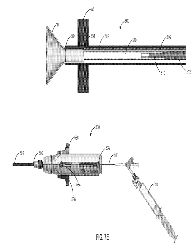
[0088] Referring now to FIGS. 5A and 5B, exemplary delivery device
500 operatively
coupled to handle 530 for delivering interatrial shunt device 10 to the atrial
septum is provided_
As shown in FIG. 5A, delivery device 500 includes sheath 502, a dilator, e.g.,
balloon catheter
510, slidably disposed within the lumen of sheath 502, release knot 516 for
releasably coupling
to shunt 10 at knot connection 518 within the lumen of sheath 502, and pusher
520 slidably
disposed within the lumen of sheath 502. For example, release knot 516 may be
a Dyneema
wire/cord. The lumen of sheath 502 may be sized and shaped to receive shunt 10
in its collapsed
delivery state. Distal region 504 of sheath 502 may be linear or may have
geometry that
facilitates a step-free transition between distal region 504 and balloon 512
to form a smooth and
continuous dilator when balloon 512 is in its expanded state, as described in
further detail below.
For example, distal region 504 of sheath 502 may be curved radially inward to
engage with the
outer surface of balloon 512. Moreover, delivery device 500 may include
guidewire lumen 514
extending through balloon catheter 510, sized and shaped to receive a
guidewire therethrough.
[0089] Each of sheath 502, balloon catheter 510, release knot 516,
and pusher 520 may be
operatively coupled to handle 530, such that they are all independently
actuatable relative to each
other. For example, as shown in FIG. 5B, proximal region 506 of sheath 502 may
be coupled to
handle 530, balloon catheter 510 may be operatively coupled to actuator 532 of
handle 530
which may be actuated to move balloon catheter 510 axially relative to sheath
502, pusher 520
may be operatively coupled to actuator 534 of handle 530 which may be actuated
to move pusher
520 axially relative to sheath 502, a first end of release knot 516 may be
operatively coupled to
CA 03237108 2024- 5-2
WO 2023/079498
PCT/1B2022/060621
28
release actuator 536 of handle 530 which may be actuated to disassemble knot
connection 518
and disengage release knot 516 from shunt 10, and a second end of release knot
516 may be
operatively coupled to retrieval actuator 538 of handle 530 which may be
actuated to retract
release knot 516, and accordingly shunt 10 via knot connection 518, within the
lumen of sheath
502_ In some embodiments, to prevent accidental disengagement between release
knot 516 and
shunt 10, release actuator 536 may include lock 537 which may be actuated to
transition between
a locked configuration where release actuator 536 may not be actuated relative
to handle 530,
and an unlocked configuration where release actuator 536 may be actuated and
moved along
handle 530. Moreover, to prevent accidently half-way retrieval of shunt 10,
retrieval actuator
538 may include lock 539 which may be actuated to transition between a locked
configuration
where retrieval actuator 538 may not be actuated relative to handle 530, and
an unlocked
configuration where retrieval actuator 538 may be actuated and moved along
handle 530.
[0090] Referring again to FIG. 5A, balloon catheter 510 may include
inflatable balloon 512
disposed at its distal region. Balloon 512 is configured to transition between
a deflated,
compressed state and an inflated, expanded state. Accordingly, balloon
catheter 510 may include
a fluid lumen fluidically coupled to a fluid source for introducing fluid to
balloon 512 to
inflate/deflate balloon 512. In addition, balloon 512 may be formed having a
tapered cone shape
at its distal end. Balloon 512 may have a symmetric profile, such that both
its proximal and
distal ends have a tapered cone shape when balloon 512 is in its expanded
state. When balloon
512 is positioned adjacent to distal region 504 of sheath 502, in its expanded
state, the outer
surface of balloon 512 may form a step-free transition between distal region
504 and balloon 512
to form a smooth and continuous dilator.
[0091] Moreover, pusher 520 may be a multi-lumen catheter slidably
disposed within the
lumen of sheath 502, having a distal end configured to engage with shunt 10 in
its collapsed
delivery state within the lumen of sheath 502, e.g., via actuation of actuator
534. For example,
pusher 520 may have a first lumen sized and shaped to slidably receive balloon
catheter 510
(including balloon 512 in its collapsed state) therethrough, and one or more
lumens sized and
shaped to slidably receive one or both ends of release knot 516 therethrough.
For example,
pusher 520 may have a single lumen sized and shaped to slidably receive both
ends of release
knot 516 therethrough, or alternatively, pusher 520 may have one lumen sized
and shaped to
CA 03237108 2024- 5-2
WO 2023/079498
PCT/1B2022/060621
29
slidably receive a first end of release knot 516 operatively coupled to
release actuator 536
therethrough, and another lumen sized and shaped to slidably receive a second
end of release
knot 516 operatively coupled to retrieval actuator 538 therethrough. The lumen
of pusher 520
that slidably receives balloon catheter 510 may be coaxial with the
longitudinal axis of sheath
502, and the one or more lumens that slidably receive the ends of release knot
516 may not he
coaxial with the longitudinal axis of sheath 502. Accordingly, pusher 520 may
be advanced
distally relative to sheath 502 and balloon catheter 510 via actuation of
actuator 534 to push
shunt 10 distally through the lumen of sheath 502 until at least the distal
portion of shunt 10 is
exposed beyond distal region 504 and transitions to its expanded deployed
state, e.g., within the
left atrium.
[0092] As described above, release knot 516 may be releasably
coupled to shunt 10 in its
collapsed delivery state within the lumen of sheath 502 via knot connection
518. FIG. 6A
illustrates an exemplary knot mechanism for releasably coupling release knot
516 to shunt 10.
As shown in FIG. 6A, release knot 516 may be formed of a single force
transmission element,
e.g., a Dyneema wire/cord, having a release wire portion, e.g., release end
517, knot connection
518, and a standing portion, e.g., retrieval end 519. For example, release
knot 516 may be tied to
shunt 10, e.g., at the proximal portion of shunt 10, to form knot connection
518, such that release
end 517 and retrieval end 519 extends therefrom. Knot connection 518 may be a
knot such as a
painters hitch or Quick Tie and Release (QTaR) hitch, such that application of
a retraction force
to release end 517, e.g., via release actuator 536, causes release end 517 to
pull knot connection
518 in a manner that causes knot connection 518 to disassemble and disengage
release knot 516
from shunt 10, whereas application of a retraction force to retrieval end 519,
e.g., via retrieval
actuator 538, causes retrieval end 519 to pull knot connection 518 to thereby
pull shunt 10 within
the lumen of sheath 502. Accordingly, release knot 516 may have a
predetermined amount of
excess length, e.g., slack, disposed within device 800, e.g., within sheath
502/pusher 520 distal to
handle 520, such that when shunt 10 is moved distally within sheath 502 via
pusher 520, release
knot 516, which is coupled to shunt 10 via knot connection 518, also may move
distally within
sheath 502 without applying force to release actuator 536 or retrieval
actuator 538. Thus,
actuation of release actuator 536 and/or retrieval actuator 538 may not
disengage or halfway
retrieve shunt 10 until shunt 10 is halfway deployed from sheath 502, e.g.,
when the slack of
release knot 516 is removed.
CA 03237108 2024- 5-2
WO 2023/079498
PCT/1132022/060621
[0093] As will be understood by a person having ordinary skill in
the art, the knot
configuration illustrated in FIG. 6A is one example of numerous knot
configurations suitable for
use with the delivery devices described herein, in accordance with the
principles of the present
disclosure. For example, FIG. 6B illustrates another exemplary release knot
516' having a
release wire portion, e.g., release end 517', knot connection 518', and a
standing portion, e.g.,
retrieval end 519'. Knot connection 518' may have a configuration similar to
that of knot
connection 518, except that knot connection 518' includes an additional loop
to provide
additional securement between release knot 516' and the shunt.
[0094] Moreover, the release knots described herein may be used to
facilitate transitioning of
shunt 10 from its expanded deployed state toward its collapsed delivery state,
for example, as
described in U.S. Patent No. 10,940,296 to Keren, assigned to the assignee of
the present
invention, the entire contents of which are incorporated herein by reference.
For example, in
some embodiments, the release knot may be coupled to a proximal portion of
shunt 10 and both
the retrieval and release ends may be woven through two or more loops of the
proximal end of
shunt 10, e.g., at points evenly spaced around the circumference of the
proximal end of shunt 10,
such that retraction of the retrieval end of the release knot, e.g., through
the lumen of pusher 520,
applies an inward force to the proximal portion of shunt 10 to thereby
collapse the proximal
portion of shunt 10 radially inward to its collapsed delivery state; whereas,
retraction of the
release end of the release knot causes the knot connection to disassemble and
disengages the
release knot from shunt 10. Accordingly, the release knot may remain coupled
to shunt 10 when
shunt 10 is fully deployed at the atrial septum, e.g., by having enough slack
within delivery
device 500 distal to handle 530. Once shunt 10 is satisfactorily deployed, the
release knot may
be disengaged from shunt 10 by retraction of the release end of the release
knot, and the entire
release knot, including both the release and retrieval ends, may he pulled
back into delivery
sheath 502. If the deployment of shunt 10 is unsatisfactory, the retrieval end
of the release knot
may be retracted proximally to transition the proximal portion of shunt 10
toward its collapsed
delivery state, and shunt 10 may be further retracted back into the lumen of
sheath 502 via
retraction of the retrieval end of the release knot for redeployment or
removal.
[0095] Additionally or alternatively, the release knot may be
coupled to a middle portion of
shunt 10, e.g., the neck region between the proximal and distal flared end
regions of shunt 10,
CA 03237108 2024- 5- 2
WO 2023/079498
PCT/1B2022/060621
31
and both the retrieval and release ends may be looped around the outer surface
of the middle
portion of shunt 10 toward the knot connection. Once shunt 10 is
satisfactorily deployed, the
release knot may be disengaged from shunt 10 by retraction of the release end
of the release
knot, and the entire release knot, including both the release and retrieval
ends, may be pulled
hack into delivery sheath 502_ If the deployment of shunt 10 is
unsatisfactory, shunt 10 may be
retrieved back into delivery sheath 502 by retraction of the retrieval end of
the release knot
proximally, e.g., through the lumen of pusher 520, which applies an inward
force to the middle
portion of shunt 10 to thereby collapse the middle portion of shunt 10
radially inward toward its
collapsed delivery state. Collapsing the middle portion of shunt 10 also may
cause the proximal
portion of shunt 10 to at least partially collapse, such that shunt 10 may
then be further retracted
back into the lumen of sheath 502 via retraction of the retrieval end of the
release knot for
redeployment or removal.
[0096] Alternatively, the release knot may be coupled to a proximal
portion of shunt 10 and
both the retrieval and release ends may be passed through an initial loop at
the proximal end of
shunt 10, then through the central passageway of shunt 10, and out of the
central passageway and
looped around the outer surface of the middle portion of shunt 10, and back
towards the knot
connection, such that retraction of the retrieval end of the release knot,
e.g., through the lumen of
pusher 520, applies an inward force to the middle portion of shunt 10 to
thereby collapse the
middle portion of shunt 10 radially inward toward its collapsed delivery
state; whereas, retraction
of the release end of the release knot causes the knot connection to
disassemble and disengages
the release knot from shunt 10. As will be understood by a person having
ordinary skill in the
art, more than one release knot may be coupled to the shunt, e.g., at
locations evenly spaced
around the circumference of the shunt, to facilitate transitioning of shunt 10
toward its collapsed
delivery state for full and/or halfway retrieval.
[0097] Referring now to FIGS. 7A to 7H, exemplary method steps for
delivering shunt 10 to
an implantation site within the atrial septum via delivery device 500 are
provided. As shown in
FIG. 7A, fluid source 540, e.g., a syringe pump, fluidically coupled to
balloon 512 via fluid
lumen 511 of balloon catheter 510 may be actuated to move a fluid into balloon
512 to thereby
inflate balloon 512 from its deflated collapsed state to its inflated expanded
state, such that in its
expanded state, an outer surface of balloon 512 engages with the inner surface
of the lumen of
CA 03237108 2024- 5-2
WO 2023/079498
PCT/1B2022/060621
32
sheath 502 at distal region 504 of sheath 502, thereby forming a smooth and
continuous dilator
for delivering delivery device 500 to the atrial septum.
[0098] Device 500 with balloon 512 in its expanded state at distal
region 504 of sheath 502
may be advanced over guidewire 101 via guidewire lumen 514 until the distal
portion of balloon
512 comes into contact with the puncture of atrial septum AS. Device 500 may
be further
advanced such that balloon 512 enlarges/dilates the puncture of atrial septum
AS as the tissue
surrounding the puncture is smoothly guided over the distal portion of balloon
512, followed by
distal region 504 of sheath 502, as shown in FIG. 7B. Under visualization
methods such as
fluoroscopy and/or ultrasound imaging such as trans-esophageal echo (TEE) or
intracardiac echo
(ICE), the target position of device 500 relative to atrial septum AS may be
verified, e.g., via a
radiopaque marker on sheath 502. Preferably, device 500 is positioned relative
to atrial septum
AS such that distal region 504 is spaced at least a predetermined distance
from atrial septum AS
within the left atrium to ensure full deployment of the distal portion of
shunt 10 within the left
atrium, as described in further detail below.
[0099] Next, as shown in FIG. 7C, balloon 512 may be deflated via
fluid source 540, e.g., by
actuating the syringe pump to withdraw fluid from balloon 512, such that
balloon 512 transitions
to its deflated, collapsed state. In some embodiments, shunt 10 may be
maintained stationary
relative to the atrial septum using devices within sheath 502 such as those
described in
W02020202046. As shown in FIG. 7D, actuator 532 may then be actuated, e.g.,
moved from a
first position on handle 530 proximally along handle 530 to a second position
on handle 530, to
thereby retract balloon catheter 510 and balloon 512 in its deflated state
proximally relative to
sheath 502. For example, balloon catheter 510 may be retracted proximally
through shunt 10 in
its collapsed, delivery state within sheath 502, until balloon 512 is disposed
within the lumen of
pusher 520, as shown in FIG. 7D. Accordingly, the lumen of sheath 502 may be
unobstructed
between shunt 10 and distal region 504.
[0100] Next, actuator 534 may be actuated, e.g., moved from a first
position on handle 530
distally along handle 530 to a second position on handle 530, to thereby move
pusher 520, and
accordingly shunt 10, distally relative to sheath 502 until the distal portion
of shunt 10 is exposed
beyond distal region 504 of sheath 502 and deploys within the left atrium, as
shown in FIG. 7E.
CA 03237108 2024- 5-2
WO 2023/079498
PCT/1B2022/060621
33
For example, as pusher 520 is advanced distally within the lumen of sheath
502, the distal end of
pusher 520 engages with the proximal end of shunt 10 in its collapsed delivery
state, and pushes
shunt 10 distally through the lumen of sheath 502. As described above, distal
region 504 may be
spaced from atrial septum AS at least a predetermined distance such that the
distal portion of
shunt 10 may fully deploy within the left atrium. Accordingly, device 500 may
then he retracted
relative to atrial septum AS, e.g., by moving handle 530 proximally, until the
deployed distal
portion of shunt 10 contacts atrial septum AS, as shown in FIG. 7F. The
desired position of
shunt 10 relative to atrial septum AS may be observed by the physician, e.g.,
via force feedback
applied to device 500 via atrial septum AS, and/or may be visually verified
via, e.g., fluoroscopy
and/or ultrasound imaging such as trans-esophageal echo (TEE) or intracardiac
echo (ICE).
[0101] As shown in FIG. 7G, release actuator 536 may be actuated,
e.g., moved from a first
position on handle 530 proximally along handle 530 to a second position on
handle 530, to
thereby retract release end 517 of release knot 516, which causes knot
connection 518 to
disassemble and disengage release knot 516 from shunt 10 within sheath 502. As
described
above, in some embodiments, lock 537 of release actuator 536 may be required
to transition from
its locked configuration to its unlocked configuration prior to moving release
actuator 536 from
the first position to the second position. Next, when shunt 10 is disengaged
from release knot
516, device 500 may be further retracted proximally relative to atrial septum
AS, e.g., by moving
handle 530 proximally, such atrial septum AS applies a force to the deployed
distal portion of
shunt 10 to maintain shunt 10 in position relative to atrial septum AS as
device 500 is retracted
proximally until the proximal portion of shunt 10 is exposed beyond distal
region 504 of sheath
502 and deploys within the right atrium, thus completing full deployment of
the shunt, as shown
in FIG. 7H.
[0102] After shunt 10 is fully deployed at atrial septum AS, device
500 may be removed
from the patient, leaving shunt 10 implanted at the atrial septum. In some
embodiments, prior to
removal, actuator 532 may be actuated to move balloon catheter 510 distally
within sheath 502,
such that balloon 512 is positioned within sheath 502 adjacent to distal
region 504, and balloon
512 may be inflated to its expanded state, e.g., via fluid source 540, to form
a continuous dilator
with distal region 504, as described above.
CA 03237108 2024- 5-2
WO 2023/079498
PCT/1132022/060621
34
[0103] Referring now to FIGS. 71 to 7K, exemplary method steps for
halfway retrieval of
shunt 10 during delivery of shunt 10 at atrial septum AS are provided. For
example, after the
distal portion of shunt 10 is deployed, e.g., within the left atrium as shown
in FIG. 7E or in
another improper location, it may be desirable to transition shunt 10 back to
its collapsed
delivery state within sheath 502 and retrieve shunt 10_ Accordingly, as shown
in FIG. 71,
actuator 534 may be actuated, e.g., moved from the second position on handle
530 proximally
along handle 530 to the first position on handle 530, to thereby move pusher
520 proximally
relative to sheath 502, and provide an unobstructed pathway within the lumen
of sheath 502 for
shunt 10 to be disposed therein in its collapsed delivery state. Next,
retrieval actuator 538 may
be actuated, e.g., moved from a first position on handle 530 proximally along
handle 530 to a
second position on handle 530, to thereby retract retrieval end 519 of release
knot 516, which
pulls shunt 10 proximally via knot connection 518 within the lumen of sheath
502, as shown in
FIG. 7J. As shunt 10 is pulled proximally within sheath 502, distal region 504
of sheath 502
applies a force against the deployed distal portion of shunt 10, which causes
the distal portion to
transition to its collapsed delivery state within the lumen of sheath 502. As
described above, in
some embodiments, lock 539 of retrieval actuator 538 may be required to
transition from its
locked configuration to its unlocked configuration prior to moving retrieval
actuator 538 from
the first position to the second position.
[0104] When shunt 10 is completely in its collapsed delivery state
within sheath 502, device
500 may be removed from the patient, e.g., by moving handle 530 proximally, as
shown in FIG.
7K. Alternatively, device 500 may be repositioned relative to atrial septum
AS, such that shunt
may be implanted atrial septum AS in accordance with the delivery methods
described above
with regard to FIGS. 7E to 7H.
[0105] Alternatively, as described above, release knot 516 may
remain coupled to shunt 10
during full deployment of shunt 10 at atrial septum AS. Accordingly, release
actuator 536 may
not be actuated prior to retracting device 500 proximally relative to atrial
septum AS to thereby
complete full deployment of the shunt by deploying the proximal portion of
shunt 10 within the
right atrium. In this embodiment, upon satisfactory full deployment of shunt
10 at atrial septum
AS, release actuator 536 may then be actuated to disassemble knot connection
518 and disengage
release knot 516 from shunt 10. If deployment is unsatisfactory, retrieval end
519 may be
CA 03237108 2024- 5-2
WO 2023/079498
PCT/1B2022/060621
retracted proximally, e.g., via actuation of retrieval actuator 538, to
facilitate transition of the
proximal portion and/or the middle portion of shunt 10 toward the collapsed
delivery state, as
described above, such that shunt 10 may be retracted back into the lumen of
sheath 502 for
redeployment or removal.
[0106] Referring now to FIGS. 8A to 81, exemplary method steps for
delivering interatrial
shunt device 10 to the atrial septum via exemplary delivery device 800
operatively coupled to
handle 830 are provided. Device 800 may be constructed similar to delivery
device 500. For
example, sheath 802 having distal region 804 and proximal region 806
corresponds to sheath 502
having distal region 504 and proximal region 506, balloon catheter 810 having
inflatable balloon
812 and fluid lumen 811 corresponds to balloon catheter 510 having inflatable
balloon 512 and
fluid lumen 511, pusher 820 corresponds with pusher 520, and release knot 816
having release
end 817, knot connection 818, and retrieval end 819 corresponds with release
knot 516 having
release end 517, knot connection 518, and retrieval end 519. Device 800
differs from delivery
device 500 in that, rather than extending through the length of pusher 820
from knot connection
818 to a retrieval actuator of handle 830, retrieval end 819 may be coupled to
the distal portion
of pusher 820. Accordingly, handle 830, which may be constructed similar to
handle 530 such
that handle 830 is coupled to proximal region 806 of sheath 802, balloon
catheter actuator 832
corresponds to balloon catheter actuator 532, pusher actuator 834 corresponds
to pusher actuator
534, release actuator 836 corresponds to release actuator 536, and fluid
source 840 corresponds
to fluid source 540, does not need a separate retrieval actuator. For example,
actuation of
actuator 832, e.g., moving actuator 832 proximally along handle 830, causes
movement of
pusher 820, and accordingly shunt 10 and retrieval end 819 coupled thereto,
proximally within
the lumen of sheath 802. Moreover, pusher 820 does not have a separate lumen
for receiving
retrieval end 819 therethrough.
[0107] Like release end 517, as described above, release end 817
may have slack within
device 800, such that actuation of release actuator 836 may not cause
disassembly of knot
connection 818 until shunt 10 is halfway deployed from sheath 802.
Alternatively, in some
embodiments, release actuator 836 may be releasably coupled to pusher actuator
834, such that
release actuator 836 moves along with pusher actuator 834 when pusher actuator
834 is actuated
to move pusher 820, and accordingly shunt 10, distally within sheath 802.
Accordingly, release
CA 03237108 2024- 5-2
WO 2023/079498
PCT/1B2022/060621
36
end 817 may not have slack within device 800 as release end 817 moves distally
along with
pusher 820 via movement of release actuator 836. Release actuator 836 may be
decoupled from
pusher actuator 834, e.g., via lock 837 or another locking mechanism coupling
release actuator
836 and pusher actuator 834, and then independently actuated to pull release
end 817 to
disassemble knot connection 818 and disengage release knot 816 from shunt 10,
as described
above.
[0108] In some embodiments, release actuator 836 may include a rope
clutch mechanism
through which release end 817 may be passed through. For example, the rope
clutch mechanism
may be in an open state during actuation of pusher actuator 834, such that
release end 817 moves
through the rope clutch mechanism as pusher 820, and accordingly retrieval end
819 coupled
thereto, are moved distally through sheath 802. If halfway retrieval of shunt
10 is desirable, as
described above, pusher actuator 834 may be actuated to retract pusher 820,
and accordingly
shunt 10 via knot connection 818, within sheath 802. To disengage release knot
816 from shunt
10, the rope clutch mechanism may be transitioned to a closed state, e.g., via
lock 837 or another
closing mechanism operatively coupled to the rope clutch mechanism, to fix
release end 817 to
release actuator 836, such that actuation of release actuator 836 pulls on
release end 817 to
disassemble knot connection 818.
[0109] Like the method steps for delivery shunt 10 via device 500
described above with
regard to FIGS. 7A-7H, as shown in FIG. 8A, device 800 with balloon 812 in an
inflated
expanded state adjacent distal region 804 of sheath 802 may be delivered
through a hole in atrial
septum AS, such that balloon 812 enlarges/dilates the puncture of atrial
septum AS as the tissue
surrounding the puncture is smoothly guided over the distal portion of balloon
812, followed by
distal region 804 of sheath 802. Further, balloon 812 may be deflated, as
shown in FIG. 8B, and
retracted proximally within a lumen of pusher 820 via actuator 832, as shown
in FIG. 8C. As
shown in FIG. 8D, pusher 820 may be advanced distally within sheath 802 via
actuator 834, such
that the distal end of pusher 820 engages with the proximal end of shunt 10
and moves shunt 10
distally within sheath 802 until the distal portion of shunt 10 extends beyond
distal region 804
and deploys within the left atrium.
[0110] As shown in FIG. 8E, device 800 may then be moved
proximally, e.g., by moving
CA 03237108 2024- 5-2
WO 2023/079498
PCT/1B2022/060621
37
handle 830 proximally, until the distal portion of shunt 10 contacts atrial
septum AS. Next,
release end 817 may be pulled proximally via release actuator 836 to
disassemble knot
connection 818 and disengage release knot 818 from shunt 10, while retrieval
end 819 remains
coupled to the distal portion of pusher 820, as shown in FIG. 8F. In some
embodiments, lock
837 of release actuator 836 may he required to transition from its locked
configuration to its
unlocked configuration prior to actuating release actuator 836. As shown in
FIG. 8G, device 800
may be retracted proximally while atrial septum AS maintains shunt 10 in place
until the
proximal portion of shunt 10 is exposed from sheath 802 and deploys within the
right atrium.
Delivery device 800 may then be removed from the patient, leaving shunt 10
implanted. As
described above, balloon 81 may be re-inflated adjacent to distal region 804
prior to removal of
device 800 from the patient.
[0111] As described above, after the distal portion of shunt 10 is
deployed, e.g., within the
left atrium as shown in FIG. 8D or in another improper location, it may be
desirable to transition
shunt 10 back to its collapsed delivery state within sheath 802 and retrieve
shunt 10.
Accordingly, as shown in FIG. 8H, actuator 832 may be actuated, e.g., moved
proximally along
handle 530, to thereby move pusher 820 proximally within the lumen of sheath
802, which pulls
retrieval end 819 and shunt 10 proximally via knot connection 818 within the
lumen of sheath
802 until the distal portion of shunt 10 transitions to its collapsed delivery
state within sheath
802. Device 800 may then be removed from the patient, as shown in FIG. 81, or
alternatively,
device 800 may be repositioned relative to atrial septum AS, such that shunt
10 may be
implanted atrial septum AS in accordance with the delivery methods described
above with regard
to FIGS. 8D to 8G.
[0112] While various illustrative embodiments of the invention are
described above, it will
be apparent to one skilled in the art that various changes and modifications
may be made therein
without departing from the invention. The appended claims are intended to
cover all such
changes and modifications that fall within the true scope of the invention.
CA 03237108 2024- 5-2