Note: Descriptions are shown in the official language in which they were submitted.
CA 02337830 2001-O1-16
WO 00/05739 PCT/US99/16412
-1-
ELECTRO-OPTICAL MECHANICAL INSTRLfMENT
BACKGROUND OF THE INVENTION
The present invention relates to optical scanners, and more particularly
to a quasi con-focal microscope scanner in which the specimen and the scanner
are simultaneously moved relative to each other.
Micro array biochips are currently being developed by several
biotechnology companies. Micro array biochips are small substrates
containing thousands of DNA sequences that represent the genetic codes of a
variety of living organisms including human, plant, animal, and pathogens.
They provide researchers with volumes of information in a more efficient
format. Experiments can be conducted with significantly higher throughput
than previous technologies offered. Biochip technology is used for genetic
expression, DNA sequencing of genes, food and water testing for harmful
pathogens, and diagnostic screening. Biochips may be used in
pharmacogenomics and proteomics research aimed at high throughput
screening for drug discovery. High-speed automated biochemistry may lead
to drugs for treating illnesses including HIV, cancer, heart disease and
others.
DNA sequences are extracted from a sample and are tagged with a
fluorescent probe, a molecule that, when "excited" by a laser, will emit light
of various colors. These fluorescently tagged DNA sequences are then spread
over the chip. A DNA sequence will bind to its complementary (cDNA)
sequence at a given array location. A typical biochip contains a two
dimensional array of thousands of cDNA sequences, each one unique to a
specific gene. These cDNA sequences may be "printed" on the chip in several
ways. Once the biochip is printed, it represents thousands of experiments in
an area usually smaller than a postage stamp.
The chip is then ready to be scanned and analyzed with a scanning laser
microscope using a dichromic beam splitter. However. The dichromic beam
sputter has two drawbacks. Each time a specimen with a different dye is to
be read, the beam splitter must be changed to match the different wavelengths
CA 02337830 2001-O1-16
WO 00/05739 PCT/US99/16412
-2-
of operation of the new dye and the number of multiple dyes that can be
simultaneously interrogated is usually limited to two.
The microscope collects data from successive "pixels" which are best
dimensioned in microns. There are essentially two types of optical scanner,
namely scanners that move scan heads and associated optics over stationary
specimens, and scanners that move the specimens relative to stationary optics.
Known scanning microscopes must therefore precisely align the optics of a
moving scan head with the beam of a stationary laser, or alternatively carry
the laser on the moving scan. A stationary laser can be aligned with a moving
scan head only at relatively slow speeds, and therefore the scan speed of the
system is inherently limited. The alternate system requires a relatively large
scan head to carry the associated optics whereby the relatively great size and
weight also effectively limits the scan speed.
SUMMARY OF THE INVENTION
The present invention provides a scanning laser microscope which can
be used to scan biochips and display the information embodied in the
fluorescent energy emitted by the individual dots as a pictorial
representation
of the array on a T.V. monitor. The means of interrogation is laser light
(the excitation energy). The laser light excites the fluorescein that is
contained
in the fluorescent dyes. The fluorophores will subsequently emit light of a
wavelength that is longer than the wavelength of the excitation energy. Thus,
by using a beam splitting minor, the number of different dyes that can be
interrogated simultaneously is unlimited.
The optical diagram is a quasi con-focal microscope, i. e. , only an area
the size of approximately one pixel is illuminated (excited) and observed
(detected) at a time, however the size of the illuminating spot is not nearly
as
closely matched to that of the detected spot as it is in a pure con-focal
microscope, in fact the former is about lOX larger in diameter than the
latter.
CA 02337830 2001-O1-16
WO 00/05739 PCT/US99116412
-3-
The emitted light is conducted by lenses, mirrors, and optical filters to a
detector, where it is converted into computer readable data.
The horizontal, or line scan (the X scan) is mechanized by moving the
objective lens of the system rapidly back and forth in the X direction across
the shorter length of a microscope slide specimen collecting data in each
direction. The slide specimen does not move in the X direction as the
vertical,
or page scan (the Y scan) is mechanized by moving the slide specimen in the
Y direction, incrementally advancing the slide each time the X scan is about
to start a line.
The information is preferably processed so that it may be displayed in
a convenient format such as tables, histograms and the like. The pictorial or
image-processed information can thereafter be stored on a hard drive and sent
to a~hard copy printer, transmitted to a LAN, or transmitted over the
Internet.
BRIEF DESCRIPTION OF THE DRAWINGS
Other advantages of the present invention will be readily appreciated
as the same becomes better understood by reference to the following detailed
description when considered in connection with the accompanying drawings
wherein:
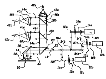
Figure 1 is a detailed perspective view of an optical instrument of the
present invention;
Figure 2 is a plan view of a slide specimen of the present invention
showing the movement of the scanning objective lens;
Figure 3 is a side view of the first drive mechanism; and
Figure 4 is a top view of the second drive mechanism.
DETAILED DESCRIPTION OF THE PREFERRED EMBODIMENT
An optical instrument 10 of the present invention is generally shown
in Figure 1. As will be further described below the optical instrument 10
CA 02337830 2001-O1-16
WO 00/05739 PCT/US99116412
-4-
generally includes a transmitter 12 that emits an optical signal 14, a beam
splitting mirror 20 having an opening 22, a reflector assembly 30 which
directs the optical signal 14 onto a specimen 90, a detector assembly 40 which
detects a reflected optical signal 44 from the specimen 90, a first drive
mechanism 50 for varying the position of the optical signal 14 on the specimen
90, and a second drive mechanism 66 for varying the position of the specimen
90 relative to the optical signal 14.
Figure 1 illustrates the main components of the optical instrument 10
and the optical signal 14 path. The means of interrogation is preferably laser
light and more than one laser can be incorporated into the system. Further,
various types of lasers may be employed, such as argon-ion, semiconductor
diode, and other similar solid state lasers. In the preferred embodiment, a
plurality of lasers I2A-C, each operating on a different wavelength, are
shown.
The optical signals 14A-C are each first transmitted through a beam
correcting lens 16A-C and then through a continuously variable neutral
density filter 18A-C, which is employed to adjust the intensity of the beam.
The variable neutral density filter 18A-C can be an addressable array of
several fixed neutral density filters of different densities, a pair of
polarizers
of which one is rotatable, or a rotating polarization retarder, in front of a
polarizer.
To direct the optical signal 14, the reflector assembly 30 includes a
plurality of turn mirrors 32A-C. Each optical signal 14A-C is folded as
appropriate by the turn minors 32A-C to a beam combiner 34A-C. The beam
combiner is preferably a know dichroic filter which transmits light of one
wavelength while blocking others. The individual optical signal are thereby
collected into a combined beam along a first path which then passes through
the opening 22 in the beam splitting mirror 20. The combined beam is then
directed to a 90 degree fold mirror 36 located immediately above the scanning
CA 02337830 2001-O1-16
WO 00/05739 PCT/US99/16412
-5-
objective lens 52. The fold mirror 36 reflects the combined optical signal 14
into the scanning objective lens 52, which in turn is focused onto the
specimen
90, thus creating a scanning illumination spot. The embodiment shown in
Figure 1 shows three laser transmitters 12A-C, however, those skilled in the
art will realize that additional lasers can be used to interrogate multiple
dyes
of different fluorescent properties in the specimen 90 simultaneously or
sequentially. The optical signal from each additional laser is located
downstream from the last laser and is brought onto the system optical axis via
reflection off a dichromic beam combiner.
When illuminated by the combined beam the fluorophores will emit
energy all around, i. e. , into 4 pi directions. It is imperative to collect
as much
of this energy as possible, so it is preferable to employ a custom designed
objective lens 52 such as one, for example only, with an NA=0.9, air-
coupled 4. The objective lens 52 preferably outputs a beam of emitted energy
concentric with the laser beam, having a diameter about lOX larger than that
of the laser beam.
After reflecting from the specimen the fold mirror 36 located above the
scanning lens 52 will fold the reflected optical signal 44 along a second
path.
The reflected optical signal 44 is again directed by 90 degrees towards the
beam splitting minor 20. The latter will fold the emission beam 90 degrees
away from the combined optical signal first path, except for a very small
central portion in the middle as determined by the opening 22 in the beam
splitting minor 20. It can be seen that a for a portion of the path the
original
combined optical signal 14 traveling along the first path, and the reflected
optical signal 44 traveling along the second path, have a common path
segment. This common path segment is shown between the beam splitting
minor 20 the fold minor 36, and the scanning lens 52.
The reflected optical signal 44 reflecting from the opposite side of the
beam splitting mirror 20 will then pass through a plurality of beam splitters
CA 02337830 2001-O1-16
WO 00/05739 PCT/US99/16412
_(r
38A-B to separate the combined signal into individual signals 44A-C. Each
individual signal 44A-C passes through an emission filter 46A-C, and will then
be focused by a detector lens 48A-C into a pinhole. The pinhole acts as the
field stop of the system, i. e. , it defines the size of the scanning
detection
aperture on the slide. Finally, the individual signals 46A-C diverts once
through the pinhole until it impinges onto a detector 42A-C.
As shown in Figure 2 the horizontal, or line scan (the X scan) is
mechanized by moving the objective lens 52 of the system rapidly (20 Hz or
so) back and forth in the X direction across the shorter length of a
microscope
slide specimen 90 (commonly 1 inch wide), collecting data in each direction.
Note that the slide specimen 90 does not move in the X direction. The
vertical or page scan (the Y scan), is mechanized by moving the slide
specimen 90 in the Y direction, incrementally advancing the slide specimen 90
each time the X scan is about to start a line scan as further described below.
Figure 3 shows the first drive mechanism 50 for varying the position
of the combined optical signal on the specimen 90. The first or X scan
mechanism preferably employs a galvanometric torque motor 54 to rotate a
sector-shaped cam 56 over an angle between+40 degrees, and -40 degrees.
The circular portion of the cam 56 is connected to the carriage 58 via a set
of
roll-up, roll-off thin, high-strength steel wires 66A-B. The scanning
objective
lens 52 is attached to the carriage 54. The radius of the cam 56 is such that
its degree rotation will cause the carriage 58 to travel a linear distance
along
a rail 60 commensurate with the length of the X scan pattern of the objective
lens 52.'
Figure 4 shows the second drive mechanism 70. The second or Y scan
mechanism employs a stepper motor 72 to drive a precision screw 74 in a
known manner. The nut 76 on the screw 74 is attached to the carriage 58, so
that any rotation of the screw 74 will cause the carriage 58 to move along a
linear rail 60. The carriage 58 in turn is equipped with a tray 76. The tray
CA 02337830 2001-O1-16
WO 00/05739 PG"T/US99/16412
_7_
76 is equipped with appropriate retainers 78 to hold a specimen slide 90 in a
position and orientation which is repeatable within an accuracy required by
optical focus and alignment criteria. The rail of the linear slide and the
stepper motor 72 are attached to the frame of the Y scan mechanism.
In an alternate embodiment, the carriage S8 is pivotally mounted such
that the carriage 58, and thus the objective lens S2, move in an arcuate
motion. The arcuate motion is thereafter converted into linear motion by
lrnow computer mapping programs.
The frame of the Y scan mechanism is further attached to the carriage
of a verkically oriented linear slide. The rail of the slide is mounted to the
main frame of the reader system. The carriage is supported by a precision
screw, the nut of which is attached to the frame. The screw is turned causing
the Y scan mechanism, and with it the slide holding the specimen, to move
toward or away from the objective lens, thus affecting a focusing sequence.
1S The foregoing description is exemplary rather than limiting in nature.
Obviously, many modifications and variations of the present invention are
possible in light of the above teachings. It is, therefore, to be understood
that
within the scope of the appended claims the invention may be practiced
otherwise than as specifically described.
