Sélection de la langue

Search

Sommaire du brevet 2337830 

Énoncé de désistement de responsabilité concernant l'information provenant de tiers

Une partie des informations de ce site Web a été fournie par des sources externes. Le gouvernement du Canada n'assume aucune responsabilité concernant la précision, l'actualité ou la fiabilité des informations fournies par les sources externes. Les utilisateurs qui désirent employer cette information devraient consulter directement la source des informations. Le contenu fourni par les sources externes n'est pas assujetti aux exigences sur les langues officielles, la protection des renseignements personnels et l'accessibilité.

Disponibilité de l'Abrégé et des Revendications

L'apparition de différences dans le texte et l'image des Revendications et de l'Abrégé dépend du moment auquel le document est publié. Les textes des Revendications et de l'Abrégé sont affichés :

  • lorsque la demande peut être examinée par le public;
  • lorsque le brevet est émis (délivrance).
(12) Demande de brevet: (11) CA 2337830
(54) Titre français: INSTRUMENT MECANIQUE ELECTRO-OPTIQUE
(54) Titre anglais: ELECTRO-OPTICAL MECHANICAL INSTRUMENT
Statut: Réputée abandonnée et au-delà du délai pour le rétablissement - en attente de la réponse à l’avis de communication rejetée
Données bibliographiques
(51) Classification internationale des brevets (CIB):
  • H01J 03/14 (2006.01)
  • G02B 21/00 (2006.01)
(72) Inventeurs :
  • DEWEERD, HERMAN (Etats-Unis d'Amérique)
  • BEACH, MICHAEL (Etats-Unis d'Amérique)
  • HERNANDEZ, JOSE (Etats-Unis d'Amérique)
(73) Titulaires :
  • VIRTEK BIOTECH CANADA, INC.
(71) Demandeurs :
  • VIRTEK BIOTECH CANADA, INC. (Canada)
(74) Agent: GOWLING WLG (CANADA) LLP
(74) Co-agent:
(45) Délivré:
(86) Date de dépôt PCT: 1999-07-20
(87) Mise à la disponibilité du public: 2000-02-03
Requête d'examen: 2004-06-23
Licence disponible: S.O.
Cédé au domaine public: S.O.
(25) Langue des documents déposés: Anglais

Traité de coopération en matière de brevets (PCT): Oui
(86) Numéro de la demande PCT: PCT/US1999/016412
(87) Numéro de publication internationale PCT: US1999016412
(85) Entrée nationale: 2001-01-16

(30) Données de priorité de la demande:
Numéro de la demande Pays / territoire Date
60/093,882 (Etats-Unis d'Amérique) 1998-07-23

Abrégés

Abrégé français

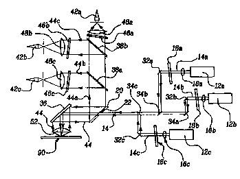
L'invention concerne un microscope laser à balayage que l'on peut utiliser pour scanner des biopuces et qui comprend un émetteur, y compris des lasers (12A-C), qui émet un signal optique (14), un miroir diviseur de faisceau (20) comportant une ouverture (22), un réflecteur (36) qui dirige un signal optique (14) sur un échantillon (90), un ensemble détecteur, y compris les détecteurs (42A-C), qui détecte un signal optique (44) réfléchi depuis l'échantillon (90), un premier mécanisme d'entraînement destiné à modifier la position du signal optique (14) sur l'échantillon (90) et un deuxième mécanisme d'entraînement destiné à modifier la position de l'échantillon (90) par rapport au signal optique (14).


Abrégé anglais


A scanning laser microscope which can be used to scan biochips includes a
transmitter, comprising lasers (12A-C), that emits an optical signal (14), a
beam slitting mirror (20) having an opening (22), a reflector (36) which
directs the optical signal (14) onto a specimen (90), a detector assembly,
including detectors (42A-C), which detects a reflected optical signal (44)
from the specimen (90), a first drive mechanism for varying the position of
the optical signal (14) on the specimen (90) and a second drive mechanism for
varying the position of the specimen (90) relative to the optical signal (14).

Revendications

Note : Les revendications sont présentées dans la langue officielle dans laquelle elles ont été soumises.


-8-
CLAIMS
What is claimed is:
1. An optical instrument comprising:
a transmitter that emits an optical signal;
a reflector assembly which directs said optical signal onto a specimen;
a detector assembly which detects a reflected optical signal from the
specimen;
a first drive mechanism for varying the position of said optical signal
on the specimen; and
a second drive mechanism for varying the position of the specimen
relative to said optical signal.
2. The optical instrument of claim 1, wherein said reflector
assembly directs said optical signal along a first path onto the specimen and
directs said reflected optical signal along a second path to said detector,
said
first path and said second path having a common path segment.
3. The optical instrument of claim 2, further comprising a beam
splitting mirror having an opening, said beam splitting mirror defining one
end
of said common path segment.
4. The optical instrument of claim 3, wherein said first path and
said second path approach said beam splitting mirror from a first direction
and
a second direction respectively.
5. The optical instrument of claim 4, wherein said beam splitting
mirror allows passage of said first path through said opening and reflects
said
second path.

-9-
6. The optical instrument of claim 1, wherein said second drive
mechanism moves the specimen in a linear manner.
7. The optical instrument of claim 1, wherein said second drive
mechanism moves the specimen in an arcuate manner.
8. The optical instrument of claim 1, wherein said first drive
mechanism moves said optical signal substantially perpendicular to movement
of the specimen.
9. The optical instrument of claim 1, wherein said first drive
mechanism includes a scanning lens to focus said optical signal onto the
specimen.
10. The optical instrument of claim 1, wherein said first drive
mechanism includes a galvanometric torque motor.
11. An optical instrument comprising:
a transmitter that emits an optical signal;
a beam splitting mirror having an opening;
a reflector assembly which directs said optical signal along a first path
passing through said opening and onto a specimen;
a detector assembly which detects a reflected optical signal from the
specimen, said reflected optical signal defining a second path directed by
said
beam splitting mirror;
a first drive mechanism including a scanning lens for varying the
position of said optical signal on the specimen; and
a second drive mechanism for varying the position of the specimen
relative to said optical signal.

-10-
12. The optical instrument of claim 11, wherein said transmitter
includes a plurality of lasers having optical signal of different wavelengths.
13. The optical instrument of claim 12, further comprising a
dichroic beam combiner to combine said plurality of optical signals along said
first path.
14. The optical instrument of claim 11, wherein said first drive
mechanism includes:
a galvanometric torque motor having a sector shaped cam;
a carriage;
a retainer to fixedly hold a specimen to said carriage; and
a first and second wire attached between said cam and said carriage,
whereby rotation of said cam is translated into linear movement of the
specimen.
15. The optical instrument of claim 11, wherein said secondt drive
mechanism includes:
a precision stepper motor having a screw;
a carriage having a nut engaging said screw;
a retainer to fixedly hold a specimen to said carriage; and
said stepper motor operable to rotate said screw, whereby rotation of
said screw is translated into linear movement of the specimen.
16. A method of scanning fluorescent samples comprising the steps
of:
(a) exciting the samples with an optical signal of a known first
wavelength;
(b) detecting an optical signal of a second wavelength;

-11-
(c) translating said optical signal in a first and second direction; and
(d) translating the sample in a third direction substantially
perpendicular to said first and said second direction.
17. A method as recited in claim 16, wherein step (a) further
comprises combining a plurality of optical signals prior to exciting of the
sample.
18. A method as recited in claim 16, wherein step (b) further
comprises splitting said optical signal into a plurality of optical signals
prior
to detecting.
19. A method as recited in claim 16, wherein a portion of said
exciting optical signal and a portion of said detecting optical signal have a
common path.

Description

Note : Les descriptions sont présentées dans la langue officielle dans laquelle elles ont été soumises.


CA 02337830 2001-O1-16
WO 00/05739 PCT/US99/16412
-1-
ELECTRO-OPTICAL MECHANICAL INSTRLfMENT
BACKGROUND OF THE INVENTION
The present invention relates to optical scanners, and more particularly
to a quasi con-focal microscope scanner in which the specimen and the scanner
are simultaneously moved relative to each other.
Micro array biochips are currently being developed by several
biotechnology companies. Micro array biochips are small substrates
containing thousands of DNA sequences that represent the genetic codes of a
variety of living organisms including human, plant, animal, and pathogens.
They provide researchers with volumes of information in a more efficient
format. Experiments can be conducted with significantly higher throughput
than previous technologies offered. Biochip technology is used for genetic
expression, DNA sequencing of genes, food and water testing for harmful
pathogens, and diagnostic screening. Biochips may be used in
pharmacogenomics and proteomics research aimed at high throughput
screening for drug discovery. High-speed automated biochemistry may lead
to drugs for treating illnesses including HIV, cancer, heart disease and
others.
DNA sequences are extracted from a sample and are tagged with a
fluorescent probe, a molecule that, when "excited" by a laser, will emit light
of various colors. These fluorescently tagged DNA sequences are then spread
over the chip. A DNA sequence will bind to its complementary (cDNA)
sequence at a given array location. A typical biochip contains a two
dimensional array of thousands of cDNA sequences, each one unique to a
specific gene. These cDNA sequences may be "printed" on the chip in several
ways. Once the biochip is printed, it represents thousands of experiments in
an area usually smaller than a postage stamp.
The chip is then ready to be scanned and analyzed with a scanning laser
microscope using a dichromic beam splitter. However. The dichromic beam
sputter has two drawbacks. Each time a specimen with a different dye is to
be read, the beam splitter must be changed to match the different wavelengths

CA 02337830 2001-O1-16
WO 00/05739 PCT/US99/16412
-2-
of operation of the new dye and the number of multiple dyes that can be
simultaneously interrogated is usually limited to two.
The microscope collects data from successive "pixels" which are best
dimensioned in microns. There are essentially two types of optical scanner,
namely scanners that move scan heads and associated optics over stationary
specimens, and scanners that move the specimens relative to stationary optics.
Known scanning microscopes must therefore precisely align the optics of a
moving scan head with the beam of a stationary laser, or alternatively carry
the laser on the moving scan. A stationary laser can be aligned with a moving
scan head only at relatively slow speeds, and therefore the scan speed of the
system is inherently limited. The alternate system requires a relatively large
scan head to carry the associated optics whereby the relatively great size and
weight also effectively limits the scan speed.
SUMMARY OF THE INVENTION
The present invention provides a scanning laser microscope which can
be used to scan biochips and display the information embodied in the
fluorescent energy emitted by the individual dots as a pictorial
representation
of the array on a T.V. monitor. The means of interrogation is laser light
(the excitation energy). The laser light excites the fluorescein that is
contained
in the fluorescent dyes. The fluorophores will subsequently emit light of a
wavelength that is longer than the wavelength of the excitation energy. Thus,
by using a beam splitting minor, the number of different dyes that can be
interrogated simultaneously is unlimited.
The optical diagram is a quasi con-focal microscope, i. e. , only an area
the size of approximately one pixel is illuminated (excited) and observed
(detected) at a time, however the size of the illuminating spot is not nearly
as
closely matched to that of the detected spot as it is in a pure con-focal
microscope, in fact the former is about lOX larger in diameter than the
latter.

CA 02337830 2001-O1-16
WO 00/05739 PCT/US99116412
-3-
The emitted light is conducted by lenses, mirrors, and optical filters to a
detector, where it is converted into computer readable data.
The horizontal, or line scan (the X scan) is mechanized by moving the
objective lens of the system rapidly back and forth in the X direction across
the shorter length of a microscope slide specimen collecting data in each
direction. The slide specimen does not move in the X direction as the
vertical,
or page scan (the Y scan) is mechanized by moving the slide specimen in the
Y direction, incrementally advancing the slide each time the X scan is about
to start a line.
The information is preferably processed so that it may be displayed in
a convenient format such as tables, histograms and the like. The pictorial or
image-processed information can thereafter be stored on a hard drive and sent
to a~hard copy printer, transmitted to a LAN, or transmitted over the
Internet.
BRIEF DESCRIPTION OF THE DRAWINGS
Other advantages of the present invention will be readily appreciated
as the same becomes better understood by reference to the following detailed
description when considered in connection with the accompanying drawings
wherein:
Figure 1 is a detailed perspective view of an optical instrument of the
present invention;
Figure 2 is a plan view of a slide specimen of the present invention
showing the movement of the scanning objective lens;
Figure 3 is a side view of the first drive mechanism; and
Figure 4 is a top view of the second drive mechanism.
DETAILED DESCRIPTION OF THE PREFERRED EMBODIMENT
An optical instrument 10 of the present invention is generally shown
in Figure 1. As will be further described below the optical instrument 10

CA 02337830 2001-O1-16
WO 00/05739 PCT/US99116412
-4-
generally includes a transmitter 12 that emits an optical signal 14, a beam
splitting mirror 20 having an opening 22, a reflector assembly 30 which
directs the optical signal 14 onto a specimen 90, a detector assembly 40 which
detects a reflected optical signal 44 from the specimen 90, a first drive
mechanism 50 for varying the position of the optical signal 14 on the specimen
90, and a second drive mechanism 66 for varying the position of the specimen
90 relative to the optical signal 14.
Figure 1 illustrates the main components of the optical instrument 10
and the optical signal 14 path. The means of interrogation is preferably laser
light and more than one laser can be incorporated into the system. Further,
various types of lasers may be employed, such as argon-ion, semiconductor
diode, and other similar solid state lasers. In the preferred embodiment, a
plurality of lasers I2A-C, each operating on a different wavelength, are
shown.
The optical signals 14A-C are each first transmitted through a beam
correcting lens 16A-C and then through a continuously variable neutral
density filter 18A-C, which is employed to adjust the intensity of the beam.
The variable neutral density filter 18A-C can be an addressable array of
several fixed neutral density filters of different densities, a pair of
polarizers
of which one is rotatable, or a rotating polarization retarder, in front of a
polarizer.
To direct the optical signal 14, the reflector assembly 30 includes a
plurality of turn mirrors 32A-C. Each optical signal 14A-C is folded as
appropriate by the turn minors 32A-C to a beam combiner 34A-C. The beam
combiner is preferably a know dichroic filter which transmits light of one
wavelength while blocking others. The individual optical signal are thereby
collected into a combined beam along a first path which then passes through
the opening 22 in the beam splitting mirror 20. The combined beam is then
directed to a 90 degree fold mirror 36 located immediately above the scanning

CA 02337830 2001-O1-16
WO 00/05739 PCT/US99/16412
-5-
objective lens 52. The fold mirror 36 reflects the combined optical signal 14
into the scanning objective lens 52, which in turn is focused onto the
specimen
90, thus creating a scanning illumination spot. The embodiment shown in
Figure 1 shows three laser transmitters 12A-C, however, those skilled in the
art will realize that additional lasers can be used to interrogate multiple
dyes
of different fluorescent properties in the specimen 90 simultaneously or
sequentially. The optical signal from each additional laser is located
downstream from the last laser and is brought onto the system optical axis via
reflection off a dichromic beam combiner.
When illuminated by the combined beam the fluorophores will emit
energy all around, i. e. , into 4 pi directions. It is imperative to collect
as much
of this energy as possible, so it is preferable to employ a custom designed
objective lens 52 such as one, for example only, with an NA=0.9, air-
coupled 4. The objective lens 52 preferably outputs a beam of emitted energy
concentric with the laser beam, having a diameter about lOX larger than that
of the laser beam.
After reflecting from the specimen the fold mirror 36 located above the
scanning lens 52 will fold the reflected optical signal 44 along a second
path.
The reflected optical signal 44 is again directed by 90 degrees towards the
beam splitting minor 20. The latter will fold the emission beam 90 degrees
away from the combined optical signal first path, except for a very small
central portion in the middle as determined by the opening 22 in the beam
splitting minor 20. It can be seen that a for a portion of the path the
original
combined optical signal 14 traveling along the first path, and the reflected
optical signal 44 traveling along the second path, have a common path
segment. This common path segment is shown between the beam splitting
minor 20 the fold minor 36, and the scanning lens 52.
The reflected optical signal 44 reflecting from the opposite side of the
beam splitting mirror 20 will then pass through a plurality of beam splitters

CA 02337830 2001-O1-16
WO 00/05739 PCT/US99/16412
_(r
38A-B to separate the combined signal into individual signals 44A-C. Each
individual signal 44A-C passes through an emission filter 46A-C, and will then
be focused by a detector lens 48A-C into a pinhole. The pinhole acts as the
field stop of the system, i. e. , it defines the size of the scanning
detection
aperture on the slide. Finally, the individual signals 46A-C diverts once
through the pinhole until it impinges onto a detector 42A-C.
As shown in Figure 2 the horizontal, or line scan (the X scan) is
mechanized by moving the objective lens 52 of the system rapidly (20 Hz or
so) back and forth in the X direction across the shorter length of a
microscope
slide specimen 90 (commonly 1 inch wide), collecting data in each direction.
Note that the slide specimen 90 does not move in the X direction. The
vertical or page scan (the Y scan), is mechanized by moving the slide
specimen 90 in the Y direction, incrementally advancing the slide specimen 90
each time the X scan is about to start a line scan as further described below.
Figure 3 shows the first drive mechanism 50 for varying the position
of the combined optical signal on the specimen 90. The first or X scan
mechanism preferably employs a galvanometric torque motor 54 to rotate a
sector-shaped cam 56 over an angle between+40 degrees, and -40 degrees.
The circular portion of the cam 56 is connected to the carriage 58 via a set
of
roll-up, roll-off thin, high-strength steel wires 66A-B. The scanning
objective
lens 52 is attached to the carriage 54. The radius of the cam 56 is such that
its degree rotation will cause the carriage 58 to travel a linear distance
along
a rail 60 commensurate with the length of the X scan pattern of the objective
lens 52.'
Figure 4 shows the second drive mechanism 70. The second or Y scan
mechanism employs a stepper motor 72 to drive a precision screw 74 in a
known manner. The nut 76 on the screw 74 is attached to the carriage 58, so
that any rotation of the screw 74 will cause the carriage 58 to move along a
linear rail 60. The carriage 58 in turn is equipped with a tray 76. The tray

CA 02337830 2001-O1-16
WO 00/05739 PG"T/US99/16412
_7_
76 is equipped with appropriate retainers 78 to hold a specimen slide 90 in a
position and orientation which is repeatable within an accuracy required by
optical focus and alignment criteria. The rail of the linear slide and the
stepper motor 72 are attached to the frame of the Y scan mechanism.
In an alternate embodiment, the carriage S8 is pivotally mounted such
that the carriage 58, and thus the objective lens S2, move in an arcuate
motion. The arcuate motion is thereafter converted into linear motion by
lrnow computer mapping programs.
The frame of the Y scan mechanism is further attached to the carriage
of a verkically oriented linear slide. The rail of the slide is mounted to the
main frame of the reader system. The carriage is supported by a precision
screw, the nut of which is attached to the frame. The screw is turned causing
the Y scan mechanism, and with it the slide holding the specimen, to move
toward or away from the objective lens, thus affecting a focusing sequence.
1S The foregoing description is exemplary rather than limiting in nature.
Obviously, many modifications and variations of the present invention are
possible in light of the above teachings. It is, therefore, to be understood
that
within the scope of the appended claims the invention may be practiced
otherwise than as specifically described.

Dessin représentatif
Une figure unique qui représente un dessin illustrant l'invention.
États administratifs

2024-08-01 : Dans le cadre de la transition vers les Brevets de nouvelle génération (BNG), la base de données sur les brevets canadiens (BDBC) contient désormais un Historique d'événement plus détaillé, qui reproduit le Journal des événements de notre nouvelle solution interne.

Veuillez noter que les événements débutant par « Inactive : » se réfèrent à des événements qui ne sont plus utilisés dans notre nouvelle solution interne.

Pour une meilleure compréhension de l'état de la demande ou brevet qui figure sur cette page, la rubrique Mise en garde , et les descriptions de Brevet , Historique d'événement , Taxes périodiques et Historique des paiements devraient être consultées.

Historique d'événement

Description Date
Demande non rétablie avant l'échéance 2006-07-20
Le délai pour l'annulation est expiré 2006-07-20
Inactive : CIB de MCD 2006-03-12
Réputée abandonnée - omission de répondre à un avis sur les taxes pour le maintien en état 2005-07-20
Inactive : Correspondance - Transfert 2005-02-28
Lettre envoyée 2004-07-07
Requête d'examen reçue 2004-06-23
Exigences pour une requête d'examen - jugée conforme 2004-06-23
Toutes les exigences pour l'examen - jugée conforme 2004-06-23
Inactive : Lettre officielle 2004-02-10
Inactive : Transferts multiples 2003-12-31
Inactive : Supprimer l'abandon 2003-10-17
Lettre envoyée 2003-10-02
Inactive : Abandon. - Aucune rép. à lettre officielle 2003-09-08
Inactive : Correspondance - Transfert 2003-07-16
Inactive : Lettre officielle 2003-03-13
Inactive : Transferts multiples 2002-12-09
Lettre envoyée 2002-10-17
Exigences de prorogation de délai pour l'accomplissement d'un acte - jugée conforme 2002-10-17
Inactive : Supprimer l'abandon 2002-10-16
Inactive : Abandon. - Aucune rép. à lettre officielle 2002-09-06
Inactive : Prorogation de délai lié aux transferts 2002-09-05
Inactive : Renseignement demandé pour transfert 2002-06-06
Inactive : Supprimer l'abandon 2002-06-03
Inactive : Abandon. - Aucune rép. à lettre officielle 2002-04-17
Inactive : Transfert individuel 2002-04-15
Inactive : Page couverture publiée 2001-04-20
Inactive : CIB en 1re position 2001-04-10
Inactive : Lettre de courtoisie - Preuve 2001-04-03
Inactive : Notice - Entrée phase nat. - Pas de RE 2001-03-28
Demande reçue - PCT 2001-03-24
Demande publiée (accessible au public) 2000-02-03

Historique d'abandonnement

Date d'abandonnement Raison Date de rétablissement
2005-07-20

Taxes périodiques

Le dernier paiement a été reçu le 2004-07-08

Avis : Si le paiement en totalité n'a pas été reçu au plus tard à la date indiquée, une taxe supplémentaire peut être imposée, soit une des taxes suivantes :

  • taxe de rétablissement ;
  • taxe pour paiement en souffrance ; ou
  • taxe additionnelle pour le renversement d'une péremption réputée.

Les taxes sur les brevets sont ajustées au 1er janvier de chaque année. Les montants ci-dessus sont les montants actuels s'ils sont reçus au plus tard le 31 décembre de l'année en cours.
Veuillez vous référer à la page web des taxes sur les brevets de l'OPIC pour voir tous les montants actuels des taxes.

Historique des taxes

Type de taxes Anniversaire Échéance Date payée
Taxe nationale de base - générale 2001-01-16
Enregistrement d'un document 2001-01-16
TM (demande, 2e anniv.) - générale 02 2001-07-20 2001-06-28
Enregistrement d'un document 2002-04-15
TM (demande, 3e anniv.) - générale 03 2002-07-22 2002-06-18
Prorogation de délai 2002-09-05
Enregistrement d'un document 2002-12-09
TM (demande, 4e anniv.) - générale 04 2003-07-21 2003-07-02
Requête d'examen - générale 2004-06-23
TM (demande, 5e anniv.) - générale 05 2004-07-20 2004-07-08
Titulaires au dossier

Les titulaires actuels et antérieures au dossier sont affichés en ordre alphabétique.

Titulaires actuels au dossier
VIRTEK BIOTECH CANADA, INC.
Titulaires antérieures au dossier
HERMAN DEWEERD
JOSE HERNANDEZ
MICHAEL BEACH
Les propriétaires antérieurs qui ne figurent pas dans la liste des « Propriétaires au dossier » apparaîtront dans d'autres documents au dossier.
Documents

Pour visionner les fichiers sélectionnés, entrer le code reCAPTCHA :


Pour visualiser une image, cliquer sur un lien dans la colonne description du document. Pour télécharger l'image (les images), cliquer l'une ou plusieurs cases à cocher dans la première colonne et ensuite cliquer sur le bouton "Télécharger sélection en format PDF (archive Zip)" ou le bouton "Télécharger sélection (en un fichier PDF fusionné)".

Liste des documents de brevet publiés et non publiés sur la BDBC .

Si vous avez des difficultés à accéder au contenu, veuillez communiquer avec le Centre de services à la clientèle au 1-866-997-1936, ou envoyer un courriel au Centre de service à la clientèle de l'OPIC.


Description du
Document 
Date
(aaaa-mm-jj) 
Nombre de pages   Taille de l'image (Ko) 
Dessin représentatif 2001-04-19 1 11
Abrégé 2001-01-15 1 61
Description 2001-01-15 7 325
Revendications 2001-01-15 4 113
Dessins 2001-01-15 1 30
Rappel de taxe de maintien due 2001-03-26 1 111
Avis d'entree dans la phase nationale 2001-03-27 1 193
Demande de preuve ou de transfert manquant 2002-01-16 1 108
Courtoisie - Certificat d'enregistrement (document(s) connexe(s)) 2003-10-01 1 106
Rappel - requête d'examen 2004-03-22 1 116
Accusé de réception de la requête d'examen 2004-07-06 1 177
Courtoisie - Lettre d'abandon (taxe de maintien en état) 2005-09-13 1 173
Correspondance 2001-03-27 1 24
PCT 2001-01-15 7 271
Correspondance 2002-06-05 1 15
Correspondance 2002-09-04 1 33
Correspondance 2002-10-16 1 16
Correspondance 2003-03-12 1 17
Taxes 2003-07-01 1 30
Correspondance 2004-02-12 1 18
Taxes 2001-06-27 1 26
Correspondance 2005-07-13 1 10