Language selection

Search

Patent 2704350 Summary

Third-party information liability

Some of the information on this Web page has been provided by external sources. The Government of Canada is not responsible for the accuracy, reliability or currency of the information supplied by external sources. Users wishing to rely upon this information should consult directly with the source of the information. Content provided by external sources is not subject to official languages, privacy and accessibility requirements.

Claims and Abstract availability

Any discrepancies in the text and image of the Claims and Abstract are due to differing posting times. Text of the Claims and Abstract are posted:

  • At the time the application is open to public inspection;
  • At the time of issue of the patent (grant).
(12) Patent: (11) CA 2704350
(54) English Title: A METHOD FOR PERFORMING VISUAL ACUITY TESTING
(54) French Title: PROCEDE DE REALISATION DE TESTS D'ACUITE VISUELLE
Status: Granted
Bibliographic Data
(51) International Patent Classification (IPC):
  • A61B 3/113 (2006.01)
  • A61B 3/028 (2006.01)
  • A61B 3/032 (2006.01)
  • A61B 3/12 (2006.01)
(72) Inventors :
  • ROGERS, JOHN (United Kingdom)
  • MCLEAN, DUNCAN (Canada)
  • PEDRO, JUSTIN (Canada)
  • WEITZ, RISHARD (Canada)
(73) Owners :
  • OPTOS PLC (United Kingdom)
(71) Applicants :
  • OPKO INSTRUMENTATION (United States of America)
(74) Agent: MARKS & CLERK
(74) Associate agent:
(45) Issued: 2016-09-20
(86) PCT Filing Date: 2008-11-05
(87) Open to Public Inspection: 2009-05-14
Examination requested: 2013-09-06
Availability of licence: N/A
(25) Language of filing: English

Patent Cooperation Treaty (PCT): Yes
(86) PCT Filing Number: PCT/CA2008/001924
(87) International Publication Number: WO2009/059400
(85) National Entry: 2010-04-30

(30) Application Priority Data:
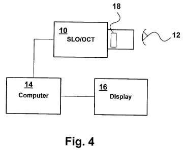
Application No. Country/Territory Date
60/985,465 United States of America 2007-11-05

Abstracts

English Abstract


A visual acuity
examination is performed on a patient by
bringing a confocal imaging apparatus
up to a patient's eye. Stimuli at various
points in the patient's field of view are
generated while the patient fixates on a
point. The patient's responses to the stimuli
are recorded with the movement of the eye
with is tracked with the aid of the confocal
imaging apparatus. The position of said
stimuli on the retina is corrected to take
into account any movement of the eye
between stimuli.


French Abstract

L'acuité visuelle d'un patient est examinée en approchant un appareil d'imagerie confocale d'un il du patient. Des stimuli sont appliqués en différents points du champ de vision du patient tandis que ce dernier fixe un point. Les réponses du patient aux stimuli sont enregistrées avec le mouvement de l'il qui est suivi au moyen de l'appareil d'imagerie confocale. La position de ces stimuli sur la rétine est corrigée pour tenir compte d'un éventuel mouvement de l'il entre les stimuli.

Claims

Note: Claims are shown in the official language in which they were submitted.


The embodiments of the invention in which an exclusive property or privilege
is
claimed are defined as follows:
1. A method of performing a visual acuity examination on a patient, the
method
comprising the steps of:
bringing a confocal imaging apparatus up to a patient's eye having a retina;
presenting a display to the patient;
displaying stimuli at various points on the target on the display in the
patient's
field of view while the patient fixates on a point on the target;
recording the patient's responses to said stimuli;
creating a succession of images of the retina with said confocal imaging
apparatus
while displaying said stimuli;
comparing said successive images to track movement of the retina; and
creating a map of the patient's acuity field from the responses to said
stimuli
taking into account any movement of the retina between successive stimuli
determined
by comparing said successive images.
2. A method as claimed in claim 1, wherein said map is overlaid on a
confocal
image of the retina obtained with said confocal imaging apparatus.
3. A method as claimed in claim 1 or 2, wherein said confocal imaging
apparatus is
a combined Scanning Laser Opthalmoscope and Optical Coherence Tomograph
(SLO/OCT) apparatus, and said map is overlaid on a tomographical image of the
retina
obtained by using the OCT portion of the apparatus.
4. A method as claimed in any one of claims 1 to 3, wherein said stimuli
comprise
light flashes, and the intensity of said light flashes is varied to determine
the patient's
response over different regions of the retina.
5. A method as claimed in any one of claims 1 to 4, wherein a shape is
displayed to
the patient on said display, and the limit of visual acuity is determined by
having the
patient report the orientation of the shape.
6. A method as claimed in claim 5, wherein said shape is a C-shape.
6

Description

Note: Descriptions are shown in the official language in which they were submitted.


CA 02704350 2015-04-22
A method for p,rforming visual acuity testing
FIELD OF THE INVENTION
This invention relates to field of ophthalmological examination, and in
particular to a
method for performing micro-perimetry and visual acuity examinations of a
patient.
BACKGROUND OF THE INVENTION
Visual field tests designed to map a person's visual field and document the
level of
peripheral vision are a mainstay in ophthalmology. Such tests are used to
assess disease
and treatment progressions. Typically, the test consists of having the patient
respond
every time a flash of light is perceived while the patient looks straight
ahead, fixating
onto a point. Visual acuity is normally tested using charts projected onto a
wall where the
patient is asked to read letters..Perimetry examination are used to test point
locations on
the retina and are commonly performed using a HumphreyTM field analyzer.
A major drawback of both of these systems is the lack of fundus tracking. For
instance, if
someone has 20/200 vision in his or her macular, but 20/40 vision in his or
her periphery,
a standard visual field test will not discern the difference, or tell the
doctor what part of
the fundus the patient is using to fixate. The fundus is the interior part of
the eye opposite
the lens.
1

CA 02704350 2010-04-30
WO 2009/059400
PCT/CA2008/001924
A standard perimetry examination provides no tracking. Projecting stimuli to
patients
with poor fixation gives meaningless results if the patient moves his eye
between stimuli.
SUMMARY OF THE INVENTION
According to the present invention there is provided a method of performing a
visual
acuity examination on a patient comprising bringing a confocal imaging
apparatus up to a
patient's eye; displaying stimuli at various points in the patient's field of
view while the
patient fixates on a point; recording the patient's responses to said stimuli;
tracking
movement of the eye with said confocal imaging apparatus; and correcting the
position of
the stimuli on the patient's retina to take into account any movement of the
eye between
successive stimuli.
Thus, in accordance with an embodiment of the invention, SLO imaging from a
combined Scanning Laser Ophthalmoscope and Optical Coherence Tomography
(SLO/OCT) system is used to image the eye while stimuli are displayed to the
patient.
Software automatically tracks the motion of the eye during the examination.
This gives
the doctor valuable information, such as what parts of the eye the patient is
using to
fixate, helps ensure the accuracy of the test, and provides the doctor with an
excellent
fundus image for use in diagnosis.
As soon as the acuity or perimetry exam is complete, the patient can be
scanned using the
OCT portion of the system so the doctor can see structural information from
the patient's
eye by performing B-Scans, C-Scans, or obtaining a 3D tomography. The
functional
(perimetry and acuity) information can then be overlaid on the structural
information so
the doctor can see which structures in the eye correlate to visual loss.
2

CA 02704350 2014-05-07
Having functional and structural capabilities built into a single machine
means less
examination time is required for a patient, less training is required for the
operator, and
more accurate information can be delivered to the doctor.
According to an aspect of the present invention, there is provided a method of
performing
a visual acuity examination on a patient, the method comprising the steps of:
bringing a confocal imaging apparatus up to a patient's eye having a retina;
presenting a display to the patient;
displaying stimuli at various points on the target on the display in the
patient's
field of view while the patient fixates on a point on the target;
recording the patient's responses to said stimuli;
creating a succession of images of the retina with said confocal imaging
apparatus
while displaying said stimuli;
comparing said successive images to track movement of the retina; and
creating a map of the patient's acuity field from the responses to said
stimuli
taking into account any movement of the retina between successive stimuli
determined
by comparing said successive images.
BRIEF DESCRIPTION OF THE DRAWINGS
The invention will now be described in more detail, by way of example only,
with
reference to the accompanying drawings, in which:
Figure 1 is a picture of the retina showing the results of a perimetry exam;
Figure 2 is an OCT topographic image (False color indicates retinal thickness,
black and
white are OCT B-Scans);
Figure 3 shows the results of a perimetry examination overlaid on the
topographic image;
and
3

CA 02704350 2014-05-07
Figure 4 is a block diagram of an apparatus for performing a visual acuity
examination in
accordance with one embodiment of the invention.
DETAILED DESCRIPTION OF THE INVENTION
A commercially available combined SLO/OCT system is described, for example, in
US
patent No. 6,769,769. Such an SLO/OCT system is capable of making OCT images
of
the eye or retina as well as SLO or confocal microscopic images.
In accordance with an embodiment of the invention, a visual acuity examination
is
performed while the patient's eye motion is tracked using high resolution SLO
scans
generated by the SLO-OCT system. As shown in Figure 4, the SLO-OCT system 10
is
brought up to the patient's eye 12, and a series of SLO images of the retina
obtained by
3a

CA 02704350 2010-04-30
WO 2009/059400
PCT/CA2008/001924
scanning. The confocal images are created by computer 14 from the SLO scans
and
displayed on display screen 16.
The SLO-OCT system 10 is able to track movement of the retina by comparing
successive images. The tracking can be performed automatically in software in
the
computer 14 using any of a number of known algorithms suited for this purpose,
such as
a combination of edge detection and Hausdroff s algorithm for image alignment.
The
actual algorithm used is not important as long as it is able to accurately
track the eye
motion.
The SLO-OCT system 10 includes a high resolution display 18, controlled by the

computer 14, that the patient can see while being scanned. The high resolution
display 18
is used to create a fixation target for the patient to fixate on. The patient
is asked to fixate
on the target while the examination is performed by generating the stimuli at
various
points in the patient's field of view and at various intensities. For example,
if the patient
is looking at a central point, a flash may be generated at a location
described by polar
coordinates r1, 01, and the patient asked to indicate whether he can observe
this flash. The
intensity of the flash can be changed to determine the patient's
responsiveness at this
point. The process can then be repeated at different points, ri, 0.
The resulting tracking data obtained from the SLO-OCT system are then used
with the
display 18 to accurately place the perimetry stimuli on the same fundus
location for each
point tested taking into account any patient movement. Thus, based on the
patient's
responses, a map of the patient's acuity field, as shown in Figure 1, can be
created. By
taking into account the eye motion detected by the SLO imaging apparatus
between
scans, the data points can be correlated to the correct positions on the
patient's retina. For
4

CA 02704350 2010-04-30
WO 2009/059400
PCT/CA2008/001924
example, if the SLO imaging apparatus detects that the retina moved an amount
8x
between successive stimuli, the position of the later stimulus point on the
retina can be
adjusted to take this movement into account.
In addition, a tomographic three dimensional image, shown in Figure 2, can be
obtained
using the OCT portion of the apparatus, in which case the stimuli points can
be marked
on the 3D image as shown in Figure 3.
The display can also be used to display a Snellen 'C' in one of four
orientations (opening
of the C up, down, left, or right). In this test, the patient uses a hand-held
controller or
joystick to report the direction of the opening. When the patient can no
longer tell the
direction of the opening, he has passed the limit of their visual acuity. Any
number of
other letters or symbols can be used for the same purpose. The tracking data
obtained by
the SLO apparatus can be recorded to show which part of the eye the patient
was using to
fixate on the fixation point.
The invention takes advantage of the functional and structural information
that the SLO-
OCT system can generate. It may desirably use automatic alignment of SLO
images from
function and structural imaging to overlay perimetry or acuity results on B-
Scans, C-
Scans or topographies in the computer 14.
Aspects of the invention thus include a method for integrating micro-perimetry
exams
into a combined imaging SLO-OCT system, and a method for integrating acuity
exams
into a combined imaging SLO-OCT system.

Representative Drawing
A single figure which represents the drawing illustrating the invention.
Administrative Status

For a clearer understanding of the status of the application/patent presented on this page, the site Disclaimer , as well as the definitions for Patent , Administrative Status , Maintenance Fee  and Payment History  should be consulted.

Administrative Status

Title Date
Forecasted Issue Date 2016-09-20
(86) PCT Filing Date 2008-11-05
(87) PCT Publication Date 2009-05-14
(85) National Entry 2010-04-30
Examination Requested 2013-09-06
(45) Issued 2016-09-20

Abandonment History

There is no abandonment history.

Maintenance Fee

Last Payment of $473.65 was received on 2023-11-02


 Upcoming maintenance fee amounts

Description Date Amount
Next Payment if standard fee 2024-11-05 $624.00
Next Payment if small entity fee 2024-11-05 $253.00

Note : If the full payment has not been received on or before the date indicated, a further fee may be required which may be one of the following

  • the reinstatement fee;
  • the late payment fee; or
  • additional fee to reverse deemed expiry.

Patent fees are adjusted on the 1st of January every year. The amounts above are the current amounts if received by December 31 of the current year.
Please refer to the CIPO Patent Fees web page to see all current fee amounts.

Payment History

Fee Type Anniversary Year Due Date Amount Paid Paid Date
Application Fee $400.00 2010-04-30
Maintenance Fee - Application - New Act 2 2010-11-05 $100.00 2010-04-30
Maintenance Fee - Application - New Act 3 2011-11-07 $100.00 2011-08-26
Maintenance Fee - Application - New Act 4 2012-11-05 $100.00 2012-09-20
Request for Examination $200.00 2013-09-06
Maintenance Fee - Application - New Act 5 2013-11-05 $200.00 2013-10-17
Maintenance Fee - Application - New Act 6 2014-11-05 $200.00 2014-10-15
Maintenance Fee - Application - New Act 7 2015-11-05 $200.00 2015-10-29
Registration of a document - section 124 $100.00 2016-07-22
Final Fee $300.00 2016-07-25
Maintenance Fee - Patent - New Act 8 2016-11-07 $200.00 2016-11-02
Maintenance Fee - Patent - New Act 9 2017-11-06 $200.00 2017-09-01
Maintenance Fee - Patent - New Act 10 2018-11-05 $250.00 2018-10-15
Maintenance Fee - Patent - New Act 11 2019-11-05 $250.00 2019-10-28
Maintenance Fee - Patent - New Act 12 2020-11-05 $250.00 2020-10-29
Maintenance Fee - Patent - New Act 13 2021-11-05 $255.00 2021-10-27
Maintenance Fee - Patent - New Act 14 2022-11-07 $254.49 2022-11-02
Maintenance Fee - Patent - New Act 15 2023-11-06 $473.65 2023-11-02
Owners on Record

Note: Records showing the ownership history in alphabetical order.

Current Owners on Record
OPTOS PLC
Past Owners on Record
MCLEAN, DUNCAN
OPKO INSTRUMENTATION
PEDRO, JUSTIN
ROGERS, JOHN
WEITZ, RISHARD
Past Owners that do not appear in the "Owners on Record" listing will appear in other documentation within the application.
Documents

To view selected files, please enter reCAPTCHA code :


To view images, click a link in the Document Description column. To download the documents, select one or more checkboxes in the first column and then click the "Download Selected in PDF format (Zip Archive)" or the "Download Selected as Single PDF" button.

List of published and non-published patent-specific documents on the CPD .

If you have any difficulty accessing content, you can call the Client Service Centre at 1-866-997-1936 or send them an e-mail at CIPO Client Service Centre.


Document
Description 
Date
(yyyy-mm-dd) 
Number of pages   Size of Image (KB) 
Description 2010-04-30 5 205
Drawings 2010-04-30 2 751
Claims 2010-04-30 2 41
Abstract 2010-04-30 1 60
Representative Drawing 2010-06-18 1 4
Cover Page 2010-07-06 2 37
Claims 2014-05-07 1 42
Description 2014-05-07 6 222
Description 2015-04-22 6 212
Claims 2015-04-22 1 45
Cover Page 2016-08-16 1 34
PCT 2010-04-30 3 133
Assignment 2010-04-30 2 98
PCT 2010-05-04 1 38
Correspondence 2010-06-17 1 18
PCT 2010-08-03 1 44
Correspondence 2010-07-28 3 104
Prosecution-Amendment 2013-09-06 1 31
Prosecution-Amendment 2015-02-26 4 216
Prosecution-Amendment 2014-05-07 5 142
Prosecution-Amendment 2015-04-22 4 112
Final Fee 2016-07-25 1 36