Note: Descriptions are shown in the official language in which they were submitted.
CA 03025761 2018-11-27
WO 2016/191244
PCT/US2016/033427
SCAN BODY FOR A DENTAL IMPRESSION
TECHNICAL FIELD
The present invention relates to dental devices and, particularly, to a scan
body
for mating to an impression coping positioned in a dental impression.
BACKGROUND ART
To replace a broken or damaged tooth a patient will typically undergo a
surgical
procedure in which a dentist places an implant in the location of the
patient's mouth missing
the tooth/teeth. Generally, the implant will incorporate an opening having a
hexagonal shape
for receiving an impression coping and, subsequently, a dental prosthesis.
After the implant
has been inserted into the patient's mouth, the dentist will typically insert
the impression
coping, having a head portion, into the implant having the opening configured
for receiving
the head portion of the impression coping.
Subsequently, the dentist will take a "pick-up" dental impression of the
patient's
mouth having the impression coping. Once the impression material has hardened
and the
dental impression is complete, the dentist can remove the "pick-up" dental
impression from
the patient's mouth and attach a female hex onto the head portion, having a
hexagonal shape,
of the impression coping. This can then be sent to a lab to create a dental
mold, in stone
form, of the patient's teeth with the female hex representing the position of
the implant in the
patient's mouth. It is to be noted that the "pick-up" dental impression is
used only for
implant cases.
A scan body is screwed into the female hex positioned inside the dental mold
and
the dental mold is scanned using a CADCAM computer program so as to design the
prosthetic tooth virtually. The scan body can have indexing means that can
allow the
CADCAM program to determine the orientation and angle of the scan body
relative to the
hex shape on the dental mold, so that the CADCAM program can design the
implant
prosthetic correctly. The conventional scan body has an opening through which
a screw
extends. This hole causes slight distortion in the screw.
While most CADCAM scanners have the ability to scan both dental impressions
and dental molds, a majority of the dentists are currently scanning the dental
molds instead of
using the dental impressions created directly from the patient's mouth.
Relying on the dental
molds, however, can negatively impact the accuracy of the design of the
prosthetic tooth
CA 03025761 2018-11-27
WO 2016/191244
PCT/US2016/033427
2
implanted into a patient's mouth since accuracy tends to decrease each time
information is
transferred from the patient's mouth to the dental impression, from the dental
impression to
the dental mold and from the dental mold to the scanned impression, which then
must be
restored in a digital format by the computer. Further, most scan bodies are
too large to fit
within smaller gaps between teeth.
Thus, a scan body for a dental impression solving the aforementioned problems
is
desired.
DISCLOSURE OF INVENTION
An embodiment of a scan body for a dental impression includes a substantially
hollow member with a mounting recess defined therein for mounting onto an
impression
coping positioned in a dental impression. The mounting recess can have an
internal threaded
bore configured to receive a fastener extending through the impression coping
for securing
the scan body onto the impression coping. The hollow member can have an
exterior
peripheral wall extending between the first and second ends, the exterior wall
having a first
flat surface and a second flat surface, the first flat surface and the second
flat surface having
different dimensions.
These and other features of the present invention will become readily apparent
upon further review of the following specification and drawings.
BRIEF DESCRIPTION OF DRAWINGS
Fig. 1 is an top, rear environmental view of a scan body coupled to an
impression
coping positioned in a dental impression according to the present invention.
Fig. 2A is an environmetal view of a dental mold and a scan body of the prior
art.
Fig. 2B is a perspective view of a dental mold having a scan body of the prior
art.
Fig. 3 is a front, perpective view of a dental impression having an impression
coping including a fastener, according to the present invention.
Fig. 4 is a bottom, perspective view a dental impression having a fastener,
according to the present invention.
Fig. 5A is a side view of a dental impression having an impression coping
including a head portion, according to the present invention.
Fig. 5B is a side view of a scan body coupled to an impression coping
positioned
in a dental impression, according to the present invention.
CA 03025761 2018-11-27
WO 2016/191244
PCT/US2016/033427
3
Similar reference characters denote corresponding features consistently
throughout the attached drawings.
BEST MODES FOR CARRYING OUT THE INVENTION
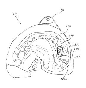
Referring to Figs. 1, 3, and 5B, a scan body 100 that can mate to an
impression
coping 110 inserted in a dental impression 120 of a patient's teeth is
generally illustrated.
The scan body 100 includes a hollow or substantially hollow member 105 having
a mounting
recess 300 defined therein. The scan body 100 is configured for mounting onto
the
impression coping 110 such as by engaging a head portion 320 of the impression
coping 110
positioned in the dental impression 120. A fastener 310a can be inserted
through the head
portion 320 from a back side of the dental impression 120 (Fig. 4) such that
the tip of the
fastener 310a protrudes above the head portion 320. The mounting recess 300
can have an
internal threaded bore 305 configured to receive the tip of the fastener 310a,
such as a screw,
to secure the scan body 100 onto the impression coping 110 positioned in the
dental
impression 120, as illustrated in Figs. 1 and 5B. For example, once the scan
body 100 is
mounted onto the impression coping 110, the fastener 310a, can be inserted
into the scan
body 100 from the bottom, as illustrated in Fig. 4, to secure the scan body
100 thereto. The
ability to secure the scan body 100 from the bottom can simplify securing the
scan body 100
between adjacent existing teeth.
The hollow member 105 can have an exterior wall 115 (Figs. 1 and 4) that is
configured to allow Computer-Aided Design and Computer-Aided Manufacturing
(CAD/CAM) technology to determine the orientation of the scan body 100
relative to the
impression coping 110 in the dental impression 120. For example, the external
wall 115 can
include a first flat exterior surface 125a and a second flat exterior surface
125b. The first flat
surface 125a can have a different length and/or height than the second flat
surface 135. The
positioning of the first flat exterior surface 125a and the second flat
exterior surface 125b can
then be used by the CAD/CAM system to determine the orientation of the scan
body 100
relative to the impression coping 110.
The scan body 100 can have any suitable shape, such as a substantially
rectangular, square, or cylindrical shape, and can be formed from any suitable
medical grade
material, such as titanium. The scan body 100 can be coated with any suitable
coating, to
make the exterior portion 115 of the scan body 100 opaque, which can make the
scan body
100 capable of being scanned, by a scanner, such as a white light scanner. The
fastener 310a
CA 03025761 2018-11-27
WO 2016/191244
PCT/US2016/033427
4
can be made from any suitable medical grade material, such as titanium and can
include a
head 310b having any suitable type of configuration, such as a substantially
circular
configuration, as illustrated in Fig. 4.
The mounting recess 300 of the scan body 100 can have a hexagonal shape, as
illustrated in Figs. 3 and 4. The mounting recess 300 can receive the head
portion 320,
having a corresponding hexagonal shape. It is to be noted that the shape of
the mounting
recess 300 is not limited to a hexagonal shape, as illustrated in Figs. 3 and
4, but can have any
shape, such as a circular shape, a triangular shape, or a pentagonal shape,
suitable to receive a
head portion of any type of impression coping having the corresponding shape.
By way of operation, after placing an implant (not shown) into the jaw bone of
a
patient's mouth, the dentist can insert the head portion 320 of the impression
coping 110 into
the opening of the implant. Once the impression coping 110 has been attached
to the implant
(positioned inside the patient's jaw bone) the impression coping 110 can be
secured to the
implant, such as by a thumb screw (not shown) inserted into the impression
coping 110,
which can then be tightened.
The dentist can then use impression material, such as ImpregumTM, to cover the
patient's teeth and the impression coping 110 and an impression tray 160 to
hold the
impression material against the patient's teeth and the impression coping 110
until the
impression material hardens so as to form the dental impression 120 of the
patient's teeth.
The impression material hardens to form the dental impression 120, as
illustrated Fig. 5A.
The dentist can remove the thumb screw attaching the impression coping 110 to
the implant
and, subsequently, remove the impression tray 160 including the hardened
impression
material and the impression coping 110 from the patient's mouth.
Once the dental impression 120 having the impression coping 110 is removed
from the patient's mouth, the scan body 100 can be attached to the head
portion 320 of the
impression coping 110, e.g., by inserting the head portion 320 into the
mounting recess 300
of the scan body 100, as illustrated in Figs. 1 and 5B. The dental impression
120 having the
scan body 100 attached to the impression coping 110, as illustrated in Figs. 1
and 5B, can
then be scanned. The scanner, based on the orientation of the exterior wall
115 of the scan
body 100 can determine the orientation and alignment of the scan body 100 in
relation to the
impression coping 110. This data can be transferred to the CAD/CAM system for
use by a
dentist and/or a lab technician to create a dental prosthesis (not shown), to
replace broken or
damaged tooth. Scanning the dental impression 120 and the scan body 100
directly, without
having to make a dental mold 200 (Figs. 2A and 2B), can decrease the number of
steps
CA 03025761 2018-11-27
WO 2016/191244
PCT/US2016/033427
required to create the dental prosthesis. As such, more accurate data and a
faster and easier
workflow can be achieved. Additionally, errors on the part of the dental lab
when creating
the dental prosthesis can be minimized.
It is to be understood that the present invention is not limited to the
embodiments
5 described above, but encompasses any and all embodiments within the scope
of the following
claims.