Sélection de la langue

Search

Sommaire du brevet 3025761 

Énoncé de désistement de responsabilité concernant l'information provenant de tiers

Une partie des informations de ce site Web a été fournie par des sources externes. Le gouvernement du Canada n'assume aucune responsabilité concernant la précision, l'actualité ou la fiabilité des informations fournies par les sources externes. Les utilisateurs qui désirent employer cette information devraient consulter directement la source des informations. Le contenu fourni par les sources externes n'est pas assujetti aux exigences sur les langues officielles, la protection des renseignements personnels et l'accessibilité.

Disponibilité de l'Abrégé et des Revendications

L'apparition de différences dans le texte et l'image des Revendications et de l'Abrégé dépend du moment auquel le document est publié. Les textes des Revendications et de l'Abrégé sont affichés :

  • lorsque la demande peut être examinée par le public;
  • lorsque le brevet est émis (délivrance).
(12) Brevet: (11) CA 3025761
(54) Titre français: CORPS DE BALAYAGE POUR EMPREINTE DENTAIRE
(54) Titre anglais: SCAN BODY FOR A DENTAL IMPRESSION
Statut: Octroyé
Données bibliographiques
(51) Classification internationale des brevets (CIB):
  • A61C 9/00 (2006.01)
  • A61C 8/00 (2006.01)
  • A61C 19/04 (2006.01)
(72) Inventeurs :
  • CHUNG, FELIX (Etats-Unis d'Amérique)
  • TOWNSEND, JOSHUA (Etats-Unis d'Amérique)
(73) Titulaires :
  • CHUNG, FELIX (Etats-Unis d'Amérique)
  • TOWNSEND, JOSHUA (Etats-Unis d'Amérique)
(71) Demandeurs :
  • CHUNG, FELIX (Etats-Unis d'Amérique)
  • TOWNSEND, JOSHUA (Etats-Unis d'Amérique)
(74) Agent: MBM INTELLECTUAL PROPERTY AGENCY
(74) Co-agent:
(45) Délivré: 2021-11-09
(86) Date de dépôt PCT: 2016-05-20
(87) Mise à la disponibilité du public: 2016-12-01
Requête d'examen: 2018-11-27
Licence disponible: S.O.
(25) Langue des documents déposés: Anglais

Traité de coopération en matière de brevets (PCT): Oui
(86) Numéro de la demande PCT: PCT/US2016/033427
(87) Numéro de publication internationale PCT: WO2016/191244
(85) Entrée nationale: 2018-11-27

(30) Données de priorité de la demande:
Numéro de la demande Pays / territoire Date
14/723,387 Etats-Unis d'Amérique 2015-05-27

Abrégés

Abrégé français

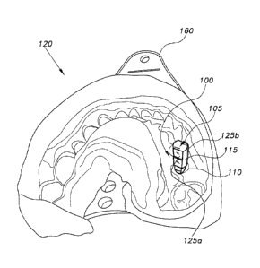
La présente invention concerne un corps de balayage (100) destiné à une empreinte dentaire (120) comprenant un élément creux (105) présentant un évidement de montage (300) conçu pour être monté sur une partie tête (320) d'une chape de transfert (110) positionnée dans une empreinte dentaire (120). L'évidement de montage (300) peut comprendre un trou fileté interne (305) permettant de recevoir un élément de fixation (310a) pour fixer le corps de balayage (100) sur la chape de transfert (110). Le corps de balayage (100) peut présenter une paroi périphérique extérieure (115) s'étendant entre les première et seconde extrémités opposées. La paroi périphérique (115) présente une première surface plate (125a) et une seconde surface plate (125b). La première surface plate (125a) et la seconde surface plate (125b) ont des dimensions différentes.


Abrégé anglais


The scan body (100) for a dental impression (120) includes a
hollow member (105) having a mounting recess (300) configured for
mounting onto a head portion (320) of an impression coping (110) positioned in
a
dental impression (120). The mounting recess (300) can have an internal
threaded bore (305) for receiving a fastener (310a) to secure the scan body
(100) onto the impression coping (110). The scan body (100) can have an
exterior peripheral wall (115) extending between opposing first and second
ends. The peripheral wall (115) has a first flat surface (125a) and a second
flat surface (125b). The first flat surface (125a) and the second flat surface

(125b) have different dimensions.

Revendications

Note : Les revendications sont présentées dans la langue officielle dans laquelle elles ont été soumises.


6
THE EMBODIMENTS OF THE INVENTION FOR WHICH AN EXCLUSIVE
PROPERTY OR PRIVILEGE IS CLAIMED ARE DEFINED AS FOLLOWS:
1. A dental impression system, comprising:
a dental impression having an impression coping; and
a scan body having a hollow member having a nonthreaded exterior peripheral
wall
extending between opposing first and second ends, the peripheral exterior wall
having at least a
first flat surface, wherein the first flat surface has an upper end, the upper
end of the first flat
surface being contiguous to the second end and extending towards the first
end, the first end
being open and in communication with a mounting recess configured for mounting
onto the
impression coping of the dental impression, the second end being closed.
2. The dental impression system according to claim 1, wherein the mounting
recess
comprises an internal threaded bore.
3. The dental impression system according to claim 1, further comprising a
fastener.
4. The dental impression system according to claim 1, wherein the scan body is
made
from titanium.
5. The dental impression system according to claim 1, wherein the exterior
peripheral
wall includes a second flat surface, and a length and height of the first flat
surface is different
from a length and height of the second flat surface.
6. A method for creating a dental prosthesis from a dental impression,
comprising the
steps of:
(a) inserting an impression coping into an opening in an implant positioned in
a patient's
jaw bone;
(b) creating a dental impression of the patient's teeth including the
impression coping;
(c) removing the dental impression from the patient's mouth;
Date Recue/Date Received 2021-03-05

7
(d) providing a scan body including a mounting recess defined therein, the
scan body
configured for mounting onto the impression coping positioned in the dental
impression, the scan
body having a nonthreaded exterior peripheral wall extending between opposing
first and second
ends, the peripheral exterior wall having at least a first flat surface,
wherein the first flat surface
has an upper end, the upper end of the first flat surface being contiguous to
the second end and
extending towards the first end, the first end being open and in communication
with a mounting
recess configured for mounting onto the impression coping of the dental
impression, the second
end being closed, the exterior peripheral wall having a second flat surface,
the first flat surface
and the second flat surface having different dimensions;
(e) attaching the scan body onto the impression coping;
(f) scanning the dental impression having the scan body attached to the
impression
coping; and
(g) designing a prosthetic based on the information transferred by the scan of
the dental
impression having the scan body attached to the impression coping.
7. The method for creating a dental prosthesis from a dental impression
according to
claim 6, further comprising the step of securing the scan body to the
impression coping.
8. The method for creating a prosthetic from a dental impression according to
claim 6,
wherein the mounting recess comprises an internal threaded bore.
Date Recue/Date Received 2021-03-05

Description

Note : Les descriptions sont présentées dans la langue officielle dans laquelle elles ont été soumises.


CA 03025761 2018-11-27
WO 2016/191244
PCT/US2016/033427
SCAN BODY FOR A DENTAL IMPRESSION
TECHNICAL FIELD
The present invention relates to dental devices and, particularly, to a scan
body
for mating to an impression coping positioned in a dental impression.
BACKGROUND ART
To replace a broken or damaged tooth a patient will typically undergo a
surgical
procedure in which a dentist places an implant in the location of the
patient's mouth missing
the tooth/teeth. Generally, the implant will incorporate an opening having a
hexagonal shape
for receiving an impression coping and, subsequently, a dental prosthesis.
After the implant
has been inserted into the patient's mouth, the dentist will typically insert
the impression
coping, having a head portion, into the implant having the opening configured
for receiving
the head portion of the impression coping.
Subsequently, the dentist will take a "pick-up" dental impression of the
patient's
mouth having the impression coping. Once the impression material has hardened
and the
dental impression is complete, the dentist can remove the "pick-up" dental
impression from
the patient's mouth and attach a female hex onto the head portion, having a
hexagonal shape,
of the impression coping. This can then be sent to a lab to create a dental
mold, in stone
form, of the patient's teeth with the female hex representing the position of
the implant in the
patient's mouth. It is to be noted that the "pick-up" dental impression is
used only for
implant cases.
A scan body is screwed into the female hex positioned inside the dental mold
and
the dental mold is scanned using a CADCAM computer program so as to design the

prosthetic tooth virtually. The scan body can have indexing means that can
allow the
CADCAM program to determine the orientation and angle of the scan body
relative to the
hex shape on the dental mold, so that the CADCAM program can design the
implant
prosthetic correctly. The conventional scan body has an opening through which
a screw
extends. This hole causes slight distortion in the screw.
While most CADCAM scanners have the ability to scan both dental impressions
and dental molds, a majority of the dentists are currently scanning the dental
molds instead of
using the dental impressions created directly from the patient's mouth.
Relying on the dental
molds, however, can negatively impact the accuracy of the design of the
prosthetic tooth

CA 03025761 2018-11-27
WO 2016/191244
PCT/US2016/033427
2
implanted into a patient's mouth since accuracy tends to decrease each time
information is
transferred from the patient's mouth to the dental impression, from the dental
impression to
the dental mold and from the dental mold to the scanned impression, which then
must be
restored in a digital format by the computer. Further, most scan bodies are
too large to fit
within smaller gaps between teeth.
Thus, a scan body for a dental impression solving the aforementioned problems
is
desired.
DISCLOSURE OF INVENTION
An embodiment of a scan body for a dental impression includes a substantially
hollow member with a mounting recess defined therein for mounting onto an
impression
coping positioned in a dental impression. The mounting recess can have an
internal threaded
bore configured to receive a fastener extending through the impression coping
for securing
the scan body onto the impression coping. The hollow member can have an
exterior
peripheral wall extending between the first and second ends, the exterior wall
having a first
flat surface and a second flat surface, the first flat surface and the second
flat surface having
different dimensions.
These and other features of the present invention will become readily apparent

upon further review of the following specification and drawings.
BRIEF DESCRIPTION OF DRAWINGS
Fig. 1 is an top, rear environmental view of a scan body coupled to an
impression
coping positioned in a dental impression according to the present invention.
Fig. 2A is an environmetal view of a dental mold and a scan body of the prior
art.
Fig. 2B is a perspective view of a dental mold having a scan body of the prior
art.
Fig. 3 is a front, perpective view of a dental impression having an impression
coping including a fastener, according to the present invention.
Fig. 4 is a bottom, perspective view a dental impression having a fastener,
according to the present invention.
Fig. 5A is a side view of a dental impression having an impression coping
including a head portion, according to the present invention.
Fig. 5B is a side view of a scan body coupled to an impression coping
positioned
in a dental impression, according to the present invention.

CA 03025761 2018-11-27
WO 2016/191244
PCT/US2016/033427
3
Similar reference characters denote corresponding features consistently
throughout the attached drawings.
BEST MODES FOR CARRYING OUT THE INVENTION
Referring to Figs. 1, 3, and 5B, a scan body 100 that can mate to an
impression
coping 110 inserted in a dental impression 120 of a patient's teeth is
generally illustrated.
The scan body 100 includes a hollow or substantially hollow member 105 having
a mounting
recess 300 defined therein. The scan body 100 is configured for mounting onto
the
impression coping 110 such as by engaging a head portion 320 of the impression
coping 110
positioned in the dental impression 120. A fastener 310a can be inserted
through the head
portion 320 from a back side of the dental impression 120 (Fig. 4) such that
the tip of the
fastener 310a protrudes above the head portion 320. The mounting recess 300
can have an
internal threaded bore 305 configured to receive the tip of the fastener 310a,
such as a screw,
to secure the scan body 100 onto the impression coping 110 positioned in the
dental
impression 120, as illustrated in Figs. 1 and 5B. For example, once the scan
body 100 is
mounted onto the impression coping 110, the fastener 310a, can be inserted
into the scan
body 100 from the bottom, as illustrated in Fig. 4, to secure the scan body
100 thereto. The
ability to secure the scan body 100 from the bottom can simplify securing the
scan body 100
between adjacent existing teeth.
The hollow member 105 can have an exterior wall 115 (Figs. 1 and 4) that is
configured to allow Computer-Aided Design and Computer-Aided Manufacturing
(CAD/CAM) technology to determine the orientation of the scan body 100
relative to the
impression coping 110 in the dental impression 120. For example, the external
wall 115 can
include a first flat exterior surface 125a and a second flat exterior surface
125b. The first flat
surface 125a can have a different length and/or height than the second flat
surface 135. The
positioning of the first flat exterior surface 125a and the second flat
exterior surface 125b can
then be used by the CAD/CAM system to determine the orientation of the scan
body 100
relative to the impression coping 110.
The scan body 100 can have any suitable shape, such as a substantially
rectangular, square, or cylindrical shape, and can be formed from any suitable
medical grade
material, such as titanium. The scan body 100 can be coated with any suitable
coating, to
make the exterior portion 115 of the scan body 100 opaque, which can make the
scan body
100 capable of being scanned, by a scanner, such as a white light scanner. The
fastener 310a

CA 03025761 2018-11-27
WO 2016/191244
PCT/US2016/033427
4
can be made from any suitable medical grade material, such as titanium and can
include a
head 310b having any suitable type of configuration, such as a substantially
circular
configuration, as illustrated in Fig. 4.
The mounting recess 300 of the scan body 100 can have a hexagonal shape, as
illustrated in Figs. 3 and 4. The mounting recess 300 can receive the head
portion 320,
having a corresponding hexagonal shape. It is to be noted that the shape of
the mounting
recess 300 is not limited to a hexagonal shape, as illustrated in Figs. 3 and
4, but can have any
shape, such as a circular shape, a triangular shape, or a pentagonal shape,
suitable to receive a
head portion of any type of impression coping having the corresponding shape.
By way of operation, after placing an implant (not shown) into the jaw bone of
a
patient's mouth, the dentist can insert the head portion 320 of the impression
coping 110 into
the opening of the implant. Once the impression coping 110 has been attached
to the implant
(positioned inside the patient's jaw bone) the impression coping 110 can be
secured to the
implant, such as by a thumb screw (not shown) inserted into the impression
coping 110,
which can then be tightened.
The dentist can then use impression material, such as ImpregumTM, to cover the

patient's teeth and the impression coping 110 and an impression tray 160 to
hold the
impression material against the patient's teeth and the impression coping 110
until the
impression material hardens so as to form the dental impression 120 of the
patient's teeth.
The impression material hardens to form the dental impression 120, as
illustrated Fig. 5A.
The dentist can remove the thumb screw attaching the impression coping 110 to
the implant
and, subsequently, remove the impression tray 160 including the hardened
impression
material and the impression coping 110 from the patient's mouth.
Once the dental impression 120 having the impression coping 110 is removed
from the patient's mouth, the scan body 100 can be attached to the head
portion 320 of the
impression coping 110, e.g., by inserting the head portion 320 into the
mounting recess 300
of the scan body 100, as illustrated in Figs. 1 and 5B. The dental impression
120 having the
scan body 100 attached to the impression coping 110, as illustrated in Figs. 1
and 5B, can
then be scanned. The scanner, based on the orientation of the exterior wall
115 of the scan
body 100 can determine the orientation and alignment of the scan body 100 in
relation to the
impression coping 110. This data can be transferred to the CAD/CAM system for
use by a
dentist and/or a lab technician to create a dental prosthesis (not shown), to
replace broken or
damaged tooth. Scanning the dental impression 120 and the scan body 100
directly, without
having to make a dental mold 200 (Figs. 2A and 2B), can decrease the number of
steps

CA 03025761 2018-11-27
WO 2016/191244
PCT/US2016/033427
required to create the dental prosthesis. As such, more accurate data and a
faster and easier
workflow can be achieved. Additionally, errors on the part of the dental lab
when creating
the dental prosthesis can be minimized.
It is to be understood that the present invention is not limited to the
embodiments
5 described above, but encompasses any and all embodiments within the scope
of the following
claims.

Dessin représentatif
Une figure unique qui représente un dessin illustrant l'invention.
États administratifs

Pour une meilleure compréhension de l'état de la demande ou brevet qui figure sur cette page, la rubrique Mise en garde , et les descriptions de Brevet , États administratifs , Taxes périodiques et Historique des paiements devraient être consultées.

États administratifs

Titre Date
Date de délivrance prévu 2021-11-09
(86) Date de dépôt PCT 2016-05-20
(87) Date de publication PCT 2016-12-01
(85) Entrée nationale 2018-11-27
Requête d'examen 2018-11-27
(45) Délivré 2021-11-09

Historique d'abandonnement

Date d'abandonnement Raison Reinstatement Date
2020-03-05 R30(2) - Absence de réponse 2021-03-05

Taxes périodiques

Dernier paiement au montant de 203,59 $ a été reçu le 2022-05-13


 Montants des taxes pour le maintien en état à venir

Description Date Montant
Prochain paiement si taxe applicable aux petites entités 2023-05-23 100,00 $
Prochain paiement si taxe générale 2023-05-23 277,00 $

Avis : Si le paiement en totalité n'a pas été reçu au plus tard à la date indiquée, une taxe supplémentaire peut être imposée, soit une des taxes suivantes :

  • taxe de rétablissement ;
  • taxe pour paiement en souffrance ; ou
  • taxe additionnelle pour le renversement d'une péremption réputée.

Les taxes sur les brevets sont ajustées au 1er janvier de chaque année. Les montants ci-dessus sont les montants actuels s'ils sont reçus au plus tard le 31 décembre de l'année en cours.
Veuillez vous référer à la page web des taxes sur les brevets de l'OPIC pour voir tous les montants actuels des taxes.

Historique des paiements

Type de taxes Anniversaire Échéance Montant payé Date payée
Requête d'examen 400,00 $ 2018-11-27
Rétablissement des droits 200,00 $ 2018-11-27
Le dépôt d'une demande de brevet 200,00 $ 2018-11-27
Taxe de maintien en état - Demande - nouvelle loi 2 2018-05-22 50,00 $ 2018-11-27
Taxe de maintien en état - Demande - nouvelle loi 3 2019-05-21 50,00 $ 2019-05-13
Taxe de maintien en état - Demande - nouvelle loi 4 2020-08-31 50,00 $ 2021-02-26
Surtaxe pour omission de payer taxe de maintien en état pour demande 2021-02-26 150,00 $ 2021-02-26
Rétablissement - Omission de répondre au rapport d'examen de bonne foi 2021-03-05 204,00 $ 2021-03-05
Taxe de maintien en état - Demande - nouvelle loi 5 2021-05-20 204,00 $ 2021-05-14
Taxe finale 2021-09-20 153,00 $ 2021-09-14
Taxe de maintien en état - brevet - nouvelle loi 6 2022-05-20 203,59 $ 2022-05-13
Titulaires au dossier

Les titulaires actuels et antérieures au dossier sont affichés en ordre alphabétique.

Titulaires actuels au dossier
CHUNG, FELIX
TOWNSEND, JOSHUA
Titulaires antérieures au dossier
S.O.
Les propriétaires antérieurs qui ne figurent pas dans la liste des « Propriétaires au dossier » apparaîtront dans d'autres documents au dossier.
Documents

Pour visionner les fichiers sélectionnés, entrer le code reCAPTCHA :


Pour visualiser une image, cliquer sur un lien dans la colonne description du document. Pour télécharger l'image (les images), cliquer l'une ou plusieurs cases à cocher dans la première colonne et ensuite cliquer sur le bouton "Télécharger sélection en format PDF (archive Zip)" ou le bouton "Télécharger sélection (en un fichier PDF fusionné)".

Liste des documents de brevet publiés et non publiés sur la BDBC .

Si vous avez des difficultés à accéder au contenu, veuillez communiquer avec le Centre de services à la clientèle au 1-866-997-1936, ou envoyer un courriel au Centre de service à la clientèle de l'OPIC.


Description du
Document 
Date
(yyyy-mm-dd) 
Nombre de pages   Taille de l'image (Ko) 
Rétablissement / Modification 2021-03-05 10 392
Revendications 2021-03-05 2 90
Taxe finale 2021-09-14 3 125
Dessins représentatifs 2021-10-20 1 13
Page couverture 2021-10-20 1 48
Certificat électronique d'octroi 2021-11-09 1 2 527
Abrégé 2018-11-27 1 64
Revendications 2018-11-27 2 75
Dessins 2018-11-27 7 212
Description 2018-11-27 5 223
Dessins représentatifs 2018-11-27 1 26
Traité de coopération en matière de brevets (PCT) 2018-11-27 1 39
Rapport de recherche internationale 2018-11-27 9 373
Demande d'entrée en phase nationale 2018-11-27 8 207
Page couverture 2018-12-05 2 45
Paiement de taxe périodique 2019-05-13 1 33
Demande d'examen 2019-09-05 4 292
Lettre du bureau 2024-03-28 2 189