Note : Les descriptions sont présentées dans la langue officielle dans laquelle elles ont été soumises.
~73S54
AUTOMATED MICROBIOLOGICAL TESTING APPARATUS A~ID METHOD
Background of the Invention
This invention retates to microbiological testing apparatus and methods,
and more particularly to an improved system for facilitating the automatic incubation
and reading of microbioiogical test trays.
A number of different types of microbiological testing are carried out in
trays or strips (referred to herein collectively as "trays") which have a number of
chambers known as test wells or cupules. Such trays are used, for example, to ident-
if y a microorganism, or to determine the susceptibility of that organism to a number
10 of antimicrobics, which latter trays are called susceptibility trays. Typically, the
test wells or cupules in the identification trays contain complex chemicals or reagents
which in the presence of an active fermenting culture change color, become cloudy
or otherwise indicate that fermentation is or has taken place. Similarly, in oneknown susceptibility test called the minimum inhibiting concentration (MIC) test,
the wells contain different dilutions of various antimicrobics and a growth medium
to determine the dilution level of the antimicrobic which is sufficient to kill and/or
inhibit growth of the organism.
Conventionally, the test reagents and any growth mediurn or antimicro-
bics are placed into the test wells in the form of an aqueous solution and later20 Iyophilized. A differerlt combination of reagent or growth medium is charged into
different wells so that a great number of individual reactions are performed in a
physically small apparatus. For example, in the MIC tests, a regular pattern of
wells arranged in rows and columns could be provided, each row of wells containing
different antimicrobics. Within a row, the concentration of the antimicrobic would
increase from well to well by a factor of, for example, 2. Of course, other dilution
ratios could be used.
When a test is to be performed, a microorganism is innoculated into each
of the test chambers with sufficient water to reconstitute the reagents. The test
trays are then incubated at an appropriate temperature, such as 35-37 degrees Celsius
30 for an extended period of time. After a predetermined period, the individual cham~
bers are examined for the presence or absence o-f a reaction or indication of color
change, or a change in turbidity. Heretofore, it is believed that the inspection of
the wells for the presence or absence of a reaction or indication was done manually
at least in part. Thus, individual trays each required the use of technician's time in
the preporation, innocvlation, incubation and reading of the results. Moreover, since
~f ~
,
1X'73~
different test trays might be needed to determine different characteristics of the
rnicroorganisms, the reading of a variety of different trays could be a fairly complex
proceedure.
Systems have been provided for automating at least a portion of the
reading process. In one existing system for use in semi-automatically recording the
results of microbiological tests, a test tray having a plurality of test wells arranged
in a certain pattern is placed beneath a transparent keyboard. A light source projects
light through the tray and the keyboard so that the user can view the tray with its
test wells through the keyboard. The keys of the keyboard correspond to the testlû wells, so that the user presses the keys overlying those wells in which the certain
test results have occurred in order to record the results of the tests conducted in
the test wells. Such a method of reading the test wells requires a highly skilled
technician and a good deal of technician's time. In addition, the incubation times
for identification and susceptibility trays may be quite different, with the result
that the user will be recording the results for a particular patient or specimen at
two different times, with the possibility that the identification and susceptibility
results might not be properly assigned to the same patient. Moreover, the difference
in times of incubation for identification and susceptibility trays means that the user
or operator must return twice to the incubator for each patient.
20 Summary of the Invention
Among the various aspects and features of the present invention may be
noted the provision of an apparatus for automating the microbiological test
procedure from incubation through the actual reading of the test tray itself; the
provision of such an apparatus which eliminates to a large extent the necessity of
having a highly trained technician read test results; the provision of such an
apparatus which insures that identification and susceptibility results for the same
patient remain together; the provision of such an apparatus which is compatible with
currently available identification and susceptibility test trays; the provision of such
an apparatus that is flexible enough to use with a number of different tray
30 combinations; and the provision of such an apparatus which is relatively economical
to use.
Other aspects and features of the present invention will be in part appar-
ent and in part pointed out hereinafter.
Briefly, in a first aspect an automated microbiological testing apparatus
of the present invention includes an incubation chamber for incubating a plurality of
microbiological test trays such as susceptibility trays and identification trays, an
,_
1~73554
inspection station at which the test trays may be inspected to determine the results
of the microbiological tests, means for moving any predetermined test trays desired
from the incubation chamber to the inspection station, and means for processing the
image of the test tray at the inspection station to determine test results.
In a second aspect of the invention, an automated microbiological testing
apparatus includes an incubation chamber for incubating a plurality of microbiological
test trays such as susceptibility trays and identification trays, an inspection station
at which the test trays may be inspected by the apparatus to determine the results
of microbiological tests, means for moving any predetermined test trays desired
lû from the incubation chamber to the inspection station, and means for automatically
determining test results at the inspection station.
In a third aspect of the present invention, a carrier for a microbiological
tray includes a relatively rigid frame defining at least one central opening suitable
for holding and supporting a microbiological tray, the tray having a pair of opposed,
parallel shoulders suitable for riding on a pair of parallel rails, and receiving means
integrally formed in the frame by means of which an external driving force may be
applied to the frame to move it along the rails.
In a fourth aspect of the present invention, a diagnostic microbiological
testing apparatus for obtaining test results from microbiological test trays and strips
20 such as susceptibility trays and identification trays, each tray having a plurality of
wells, comprises an inspection station at which the test trays may be inspected to
determine the results of the microbiological tests, a video camera disposed to form
images of the test trays at the inspection station, and processing means for receiving
the images from the video camera and processing them to determine test results.
In a fifth aspect of the present invention, a method of automatically
reading the results from microbiological test trays and strips such as susceptibility
trays and identification trays, each tray having a plurality of wells, comprises the
steps of making an image with a video camera of a tray to be read, electronically
analyzing only predetermined areas of interest in the image made by the camera,
30 which areas of interest are substantially within the outlines of the tray wells in the
image, electronically determining for each well of interest the~umber of pixels in
B each area of interest having an associated value that ~a predetermined thres-
hold for that area of interest, and electronically assigning a binary partial result to
each well based upon the number of pixels which exceeded the predetermined thres-
hold for each corresponding area of interest.
1~ 7~ 54
In a sixth aspect of the present invention, a method of automatically
reading the results from rnjcrobjological test trays and strips such as susceptibility
trays and indentification trays comprises the steps of moving a tray to be read to an
inspection station, and electronically reading the tray at the inspection station with
a camera which remains substantially stationary with respect to the tray while the
reading of the tray is occurring.
Brief Description of the Drawings
Fig. I is a front elevation, with parts broken away for clarity, of micro-
biological testing apparatus of the present invention;
Fig. 2 is a side elevation with parts broken away of the apparatus of Fig.
I; .
Fig. 3 is a schematic of the internal components of the apparatus of Fig.
I; .
Fig. 4 is a top plan, with parts broken away for clarity, of the apparatus
of Fig. I;
Fig. 5 is a perspective illustrating a tray carrier and transporting means
of the present invention;
Fig. 6 is a top plan of the tray carrier of Fig. 5 showing portions of ident-
if ication and susceptibiltiy trays in place;
Fig. 7 is a sectional view taken along line 7--7 of Fig. 6;
Fig. 8 is a front elevation of the carrier of Fig. 6;
Fig. 9 is a rear elevation of the carrier of Fig. 6;
Fig. 10 is a top plan of an identification tray suitable for use with the
apparatus of the present invention;
Fig. I l is a sectional view taken along line 11--1 1 of Fig. 10;
Fig. 12 is a top plan of a susceptibility tray suitable for use with the
apparatus of the present invention;
Fig. 13 is an elevation of the tray of Fig. 12;
Fig. 14 is a schematic illustrating the reagent handling and identification
30 tray removal subassemblies of the apparatus of Fig. I;
Fig. 15 is a perspective of the reagent reservoir of the apparatus of the
present invention;
Fig. 16 is a schematic of carrier presence sensing apparatus of the present
invention;
3~t~
Fig. 17 is a schematic of elevator position sensing apparatus of the present
invention; and
Fig. 18 is a schematic of carrier position sensing apparatus of the present
invention.
Similar reference characters indicate similar parts throughout the several
views of the drawings.
Description of the Preferred Embodiment
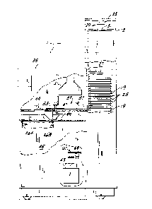
Referring now to Fig. I, there is shown an automated microbiological
apparatus 11 of the present invention which includes an incubation chamber 13 for
incubating a plurality of microbiological test trays, such as susceptibility and ident-
if ication trays 15 and 17 (see Figs. 10 and 12), carried in a common carrier 19 (Fig.
1). As shown in Figs. 10 and 12, susceptibility trays 15 and identification trays 17
each include a plurality of wells or cupules 21 and 23 respectively arranged in rows
and columns. Referring back to Fig. 1, common carriers 19 are manually placed
through an access door (not shown) in a plurality of slots 25 in incubation chamber
13. Slots 25 are vertically disposed in an elevator 27 which is movable vertically in
incubation chamber 13 by a belt driven screw drive 29, of which tef lon coated drive
screw 31 and precision stepper motor 33 are shown in Fig. 1. Elevator 27 may include,
by way of example, two rows of thirty slots so that it may accomodate up to sixty
common carriers 19. By means of drive 29, any one of the slots 25 may be moved to
the level of the lowermost slot shown in Fig. I so that the common carrier 19 there-
in may be removed through an access port from the incubator for processing as dis-
cussed below. Temperature and humidity within incubation chamber 13 are tightly
controlled by means of a number of sensors and a heater (not shown) and the humidi-
fier discussed below.
More particularly, apparatus 11 also includes a housing 35 in communica-
tion via the access port with the interior of incubation chamber 13. Housing 35
houses an inspection station 37 and means 39 for transporting common carriers from
slots 25 through the access port to the inspection station 37 and beyond as described
below. A light source 41 is disposed above inspection station 37 and a pair of video
cameras 43 are disposed below the inspection station. Alternatively, a pair of light
sources may be used, one above each camera. A waste bin 45 is also provided inside
housing 35 having a sensor system including a photodiode 46A and a photodetector46B for detecting when bin 45 is full. Housing 35 also houses a dispensing head 47
for dispensing reagent into identification trays 23, and a flipper system including a
' - -
'
1;~73554
pair of flipper forks 49 for removing identification trays or strips from commoncarriers 19.
Turning to Fig. 2, the two rows of slots 25 in elevator 27 are seen to be
disposed side by side in incubation chamber 13. Carrier transporting means 39 includes
a pair of tracks 51 upon each of which ride a separate mùtor driven carriage 53.Each carriage 53 carries a generally L-shaped rod SS which is movable into a corre-
sponding recess (see Fig. 4) in common carrier 19 to move any desired carrier from
its slot 25 in the incubation chamber through one of the pair of access ports 56 to
inspection station 37. Carriers 19 are moved from their slots to the inspection sta-
tion along a second pair of tracks 57.
Dispensing head 47 which is disposed above tracks 57 on the opposite side
of the inspection station 37 from incubation chamber 13, is carried by a carriage 59
along a track 61 by a belt drive 63 including a belt drive stepper motor 65. More
particularly, dispensing head 47 is movable between the extreme position shown
above the rightmost track 57 to a corresponding position generally to the left of the
leftmost track 57 so that any reagent may be dispensed into any cupule of the ident-
if ication tray of a common carrier on either track.
B s bJ Although there are a pair of tracks 57 and a pair of cameras 43~ it is
p~ a single light source 41 so long as cool and even illumation of the inspec-
tion area is achieved. It has been found that a cold cathode grid lamp equipped with
a diffuser plate provides such illumination. Alternatively, a pair of such lamps equip-
ped with diffuser plates may be used. For convenience, the inspection station can
be divided into left and right halves 37A and 37B, respectively. Below inspection
station 37A qnd between that inspection station and the corresponding camera 43 is
a set of filters 67 suitably mounted for moving any of a plurality of filters to cover
the field of view of camera 43. A similar set of filters is provided between inspec-
tion station 37B and rightmost camera 43. These filters can be mounted, for example,
on a wheel 69 which is rotatable about its axis by a motor 71 so that the desired
filter can be rotated into place as necessary. The filters can include color separa-
tion filters, neutral density filters, and calibration devices. The placement ofcameras 43 and filter wheels 69 is selected so that the largest tray likely to be en-
countered (e.g., a susceptibility tray) lies completely within the viewing field of the
camera, and requires no further motion once it is positioned within the viewing field.
Camera lens and camera to tray distance are optimized to maximize the size of the
tray in the field and minimize optical distortion.
~ ~'73554
Turning now to Fig. 3, in addition to the components of apparatus
11 mentioned above there is shown a signal processing and
controlling unit 73 for processing the images from cameras 43 and
controlling the various functions of apparatus 11. The signal
processing part of unit 73 may include image processors such as
those under the trademark System ~O,OOOH by Unitron Imagetek
Systems of Plalnview, New York; under the trademark Ip-512 by
Imaging Technology, Inc. of Woburn, Massachusetts; under the
trademark Model 1000 by Image Technology Corporation of Deer
Park, New York; under the trademark Scan 78/99 by Eikonix
corporation of Bedford, Massachusetts; or under the trademark
Model lO9RM by LogE/Spatial Data Systems of Goleta, California.
Signal processing and controlling unit 73 not only analyses the
images from cameras 43 but also, in the manner described below,
determined from that analysis a partial test result for each well
in a tray and a total test result or results for each tray.
Immediately to the right of the signal processing and controlling
unit 73 are shown two temperature controllers 75 for controlling
the temperature inside apparatus II and particularly the
temperature inside incubation chamber 13. Below signal
processing and controlling unit 73 is a reservoir 77 which
contains a plurality of (e.g., twenty) reagents as needed for
dispensing into identification trays 17. Pumping of reagent from
the reservoir to the dispensing head 47 is controlled by a set of
reagent pumps or solenoids 79. To the right of reagent solenoids
79 and suitably mounted to opposite sides of the frame of
apparatus 11 are a pair of precision stepper motors 81 for
driving the common carrier carriages 53. More specifically,
motors 81 each are operatively connected to a belt drive 83 to
drive the corresponding carrlage 53 along its track 51 as
necessary to move common carriers from the incubation chamber to
the inspection station and to the area beneath the dispensing
head 47 as necessary. A barrier or bulkhead 85 is provided
generally to the left of dispensing head 47 and inspection
station 37 in Fig. 3 to isolated waste bin 45 from the inspection
station. Bulkhead 85 includes an inclined plane directly below
- 7 -
: '
,: ' '
. . .
1~'73~.54
dispensing head 47 so that wasted reagent (such as might appear
during priming of the dispensing head) is directed into waste bin
45. ~ plurality of motor control drives 87 are provided to
control the energization of motors 81 for the common carrier
drive, of motor 33 for the elevator drive, of motor 65 for the
dispensing head drive, and of motors 71 for the filter wheels.
AS Will become apparent, signal processing and controlling unit
73 includes control circuitry for controlling the operation of
apparatus 11 and in particular for controlling motor drives 87 to
move the various components of the apparatus in a coordinated
fashion as described below. For example, unit 73 may include a
microcomputer
- 7a -
'~
~L~735~
suitably programmed to control the apparatus. Alternatively, hard-wired circuitry
could be provided to perform the same function. A humidifier 89 is also provided to
control the humidity in apparatus 11 and particularly the humidity in incubationchamber 1 3.
Turning now to Fig. 4, each track 57 is seen to include a pair of rails 91
and 93 extending from the access ports adjacent incubation chamber 13 past the
position of dispensing head 47. Rail 91 of each track extends beyond rail 93 to facil-
itate the disposal of carrier 19. Tracks 51 also extend generally from incubation
chamber 13 generally to the opposite side of apparatus 11. Each common carrier
includes a recess 95 in which a puller or grabber rod 55 may loosely rest to tow de-
sired common carrier 19 from its corresponding slot 25 in the incubation chamber to
the position shown in Fig. 4 at the inspection station. By moving the appropriate
carriqge 53 further to the left as seen in Fig. 4, common carrier 19 may be moved
underneath the dispensing head 47. And, if desired, further motion of carriage 53 to
the left in Fig. 4 results in the common carrier falling off the end of rail 93 directly
into waste bin 45.
Common carrier 19 (shown in more detail in Fig. 5) includes a generally
rectangular frame 97 having a cross-bar 99 extending thereacross to define two
cen,ral openings 101 and 103. Opening 101 is sized to receive an identification tray
such as shown in Fig. Iû while opening 103 is sized to hold one or more susceptibility
trays 15 as shown in Fig. 12. A ledge 105 about one-half way down in opening 101along the perimeter thereof is provided to support an identification tray 17 in central
opening 101. A pair of notches lû7 are provided in the front wall of frame 97 toallow the tines lû9 of fork 49 to remove an identification tray from central opening
lûl. Notches 107 extend below ledge 105 and the tines 109 are sloped rearwardly so
that as carrier 19 is moved to the position of fork 49, the tines pass under the ident-
if ication tray and lift it free of carrier 19. Between both forks 49 extends a striker
flange 111 disposed generally at the top rear of the forks.
Similarly, central opening 103 includes a ledge 1 13 for supporting one or
more susceptibility trays 15. A pair of positioning posts 115 extend up from ledge
113 to accurately and securely position a susceptibility tray in central opening 103.
Common carrier 19 also includes an offset 117 extending generally out from the
frame at the lower right-hand corner thereof as shown in Fig. 5 for the purpose of
insuring that common carrier 19 is loaded into incubation chamber 13 with the proper
orientation. Chamber 13 includes corresponding structure (not shown) which prevents
the carrier from being inserted into a slot 25 if it is turned the wrong way. Also on
35'~
the rightmost part of frame 97 is a set of recesses 119, each in the shape of the
numeral "8" which are provided to accurately define the position at which the user
writes down the patient or specimen identification information for the trays carried
by that particular carrier 19. Recesses 119 also insure that the identification number
can be easily read by the image processing system of the present invention. Fig. 5
also illustrates one of a number of alternative embodiments (this one labelled 51A)
of track 51.
Looking now at Figs. 6 and 7, frame 97 is seen to include a upwardly
sloping front surface 121 up which rod 55 may slide if necessary (although it is pre-
lû ferred that such sliding not be necessary) as it is pushed into a slot 25 in incubation
chamber 13. At the uppermost extent of ramp 121, a descending ramp 123 is provided
which terminates in recess 95. Another upwardly extending ramp 125 is disposed at
the rear of recess 95 and it terminates in a descending ramp 127 which descends to
the general level of the top of frame 97. Also shown in Fig. 6 is a portion of suscept-
ibility tray 15 in central opening lû3 and a portion of identification tray 17 in central
opening lûl. Frame 97 also includes a front lip 129 disposed generally at the bottom
of the frame. In Fig. 7, cross-bar 99 is seen to be generally C-shaped and number
recesses 119 are seen to be positioned on the upper surface of a ledge 131 of carrier
19.
2û Frarne 97 (Fig. 8) has a pair of shoulders 133 at the front which extend
out from the body of the carrier and provide the front surface from which ramp 121
inclines. Similarly, the rear view of carrier 19 (Fig. 9) reveals that frame 97 also
defines a surface or shoulder 135 for supporting carrier 19 as it is moved along tracks
57.
An identification tray 17 (Fig. Iû) suitable for being carried in central
opening 101 of carrier 19 includes a pair of rows of wells or cupules 23 arranged in
columns. The cupules may contain different reagents, some of which are dispensedtherein by dispensing head 47, for identifying various microorganisms. Each cupule
includes a generally circular open or aerobic portion 137 and a generally closed or
3û anaerobic portion 139 in fluid communication with portion 137. Cupules 23 are in
fact chambers where reactions take place between the reagent therein and the par-
ticular sample which has been innoculated into each cupule, which reactions can
identify the particular microorganism present in the sample. However, not each
reaction has the same result. In some reactions, a result would appear only in sec-
tion 137, which is exposed to air. Other reactions might occur only in the anaerobic
portion 139 of the cupule. Still oiher reactions might be present in both parts of the
;3S5~
cupule. Such reactions can involve color change, turbidity change, or the formation
of a product of some generally predetermined shape. Thus, for any given cupule it
may not be necessary to analyze the entire cupule. It may be that an area of interest
141 in section 137 would be the only area of that cupule whose image would need to
be processed. Similarly, for other reactions an area of interest 143 in the closed
section 139 of cupule 23 may be all that is required. In still other reactions, an area
of interest 145 extending through both the aerobic and anaerobic sections of thecupule may be needed. Other possible areas of interest in terms of shape, placement,
and size could be needed or desired depending upon the reactions involved.
Cupule 23 (Fig. I 1~ includes a raised neck 147 in the area of section 137
and is preferably formed out of a single layer of transparent, relatively rigid plastic
material. The base of cupule 23 is formed by a transparent substrate 149 common
to all the cupules.
Turning to Fig. 12, susceptibility tray 15 includes a large number of wells
21 arranged in rows and columns. Wells 21 are disposed in a relatively flat sheet 151
of rigid plastic material having depending shoulders 153 (Fig. 13). Along two sides,
shoulder 153 is extended outwardly and downwardly in a flange 155 having a wall
thickness sufficiently small to fit between positioning post 115 and frame 97 of the
carrier (Fig. 6). An area of interest 157 for any of wells 21 could be generally of
the same outline as the well itself, since in susceptibility testing one is normally
looking for a turbidity change which will be present throughout the entire well. Other
areas of interest coulà, of course, be used. Wells 21 (Fig. 13) have a convex bottom
surface 159 which tends to magnify the contents of well 23 when that well is viewed
from below as seen in Fig. 13.
The reagent dispensing and identification tray removing features of the
present invention are illustrated in more detail in Fig. 14. Dispensing head 47 in-
cludes two rows of reagent dispensing nozzles 161 and 163 for dispensing reagentinto cupules 23. The identification tray can be positioned by rod 55 as described
above so that the aerobic portions of cupules 23 are disposed directly underneath
3û nozzles 161 and 163. Each nozzle of each row is connected by a flexible tube 165
(only two of which are shown) through a pair of one-way valves 167 and 169 and apumping mechanism 171 to a bottle 173 of the appropriate reagent. Assuming thereare twenty nozzles, up to twenty different reagents may be dispensed into any ofthe cupules by appropriate placement of dispensing head 47.
Each reagent bottle 173 may have associated therewith a sensor 175 for
detecting when the reagent bottle is effectively empty. Such a sensor 175 is illus-
1~73~4
l l
trated in Fig. 14 as a photosensor having a light source 175A and a photodetector
175B. Other sensors could of course be used. Assuming reagent is present in reagent
bottle l 73J it is pumped by a pumping rnechanism 171 (each tube and associated
reagent has its own pumping mechanism 171) through tube 165 to the correspondingnozzle 161 or 163. Pumping mechanism 171 includes a solenoid 177 with a plunger
179 disposed on one side of tube 165 while on the other side of tube 165 is a flange
181 against which tube 165 can be compressed by plunger 179 when solenoid 177 isenergized. The operation of solenoid 177 is, of course, under the control of control
circuitry 73. Each time solenoid 177 is energized it forces a predetermined amount
lû of reagent out of tube 165. Note should be taken that tube 165 is normally open and
that dispensing of reagent occurs only when the tube is closed or compressed by
solenoid 177. This feature is made possible by the presence of the two one-way
valves 167 and 169 which may be conventional duck-bill valves. As a result of this
arrangement, an accurate amount of reagent is dispensed with each operation of
solenoid 177 and drops from tube 165 are prevented from falling from the dispensing
head after the solenoid is deenergized.
Each tube 165 is suspended above fork 49 by a strap 183 secured to carri-
age 59. Carriage S9 also includes a spacer 185 to hold tubes 165 secure as dispensing
head 47 is moved. Carrlage 59 also suitably supports a magnet 187 which moves
D 20 along with carriage ~as it is moved along track 61. Disposed adjacent track 61
but fixed with respect thereto is a sensor support 189 which fixably support a series
of Hall-effect sensors 191 (oniy one of which is shown) which determine when thedispensing head 47 is properly positioned above an identification tray (assuming the
identification tray is in the position shown in Fig. 14) to dispense the desired reagent
into the desired cupule.
The system for removing and dumping identification trays inlcudes not
only fork 49 but also a pair of solenoids 193 and 195. Fork 49 is rotatably mounted
on a rod 197 extending therethrough at its upper end. Rod 197 defines an axis about
which fork 49 is pivotable. Rod 197 is fixedly secured to a rigid beam 199 of a pair
30 of generally parallel beams 199 at one end of said beams. The other end of beams
199 is pivotably mounted on a second rod 2û 1.
The plunger of solenoid 193 is fixedly connected to one end of a lifter
bar 203 which has solenoid 195 secured to the other end thereof. Lifter bar 2û3 is
constrained to move only vertically, so that when solenoid 193 is energized, it raises
lifter bar 203 and solenoid 195 to the positions shown in phantom in Fig. 14. This
also causes fork 49 to be pushed upwardly without rotation of the fork, which results
1~3S~
in beam 199 pivoting around rod 201 to the position shown in phantom in Fig. 14. If
the tines of fork 49 are under an identification tray at the time solenoid 193 is ener-
gized, energization of that solenoid will therefore result in the fork and the identifi-
cation tray being moved upward an amount sufficient to lift the tray free of carrier
19. Subsequent energization of solenoid 195 causes its plunger to strike striker f lange
111 between the forks, forcing both forks to pivot about the axis of rod 197 to the
position shown in phantom in Fig. 14. In this position, identification tray 17 falls
free into waste bin 45. Thus, to remove an identification tray from a common carrier
19, the tray is first moved by tray transporting means 39 to the position somewhat
to the left of that shown in Fig. 14 in which the tines of fork 49 enter through the
notches in the front wall of common carrier 19 and pass under identification tray 17.
Once the common carrier is in this position, soienoid 193 is energized which lifts the
identification tray free of the carrier. Then carrier transporting means 39 is ener-
gized to move the common carrier back to the position shown in Fig. 14 or beyondand solenoid 195 is energized to discard the identification tray as shown in Fig. 14.
Turning now to Fig. 15, reagent reservoir 77 is seen to include a mounting
plate 2û5 slidingly disposed in a pair of end plates 2û7 (only one of which is shown).
A stationary alignment plate 2û9 is disposed directly above mounting plate 2û5 and
contains a plurality of openings therethrough centered directly above the reagent
bottles 173. A movable top plate 211 is also slidingly secured to end plates 2û7.
Top plate 211 has a plurality of draw tubes 213 secured thereto which fit through
the openings in alignment plate 2û9 for passage into reagent bottles 173. Each draw
tube 213 is suitably connected to its corresponding tube 165. This particular con-
struction of reagent reservoir 77 allows the reagent bottles to be removed as a module
or unit for replacement, thereby minimizing downtime of the apparatus. This remov-
al and replacement is easily accomplished by moving top plate 211 up with respect
to end plates 2û7 to the position shown in Fig. 15 and then sliding reagent mounting
plate 205 along with the reagent bottles to left as shown in Fig. 15 to remove the
entire module from the reservior. A filled reagent module can then be inserted in
3û its place and top plate 211 moved back down to the position shown generally in Fig.
14. A spring 215 is provided to bias top plate 211 upwardly and to cushion the rela-
tive motion between top plate 211 and the reagent module. In addition, a latch 217
is provided to latch the top plate in the operating position shown in Fig. 14.
Turning now to Fig. 16, a slot 25 in incubation chamber 13 is shown with
a photoelectric sensor consisting of a light source such as a photodiode 219 on one
side of the slot and a photodetector 221 on the other side of the slot. When a com-
1~735~4
13
mon carrier 19 is present in the slot as shown in phantom in Fig. 16, the light from
photodiode 219 is prevented from reaching photodetector 221, but when the commoncarrier is not present the light from the photodiode can fall on photodetector 221.
Thus, the combination of photodiode 219 and photodetector 221 comprise a sensor
for sensing the presence of a common carrier in the particular slot 25 associated
with that photodiode and photodetector. Each of the 6û slots has a photodiode/photo-
detector combination so that the control circuitry can determine at all times which
slots contain common carriers and which slots are empty.
The control circuitry must not only know which slots are empty but also
10 must know the position of elevator 27 in incubation chamber 13 since common carriers
can be removed from the incubation chamber only when there particular slot is atthe level of the corresponding access port as shown in Fig. 2. The control circuitry
determines the position of the elevator by means of a series of 30 Hall-effect sensors
223 (Fig. 18) fixedly secured to a flange 225 which is stationary with respect to the
housing of incubation chamber 13. A magnet 227 is suitably mounted on a member
229 which moves with elevator 27. There is a sensor 223 corresponding to each ofthe slots 25 in a row so that as elevator 27 is raised or lowered the Hall-effect sens-
ors detect the magnet as the magnet is moved each slot position. In this way thecontrol circuitry can accurately determine which slot is present at the access ports,
20 so that its common carrier can be removed from the incubation chamber by the
carrier transporting means 39. The use of precision stepper motor 33 also helps
provide accurate control of the position of the elevator.
Similarly, a series of Hall-effect sensors 231 are fixedly secured which
respect to track 57 and represent possible desired postions of the common carrier.
Such desired positions of the common carrier could include at the inspection station,
at the dispensing head and the position at which the identification tray is removed
by fork 49 from the carrier. A magnet 233 is suitably mounted to a flange 235 which
moves with carrier 53. The position of magnet 233 thus accurately represents the actual position of the common carrier. Accurate positioning of the carriers is also
30 achieved by the use of precision stepper motors 81.
The operation of apparatus 11 is as follows:
The user of apparatus 11 would first innoculate identification tray 17 and
susceptibility tray 15 in the ordinary manner. These trays could be placed in com-
mon carrier 19 either before or after innoculation with the sample to be tested. The
user would write an identifying number in recesses 119 on the common carrier andinsert the common carrier into an empty slot 25 in incubation chamber 13. To insert
~;~'7~554
the carrier into an empty slot the user must open the access door (not shown). All
doors of apparatus 11 are normally locked and are under the control of controlling
unit 73. To open the elevator access door, the user would press a door request switch.
In response unit 73 would unlatch all doors as soon as any critical opertion occurrina
at the time was completed. Such critical operations would include any movement of
transporting means 39, the reading of any tray by the video cameras, the addition of
B reagent to the cupules, and the developmentS~f color in the cupules. This latching
feature, implemented in the software of ~;$e 73, prevents operator errors such as
the insertion of a carrier into a slot whose occupant is at the inspection station, the
10 changing of a reagent which is about to be dispensed, or the removing of the waste
bln (biohazard bag) at a point when a carrier is about to be discarded.
Control circuitry 73 by means of the photodiode 219 and photodetector
221 associated with the slot in which the carrier was just inserted would determine
that a common carrier has been inserted into that particular slot 25. The control
circuitry thereupon directs motor 33 to drive elevator 27 to the proper level so that
the newly inserted tray 19 may be removed from slot 25 and taken to inspection
station 37. Once the desired slot reaches the access port at which it can be removed
and taken to the inspection station, it is indexed down by stepper motor 33 one-half
step so that the hook or rod of the carrier transporting means can be moved into the
2û incubation chamber and placed directly over groove 95. The elevator is then moved
one-half step upwards so that the groove in the carrier is engaged by the hook of the
transporting means. The hook is then moved to the left as shown in Fig. I, thereby
towing the desired common carrier along tracks 57 to the inspection station. Once
the carrier reaches the inspection station as precisely revealed by the appropriate
magnet 231 and Hall-effect sensor 233, the number written on the tray is read. This
reading is accomplished by processing the image of the tray made by camera 43.
More specifically, the control circuitry and image processing circuitry is program-
med so that in this mode of operation its only areas of interest correspond to the
line segments of recesses 119. The image processing system considers only those
3û particular line segments. By determining whether a particular line segment has
been written on (by examining the light level coming through that particular segment)
it can readily determine the particular identification number written in recesses
119. In a similar fashion, the control circuitry can identify the particular type of
identification and susceptibility trays present in common carrier 19 by examining
the area of interest corresponding to a product code for that particular tray. For
example, the susceptibility tray shown in Fig. 12 has the product identification led-
1 X7355~
gend "hlIC" stamped thereupon. By examining areas of interest corresponding to thevarious segments which make up this legend? control circuitry 73 can identify the
type of tray. The various kinds of identification trays can be similarly identified.
After identification of the trays and the specimen number, the carrier is returned to
its slot by transporting means 39 and the elevator is indexed down one-half step.
Moving the elevator down one-half step results in rod 55 being freed from slot 95 in
the carrier. Rod 55 is then moved out of the incubation chamber and the elevator is
indexed back up.
To process the images of the numbers written in recesses I 19 and the
10 images in the wells and cupules as discussed below, image processing and controlling
unit 73 requires uniformity in lighting over the inspection station 37. In part, this is
acheived by the particular light source 41 described above. In addition, the image
processor also has recorded the background light level at each point or pixel in the
areas of interest, which will account for any variability in the light source. In addi-
tion, in reading the results of various trays, it may be desirable at times to compare
the result of the image processing for any particular well after incubation with that
processed image taken generally before incubation occurs. For this reason, at some
initial time T-zero such as thirty minutes after the carrier is inserted into the incub-
ation chamber, the common carrier it is desired to test is again moved by the eleva-
20 tor to the corresponding access port and from there moved by transporting means 39to the inspection station. Because of the presence of the position sensing mechanism
of Fig. 18, the tray is accurately and repeatably positioned in the same position at
inspection station 37 each time. Such accuracy is also assured by the use of a stepper
motor to drive transporting means 39. Image processing and controlling unit 73
thereupon begins taking baseline readings for each of the cupules and wells contained
in the trays and carrier 19 at the inspection station. The image processing and con-
trolling unit 73 looks at each cupule and well sequentially and more particularly
considers the image only within the area of interest for each particular cupule and
well. Unit 73 can be programmed to set the particular size, shape and placement of
30 the areas of interest so that different trays with different tests may be read with
the same apparatus 11. Once the apparatus identifies the particular type of tray as
described above, it then sequentially inspects the areas of interest that have been
previously defined for that particular tray. For example, the identification tray
shown in Fig. 10 has three exemplary areas of interest shown in three different cup-
ules. Other trays might have areas of interest of different sizes and/or shapes and
these areas of interest may or may not vary from cupule to cupule for a particular
1;~73554
16
tray. Image processing and controlling unit 73 thus looks at the area of interest for
one cupule and records the resuits, then looks at the area of interest for the next
cupule and records the results, and so on until ali the cupules have been examined.
The wells of susceptibility tray 15 (Fig. 12) are sequentially examined in a similar
manner at the appropriate time.
The output of video camera 43 is a voltage for each picture element or
pixel in the areas of interest. The image processing and controlling unit is program-
med to count the number of pixels in a given area of interest whose associated volt-
age exceeds a particular threshold. For example, in an imaging system capable ofIQ resolving 256 gray levels from black to white, where black is full voltage and white
is zero voltage, there would be associated with each area of interest a threshold
voltage value which discriminates between a positive and negative result for that
particular well. The baseline measurements being taken, therefore, represent thenumber of pixels in each ;area of interest that have an associated voltage greater
than the predetermined threshold. This baseline value is used later by the image processing and controlling unit to determine the actual change in a well after incuba-
tion period. After the baseline values are taken, the carrier is returned to the incu-
bation chamber as described before and incubation of the trays is resumed.
Image processing and controlling unit 73 has preprogrammed information
20 concerning the incubation times for the various types of trays which may be used
therewith. Since the unit has identified the particular trays as to type in any carrier
as described above, it can and does set the incubation period for each tray. Typical
incubation time might be five hours for an identification tray and 24 hours for a
susceptibility tray. After the five hours of incubation for a particular identification
tray have expired, the common carrier carrying that particular tray is again moved
to the position in which the transporting means 39 ccn remove that carrier from the
incubation chamber. If in fact the identification tray is one of those which requires
the addition of reagent from dispensing head 47 after incubation, controller 73 then
moves the common carrier by means of transporting means 49 to the position shown30 in Fig. 14. If it is not already present, the dispensing head 47 is moved under the
control of controller 73 to the proper position over identification tray 17. It may be
that more than one reagent is desired in any given cupule of identification of tray
17. This is accomplished by positioning the dispensing nozzle corresponding to one
of the desired reagents above the cupule, pumping the desired reagent into the cupule,
and then moving the dispensing head again to position the dispensing nozzle of the
desired second reagent over the cupule. The second reagent is then pumped into the
~;~'735~4
cupule as well. Use of the stepper motor and Hall-sensors as discussed above allows
this precise placement of dispensing head 47. After dispensing any desired reagents
into the proper cupules of identification tray 17 and waiting an appropriate time for
any reaction(s) to occur, controller 73 causes carrier 19 to be moved by transporting
means 39 back to the inspection station. For certain tests, it may be necessary to
move the identification tray back to the inspection station after reagent is added to
each cupule. For others, it may be possible to wait until reagents have been added
to all cupules before the carrier is moved to the inspection station for reading. At
the inspection station, the image processing and controlling unit again examines the
10 areas of interest in each desired cupule of the identification tray. Because the
results of some identification tests may involve color changes, controller unit 73
may control the appropriate filter wheel 69 to examine a particular cupule one or
more times using one or more filters to determine if that color change has in fact
taken place. For each cupule and more specifically for each area of interest within
a cupule, the image processing and controlling unit determines the number of pixels
in that area of interest which have an associated voltage exceeding the predetermined
threshold for that area of interest. If that number pixels exceeds a predetermined
number, a positive result is assigned to that cupule. Alternatively, a positive result
could be assigned to a well or cupule if the average gray level (voltage) of the area
20 of interest exceeded some predetermined threshold. These cupule results are called
binary partial results since they represent only whether a particular reaction has
taken place in a particular cupule. The image processing and controlling unit then
analyzes the binary partial results from the cupules to determine the possible identity
of the microorganism in the sample. This determination is made by comparing the
partial binary results from this particuiar identification tray with prerecorded pat-
terns of results for such identification trays. If the pattern of binary partial results
does not correspond to a known pattern, i.e., if the pattern of results if illogical in
some way, the image processing and controlling unit causes the carrier to be returned
to the elevator and a warning message to be displayed to the user that that particular
30 identification tray should be read manually. On the other hand, if the pattern of
results for the identificafion tray is logical, the probable identification of the micro-
organism is recorded by image processing and controlling unit 73 and the common
carrier is moved to the position where the fork can remove the identification tray
and dispose of it into waste bin 45. The common carrier with its susceptibility tray
still intact is then returned to the incubation chamber for additional incubation.
35~4
After the incubation of the susceptibility tray is complete, the common
carrier carrying the susceptibility tray is again moved to the inspection station where
it is read in a similar manner to that of the identification trays. In the case of the
susceptibility tray, however, there will be a series of results which represent the
various antimicrobics to which the microorganism is susceptible and the requiredconcentration o~ that antimicrobic. These results are again stored by image proces-
sing and contro!ling unit 73. After the reading of the susceptibility tray, the trans-
porting means 39 pulls the common carrier and susceptibility tray along its track 57
until the common carrier with the tray fall off shorter rail 39 into waste bin 45.
Solenoid 193 is energized during this operation to remove fork 49 from the path of
the carrier.
Thus, it is seen that apparatus 11 in a fully automated way incubates
reads and disposes of identification and susceptibility trays while recording the re-
sults of those tests for later use. Other types of trays could of course be read in a
similar manner by qpparatus I I, including various biochemical trays and any other
tray which would show a predictable reaction change in an area of interest.
In view of the above, it will be seen that the various aspects and features
of the invention are acheived and other advantageous results attained. Changes,
alterations, and modifications of the apparatus will be apparent to those skilled in
20 the art and are to be considered within the spirit and scope of the invention.