Note : Les descriptions sont présentées dans la langue officielle dans laquelle elles ont été soumises.
~77565
METHOD AND TOOL FOR INSERTING AN INTRAOCULAR LENS
BACKGROUND OF THE INVE~TION
~ .
This invention relates to a method and tool for
inserting an intraocular lens in an anterior chamber of an
eye. More particularly, this invention relates to a tool
having a planar member with a pair of longitudinally
elongated channels for receiving the ends of haptics of an
intraocular lens for guiding the lens into position in an
eye. More particularly, this invention relates to such a
tool having openings therein for determining whether a
selected intraocular lens is an appropriate size for a
subject eye. Still more particularly, this invention
relates to a method for inserting an intraocular lens in an
anterior chamber of the eye by inserting a tool into an
incision in the eye, determining that the eye is
appropriately sized to received a given lens, inserting the
given lens in channels in the tool, and moving the lens
along the channels into position in the eye, and then
withdrawing the tool from the incision while retaining the
lens in the eye.
A number of surgical procedures are known for
extracting an impaired lens, such as a cataractous lens of a
human eye, removing the natural lens, and implanting an
artificial lens within the eye. As is well known, an
implant must be located either in the anterior chamber of
the eye or in its posterior chamber. In such procedures,
typically an incision is made in the eye, the affected
impaired lens removed through the incision, and the
implantable lens is inserted through the incision, and
positioned within the eye so that it may be fixed therein,
such as by suturing the lens or a haptic for the lens.
~27~S~S
A variety of lens structures are known to
facilitate fixing the lens within the eye by suturing at a
location remote from a fixed lens position or by the use of
haptic structures, such as loops, to stabilize a centralized
optical zone of the lens once it is implanted. U.S. Patent
No. 4,251,887 discloses an example of a posterior chamber
capsular lens implant and a method for implantation of the
lens by the use of a tool in the form of a plastic sheet in
the shape of a Sheet'~ Glide. A plastic sleeve is also
shown having a diameter sufficient to retain the lens
implant and rounded at one end. The sleeve is loaded with a
lens implant immediately after extracapsular cataract
extraction and the sleeve containing the lens implant is
introduced, rounded end first, into the eye between pre-
placed sutures. The plastic sleevé is then withdrawn from
1~ the eye while leaving the lens implant in place in the eye
for suturing.
~.S. Patent No. 4,349,027 is another example of a
tool for implant~ng an intraocular lens in the form of a
guide having an elongated narrow plate with each edge rolled
upwardly and inwardly to form flanges.
It has remained a problem in the art, however, to
provide a simple and convenient tool for retaining an
intraocular lens with the tool for ready insertion into an
anterior chamber of an eye. An associated difficulty occurs
in matching a given IOL size with the physical size of an
eye undergoing lens replacement. A physician is thus
interested in determining readily and easily whether an eye
of a patient is of sufficient size to receive a pre-selected
IOL having a given size.
12'7~5~
Accordingly, it is an object of this invention to
provide an insertion tool for assisting in the insertion of
an intraocular lens in an anterior chamber of an eye.
IS is another object of this invention to provide
5such a tool having mutually opposing channels structurally
adapted to receive the ends of haptics of a particular style
of intraocular lens having suitable transportable haptics
for ready insertion of the implantable lens in an eye.
It is another object of this invention to provide
10such a tool with a blunted distal end for insertion through
an incision at a location where the cornea meets the scler3
and having indicia in the form of two through-holes
positioned at pre-determined distances from the distal end
of ~he tool to represent the operable compression range of
15the haptics on the intraocular lens so that the surgeon can
determine that the eye is of a sufficient si~e to receive a
pre-selected IOL by viewing the iris through a distal
through-hole and the sclera through a proximal through-hole.
It is still another object of this invention to
20provide a method for inserting an intraocular lens into an
eye by inserting a tool into an incision in an eye,
inserting an intraocular lens in channels of a tool, moving
the intraocular lens along the channels into position in the
eye, retaining the lens in place in the eye while
25withdrawing the tool from the incision, and positioning the
proximal haptic of the lens within the scleral spur within
the eye.
It is still another object of this invention to
provide in connection with such a procedure a readily
30observable method for determining that an eye size is
~77565i
appropriate for receiving a given lens through the use of
structural features of the tool.
These and other objects will become apparent from
the following written description of the invention.
BRIEF SUMMARX OF THE INVENTION
Directed to achieving the foregoing o~jects of the
invention and overcoming problems in the art while providing
a convenient method and tool for inserting an implantable
intraocular lens into an eye, the invention comprises, in
one aspect, a tool structurally adapted for receiving
haptics of an intraocular lens. The tool includes an
elongated planar member having a blunted distal end and
defining a pair of opposed longitudinally elongated channels
for receiving the haptics of the lens. The planar member
member defines a pair of spaced openings at locations
related to the blunted distal end of the tool so that when
the tool is inserted through an incision in an eye where the
cornea meets the sclera, the through-holes are respectively
positioned at pre-determined distances from the distal end
of the tool to represent the operable compression range of
the haptics on the lenses. Thus, when the physician views
the iris of the eye through the distal through-hole and
sclera through the proxim~l through-hole, the eye
measurements are correct for ~he particular lens.
The method accordin~ to the invention comprises
the step of inserting a tool of the type described through
an incision at the point where the cornea meets the sclera,
viewing the portions of the eye through the spaced through-
holes positioned relative to the blunted distal end of the
tool to determine whether the eye is of sufficient size to
1277S~
receive a pre-selected IOL, inserting an intraocular lens in
the channels in the tool, moving the lens into position in
the eye by longitudinally translating the lens along the
channel, retaining the lens in place within the eye while
withdrawing the tool from the incision, positioning the
proximal haptic within the scleral spur within the eye, and
fixing the lens within the eye.
These and other objects and features of the
invention will become apparent from the detailed description
of the invention in its preferred embodiment which follows,
taken in conjunction with the following drawings.
BRIEF DESCRIP~ION OF THE DRAWI~;S
In the drawings:
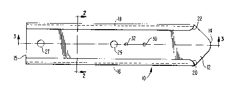
Fig. 1 is a top plan view of the insertion tool
for an intraocular lens according to the invention;
Fig. 2 is a cross-sectional view taken along line
2-2 of Fig. };
Fig. 3 is a side cross-sectional view taken along
line 3-3 of Fig. l;
Fig. 4 is a top plan view of the tool according to
the invention positioned within the eye and showing an
intraocular lens loaded in the tool;
Fig. 5 is a side cross-sectional view of the tool
according to the invention when loaded with an intraocular
lens for insertion into an anterior chamber of the eye; and
Fig. 6 is a diagrammatic si.e cross-sectional view
of a portion of the eye showing the tool located within the
eye through an incision for performing the method shown in
Figs. 4 and 5.
~7756~i
DETAILED DESCRIPTION OF THE PREFERRED EMBODIMENT
In Fig. 1, a tool for inserting an intraocular
lens into an eye is designated generally by the reference
numeral 10. The tool 10 includes an elongated planar member
12 defining a smooth upper surface 13 and a blunted,
generally V-shaped, distal end 14 for insertion into an
eye. Prefera~ly, the tool 10 is tapered slightly toward its
distal end 14. When the tool is inserted through an
incision in the eye at a point where the cornea meets the
sclera, or white portion of the eye, the blunted distal end
14 is located at the outer diameter of the scleral spur.
The elongated planar member 12 of the lens
insertion tool 10 defines a pair of opposed spaced channels
16 and 18 extending longitudinally substantially along the
15 entire length of the planar member 12. The channels are
respectively defined by curved outer members 20 and 22 which
respectively terminate in inwardly-directed, rounded end
portions 24 and 26 to define the channels 16 and 18. The
end portions 24 and 26 are located inwardly of the lateral-
most portions of the tool and extend substantially
longitudinally and in parallel to the centerline of the
tool, while spaced upwardly slightly from the upper surface
13 of the planar member 12. ~he portions 20, 22, 24, and 26
thus structurally define the channels 16 and 18 which are
~tructurally adapted for receiving the haptics of an
intraocular lens of the type shown in Figs. 4 and 5 in a
secure relationship which permits free longitudinal movement
of the haptics and optical zone of a lens along ~he surface
13, while securing the lens against lateral movement while
- 30 positioned in the channels 16, 18 of the tool 10. The
~Z77565
interiors of the channels 16 and 18 are smooth to permit the
haptics of the intraocular lens to travel sm~othly
therealong for easy insertion into an eye as appropriate.
Preferably, the tools are made by a known process,
such as injection molding, of a suitable polymer material
such as polypropylene having a length of about 40 mm and a
width of about 7 mm.
A pair of openings 27, 29 are provided in the
planar member 12, generally nearer its proximal end 15 to
permit drainage of sterile solution when a lens is placed on
the tool 10 prior to insertion.
The planar member 12 further defines a distal
through-opening 30 and a proximal through-opening 32,
respectively located at pre-determined distances from the
blunted distal end 14 of the tool 10. Preferably, the
distal through-opening 30 is located at a distance of 11.5
mm from the distal end 14, while the proximal through-
opening 32 is located at a distance of 13.5 mm from the
blunted distal end 14. The through-openings 30 and 32
provide a convenient method for determining whether the eye
is of sufficient size to receive a pre-selected IOL having a
siven size. As best seen in Fi~. 4, when the tool 10 is
inserted through an incision 34 in the eye and the blunted
distal end 14 is located adjacent the scleral spur 42 of an
2S eye 40, the through-hole 30 is located over the iris 44,
while the through-opening 32 is located over the sclera
42. Thus, when the physician views the iris 44 through the
distal opening 30, and the sclera through the proximal
opening 32, the eye is appropriately sized. Thereafter,
because the channels 16 and 18 are freely open to the
1~7756~
exterior at the proximal end 15 of the tool 10~ a lens 50
may be inserted into the channels 16 and 18 and moved in~o
position in the eye. The channels 16 and 18 are mutually
opposing and structurally adapted to receive the ends of
haptics 52 and 54 of the lens 50 for the particular style of
lens 50 shown in Fig. 4.
It may be thus understood that lenses of various
sizes and various operable compression ranges are thus
available to accommodate a wide range of eye sizes. Thus,
preferably, a tool of the type described is provided with a
lens wherein the tool is sized to accommodate that
particular lens. In that case, the dimensions of the
respectivç through-holes may vary from the preferred range
stated to fit more precisely with a particular lens in the
range of sizes of available lenses. Thus, in use, if the
physician determines that iris shows through both openings
30, 32 for a preselected lens and its particular tool 1~, he
will immediately know that the preselected lens is too s~all
and select a larger ~ize lens. When he selects the large
size lens, he may either merely insert it with its
specialiæed tool 10, adapted to that larger lens, or follow
the same procedure as outlined above to test whether the eye
is correctly sized to receive that preselected large size
lens. If, on the other hand, sclera appears through both
openings for a particular preselected lens, he will know
that the first preselected lens is too small, and select a
smaller size lens based on that observation. In either
case, an attempt to insert an incorrectly-sized lens and the
resulting possibility that it may need to be removed from
the chamber in favor of a correctly sized lens is avoided by
-- 8 --
~2'~7S~.~
the tool of the invention incorporating the gauge as
described.
The tool is particularly adapted for securing the
haptics of a CooperVision model 680 lens having an optical
zone 51 and a pair of spaced, opposed haptics, 52, 54 having
a lateral extent suitable for insertion in the spaced
channels 16, 18. However, other types of lenses and varying
haptic shapes may also be used with the tool shown, or the
size of the channels altered to accommodate other haptics.
With the lens 50 positioned in the channels 16 and
18 as shown in Figs. 4 and 5 and tool 10 positioned within
the eye as shown in Figs. 4 and 6, the lens 50 may be moved
along the channels 16 and 18 into position within the eye
60. The lens 50 is then held in place within the eye 60
while the tool 10 is withdrawn from the incision 34. The
proximal haptic 54 is then positioned within the scleral
spur 42 and the procedure completed.
In the method according to the invention, as
illustrated in Figs. 4 and 6, an incision is first made in
an eye 40 at a location where the cornea 64 meets the sclera
42. The tool 10 is then inserted with its blunted distal
end 14 forward, into the incision 34 in the eye 40 to an
extent where the distal end 14 contacts the scleral spur
42. When so positioned, the through-holes 3Q and 32 are
2~ positioned as described in connection with Fig. 4. 5ince
the distances of the holes 30 and 32 from the blunted distal
end 14 represent the operable compression range of the
haptics 52 and 54 for the intraocular lens 50, the inserting
physician then determines, using the tool, whether the eye
is of sufficient size to receive the intraocular lens 50.
~Z775~;S
When the physician sees the iris 44 beneath the distal
through-hole 30/ and the sclera 42 through the proximal
through-hole 32, the eye measurements are correct. When it
is determined that the eye measurements are correct, the
intraocular lens 50 is inserted into the channels 16 and 18
as shown in Figs. 4 and 5 and moved longitudinally along the
tool 10 into position in the eye. Thus, channels 16, 18
guide the haptics 52, 54 and thus the lens 50 into the
desired position by movement along the top surface 13 of the
tool 10. The structure of the tool 10 secures the lens 50
against lateral movement, while freely permitting
longitudinal movement. The lens 50 is then held in place in
the eye, while the tool 10 is withdrawn from the incision 34
leaving tbe lens 50 within the eye 40. The proximal haptic
54 is then positioned within the scleral spur 42 and the
procedure completed.
This invention may be embodied in other specific
forms without departing from its spirit or essential
characteristics. For example, the structure of the channels
in the preferred embodiment may be altered to accommodate
particular haptics for particular lenses. The present
embodiments are, therefore, to be considered in all respects
as illustrative and not restrictive, the scope of the
invention being indicated by the claims rather than by the
foregoing description, and all changes which come within the
meaning and range of the equivalence of the claims are
therefore intended to be embraced therein.
-- 10 --
