Note : Les descriptions sont présentées dans la langue officielle dans laquelle elles ont été soumises.
:~2~j~7~
E~HAN OE D-I~A OE OP~RATING HICROSCOP~
~AC~GROUND OF TH~ INVENTION
1. ~ield of the Invention
This invention relates to improved operating micro-
scopes, that is, to microscopes used by surgeons inperforming surgical operations.
2. Related Art
Many thousands of operating microscopes, such as
the Zeiss Company's OPMI microscopes, arP in use
throughout the world. United States Patent 4, 448, 498 to
Muller et al. shows such an operating microscope. These
microscopes provide a surgeon with a binocular stereo-
scopic image in which he can observe his surgical imple-
ments in the operating field; he can perform the entire
operation without looking directly at the subject. The
Zeiss OPMI microscopes are optical (that is, provide a
real image which is seen in an eyepiece)~ as opposed to
an operating microscope providing a video image on a
video screen or the like.
7~9
-- 2 --
Conventional image enhancement techniques allow one
to derive additional information from a visible image.
Typically, mathematical techniques such as Fourier
transformation are used to enhance or bring out detail~
of the image which are obscured in the unenhan~ed image.
Such enhancement technique~ typically involYe
producing an image signal responsive to an image o~ the
object using an image sensor such as a charge-coupled
device (CCD) image forming element. The image signal i5
then digitized, ~or example, using an analog-to-digital
converter connected to the CCD image forming element.
Data processing equipment and techniques are then used
to provide the desired image enhancement. The enhanced
image can then be displayed on a video display or the
like. Insofar as such conventional image enhance-
ment techniques have been applied to microscopes, the
only display provided of the enhanced image has been ~
video display or the like. Performance of image
enhancement tends to obscure the visible "landmarks"
used by th~ surgeon to orient himself with respe~t to
the tissues of the patient.
S~MMARY OF THE INVENTION
The present invention is an operating microscope in
which the image of the object is split using a beam-
splitter or the equivalent. A first portion of theimage is passed through to the eyepiece without modifi-
cation. This unaltered portion of the ima~e is the
ordinary visible image. The split portion of the image
is then processed using image enhancement techniques so
as to provide an enhanced image. The enhanced image is
then combined with the visible ima~e, and the combined
images are presented at the eyepiece.
In this way the surgeon is simultaneously provided
with the ordinarily visible landmarks, ~hich he is
accustomed to seeing in the visible image, and the
detail shown in the enhanced image. The advanta~es of
image enhancement are thus realiæed without the
detrimental loss of physical landmarks and "clues" found
in the visible image.
According to an aspect of a preferred embodiment of
the present invention, in which it is configured as an
aftermarket or retrofit image enhancement system for the
Zeiss OPMI microscope, image enhancement capabilities
can be readily and convenien-tly added to pre-existing
equipment at relatively low cost and without impediment
of the other functions of these microscopes.
Accordingly, the present invention provides an
improved operating microscope, comprising: optical
means for defining a first optical path for a visible
image of an object through said microscope from an
object location to an eyepiece; and image enhancement
apparatus, comprising: beamsplitter means located in
said first optical path for splitting said visible image
of said object into first and second portions; filter
~ 2~P,~ 9
-- 4
means, disposed in a second optical path connecting said
beamsplitter means with image formation means, for
removing a part of said visible image from said first
portion; ima~e formation means for forming an image of
the part of said first portion of the image not removed
by said filter means; image enhancement means for
enhancing the image formed by said image formation
means; display means for displayin~ the enhanced image;
an~ means for combining the displayed enhanced image
with the second portion of the visihle image and for
permitting viewing of the combined images by way of said
eyepiece means.
Further, the present invention provides a method
for production of an enhanced image at the eyepiece of
an operating microscope, comprising the steps of:
forming a visible image of an object; selectively
enhancing a portion of the visible image, combining the
enhanced portion of the visible image with the visible
image; and providing the combined images at said
eyepiece for view.
~RIEF DESCRIPTION OF THE DRAWINGS
The invention will be better understood if
reference is made to the accompanyin~, drawings, in
which:
Fi~ure l shows schematically the optical path of
~ 2~
-- 5
the Zeiss OPMI microscope; and
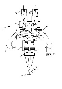
Figure 2 shows schematically ~he optical pa~h of
the microscope according to the invention, in a
preferred embodiment in which image enhancement is
S provided by a retrofit attachment to the Zeiss OPMI
microscope of Figure 1,
7~1~
-- 6
DESCRIPTION OF THE PREFER~E~ 2MBODIMENTS
In a preferred embodiment, the microscope o~ the
invention comprises a Zeiss OPMI microscope together
with a retrofit attachment which provides additional
features and functions according to the present
invention. Figure 1 shows in schematic ~orm the optical
principles of the Zeiss OPMI microscope 11. An object
10 is illuminated by visible light 13. A main objective
lens 12 provides a first focusing function. A
magnification changer 14 then provides further
magnification o~ the object 10. As shown, the
magnification changer 14 provides binocular stereoscopic
images. The images provided by the magnification
changer 14 arP then provided to a binocular tube
assembly 16, in which the images are re~lected by prisms
18 and 20 to provide the proper spacing; the visible
images thus formed are focused by eyepieces 22 for
viewing by a surgeon. Observe that in the diagram o~
Figure 1 the ray paths between the magnification
changer 14 and the binocular tube 16 are collimated;
this feature of the Zeiss OPMI microscope is desirable
but not essential to the practice of the present
invention.
The modifications made to the Zeiss OPMI microscope
according to the present invention are shown
schematically in Figure 2. According to the present
invention, an image enhancement and summing device 30 is
interposed between the magnification changer 14 and the
binocular tube 16; the microscope 11 is not otherwise
modified.
-- 7 --
In keeping with the binocular stereoscopic nature
of the Zeiss OPMI microscope 11, in the pre~erred
embodiment of the present invention the image
enhancement and summing device 30 has essentially
identical mirror-image left and right hal~es. This
enables the advantages of stereoscopic projec$ion ~that
is, three-dimensional viewing of the object 10~ to be
obtained in combination with the advantages of image
enhancement provided by the present invention. Both
halves of the image enhancement device 30 comprise
beamsplitters 32 which separate portions of the im~ges
provided by the magnification changer 14 from th~ main
optical path.
The split portions of the images will typically but
not necessarily then be filtered by narrow band pass
filters 34 of conventional design, to separate out parts
of the visible spectrum of interest. The remaining
parts of the split portions of the images are focused by
lenses 36 on sensors 38. Sensors 38 may be o~ any
conventional type, such as Newvicon video tubes, charge
coupled device (CCD) image forming arrays, or metal
oxide semiconductor (MOS) type image forming sensors,
and the like. Sensors 38 provide video signals as
output. Other devices for forming the video image, such
as one- or two-dimensional scanning systems, for
example, linear CCD arrays scanned across the image, are
within the scope of the present invention. Multiple
element sensors, each element sensitive to a different
portion of the spectrum, are also within the scope of
the invention.
The video signals output by the sensors 38 are then
digitized and processed for enhancement of various
-- 8
features of interest on $he object 10 by real time image
processing devices 40. For example, the filt~ring of
parts of the visible spectrum by filters 34 and
subsequent image processing by real time imag~
processing devices 40 may provide visual information to
the surgeon concerning variation in tissue charac-
teristics not otherwi~e visible. Any type sf
conventional image enhancement techniques can be
employed.
lo The image enhancement techniques which may be
employed include but are not limited to the Pollowing.
Freguency-domain enhancement, which will typically
comprise increasing the gain of a portion of ~pecified
frequency of the video signal provided by sensor-~ 38~
can be employed, for example to enhance a weak
fluorescence of the o~ject 10. Such frequency-domain
enhancement may be performed by Fourier transformation
of the video signal and selectively increasing the gain
applied to portions of the transformed signal in
production of the enhanced image.
Edge enhancement may also be performed. Typically
this technique involves dividing the video image into
picture elements ("pixels") and comparing the amplitude
of a particular parameter (for example, color, density,
or albedo) of each pixel to the amplitude of the same
parameter in adjacent pixels. To produce the enhanced
image, one then i~creases the gain applied to the pixels
of the displayed image corresponding to those in which
the change in the selected parameter is greatest. This
may be done by taking the derivative of the parameter
along a row (or column or both~ of pixels, to identify
the pixel of the row in which the rate of change of the
~3 ~PJ97~
parameter is greatest, and increasing the gain applied
to the corresponding pixel in a displayed video image.
The area outlined by the edge may be false colored.
Furthermore, while the converted image is literally
a ~'false-color" image, even if it is essentially a yray-
scale image, it is also within the scope of the present
invention to provide a true multi-color converted image,
in which various colors of the displayed image
correspond to differing wavelengths of the reflected
radiation. Multiple-element displays, each element
displaying a different portion of the visible spectrum,
are also within the scope of the invention.
As is well understood, edges represent high-
frequency detail in an image, inasmuch as edges are
sudden changes in a parameter. Edge enhancement thus
may also be performed by increasing the high-frequency
gain of the signal processing system of the present
invention. This in turn may be accomplished by
providing a nonlinear gain ~that is, accentuating the
high frequency response) in the video signal processing
circuitry, which may be digital or analog in nature, or
by increasing the gain applied to the high frequency
components of a Fourier-transformed signal.
Texture enhancement may also be performed according
to the present invention. This technique involves
varying the gain applied to a portion of the image which
exhibits a predetermined texture. The area exhibiting
the texture to be enhanced may be located on the image
by comparing the amplitude of a parameter of each pixel
with that of pixels spaced a number of pixels away. For
example, the signal level of each pixel may b~ compared
to the same parameter for pixels spaced three, four,
-- 10 --
five and six pixels away. An area in which a particular
repetitive pattern is detected in ~his m~nner may be
identified as having a particular tPxture. The gain
applied to the corresponding area of the displayed image
may accordingly be varied to provide ~exture
enhancement.
These and other image enhancement ~echniques and
algorithms, specifically including convolutional and
deconvolutional techniques (as these terms are
under5tood in the art), which are u~e~ul in enhancing
particular types of image structures, are within the
scope of the present invention. These techniques and
algorithms may be implemented using analog or digital
techniques.
According to the present invention, the enhanced
images provided by real time image processing units 40
are then used to drive displays 42, which may be of any
conventional type such as small cathode ray tube video
displays, liquid crystal displays, CCD displays or the
like. Multiple-element displays, each element
displaying a different portion of the spectrum, are also
within the scope of the invention. The enhanced images
provided by displays 42 are focused as needed by lenses
44, and are then combined by beam combiners 46 with the
visible images provided by the magnification changer 14.
The combined visible and enhanced images are then
provided in real time to the surgeon by way of mirrors
18 and 20 and eyepieces 22 as in the unmodified
microscope. As shown, the combined visible and enhanced
images may be recorded, separately or as combined, on a
video recorder 50, a camera (not shown), or the like.
It will be appreciated that the real time
combination of the visible image with the enhanced image
according to the present invention has several
advantages over an arrangement, for example, in which
the enhanced image alone is displayed on a display
screen which has to be viewed by the surgeon separately
from the visible image viewed through the operating
microscope. Specifically, suppose that the enhanced
image shows that an area of tissue is diseased, while
the outline of the diseased area is not visible on the
unenhanced visible image. In ~nhancing the image,
visible '~landmarks" used by the surgeon to orient
himself with respect to the tissues of the patient are
freguently obscured. ~herefore, in order for the
surgeon to correlate the location of the diszased tissue
shown in the enhanced image with the unenhanced visible
image viewed in the operating microscope ~that is, in
order for the suregon to determine where the diseased
tissue is located on the patient), a "landmark" or
another "clue" or indicia appearing in both images would
have to be provided. In addition, the surgeon must view
the enhanced and unenhanced images sequentially, which
may not always be possible or desirable.
By comparison, the real time, superimposed
combination of the enhanced and visible images provided
by the present invention automatically provides a
correlation between the enhanced image provided by the
image enhancement device and the visible image, which
includes the ordinarily-visible landmarks or clues.
As noted~ in the preferred embodiment of the
pr~sent invention the image enhancement subsyst~m 30 is
provided as a retrofit to an existing Zeiss OPMI
- 12 -
binocular stereoscopic microscope. Accordingly, two
complete image enhancement and combining devices are
provided. In this way the advantages provided by the
present invention are provided to both sides of tne
stereoscopic mi~roscope, further increasing the utility
of the microscope according to the present invention
while not interfering with its other desired attribute~
and operating advantages. Provision of image
enhancement in a monocular or non-~tereoscopic
microscope is also within the ~cope of the present
invention.
As mentioned above, one ~eature of the Zeiss OPMI
microscope which makes it especially amenable to
retrofitting with the image enhancement system o~ the
present invention is the ~act that the optical paths of
the images are collimated between the magnification
changer 14 and the binocular tube 16. This collimation
simplifies interposition of the image enhancement and
summing device 30. The fact that the Zeiss OPMI
microscope is constructed in a modular fashion also
encourages modification according to this aspect of the
present invPntion. However, it will be appreciated that
in general it will be possible to retrofit non-modular
microscopes and ones not including a comparable
collimated optical path with image enhancement and image
combination apparatus acrording to the present
invention.
It will further be appreciated that while the
present invention has been described primarily in
connection with a retrofit image enhancement system for
addition to a pre-existing microscope, the present
7~
_ 13 -
invention al50 includes a complete microscope
constructed according to the present invention.
Many modifications ~nd improvements to th~
preferred embodiment of the present invention de cribed
above will occur to those of ~kill in the art, and are
therefore within its scope. In particular, ik would be
possible to eliminate the beamsplitter 32 and filter 24
o* each optical path in favor o~ a device for ~ormlng a
video signal responsive to the image of the obj~ct, ~uch
as a CCD sensing array, an analog-to-digital conversion
device for forming a digital sign~l responsive to the
video signal, and a digital filter to select the video
information of interest for image enhancement. After
enhancement, the enhanced signal could then be digitally
combined with the unmodified diyitizsd video signal and
used to drive a video display device, to provide the
real time, superimposed combination of the visible and
enhanced images.
Therefore, while a preferred embodiment of the
present invention has been described, this should not be
taken as a limitation on the present invention, but only
as exemplary thereof; the present invention is to be
limited only by the following claimsO
