Note : Les descriptions sont présentées dans la langue officielle dans laquelle elles ont été soumises.
1 ~ 1 3 1 1 4
~escr.-lp~ion
ME~ ODS AND DEV~CE FOR ~E:TECTION Ol? MlCROORGANlSMS
The present invention relates to an improved
device fot detection and estima~ion of numbers of mi~ro-
oeganisms. The invention facilitates inoculation and
incubation of cultures and collection and visual.izat.ion of
gas from the cul~ures. More parti.cular.ly, this inventiol-
relates to a device, an apparatus, and methods for detect-
~ ing gas produced from the metabolism of certain chemical
: subs~ances by samples of pure or mixed cultures of micro-
organi.sms. The invention may a.lso be used with fluorogenic
: and chromogenic substrates ~o detect the presence o~
specific microorganisms withi.n a sample.
1.5 ~denti~icati.on of a specific microorganism or a
. group oÇ microorganisms within a na~utal sample can provide
:~- use~ul in~ormation. ~I~his iriforma~ion may be necessary ~or
decision-mak.ing in cases of possible contam.ina-ion or in~ec-
tion. The informati.on may be used to make retrospect;.ve or
predi.ctive judgments about an i.dentified samp.l.e. Ior
irlst.ance, the detection of co].iform bacteria withi.n a food,
dairy or water. sample suggests fecal contamination of ~hal:
samp.le, and in many instances, cau~i.ons against hl.~man
: consumption for fear of gastroenteritic disease. ~Iterna-
'~ 25 t.ively, isolation o~ a characteris~i.c bacterium from an
individual with a speci~ic clinj.cal presen~a~iorl usuall.y
estab~ishes a diagnosis and course of therapy ror tllat
: individual.
Samples whicll are suspec~ed of hatboring cer~ain
microorgani.sms are analyzed by prescribed me~hodologies
designed to demonstrate the ptesence of a taxonomica]ly
defined category of microorganisms. The more common charac-
'~
, `~
.. : .,
. .
:; :
~ .
- . .
1 31 31 1 4
teristi.cs which are used to d.istinguish microorganisms are
morphology and ultrastructure, staini.ng react;ons, physi-
ological. and bi.ochemical activities, and ecol.ogy. lhe
physiologica.l and biochemical reacti.ons that microorgani.sms
perform are among the most useul charac~er.istics for iden-
~ification~ Typically, a population oE mi.croorganisms is
assayed for the abili.ty to utilize a specific substrate for
growth, or to convert that substrate to a characteristic
product. Growth is usually indicated by formation of a
visible mass of cells on a solid growth med.ium or by ~he
development of turbidity in liquid grow~h medium. Forma-
tion of a characteristic product may be accompanied by a
change in color (from a change in P~! or oxidation-reduction
potential or from enzymatic cleavage of a chromogenic
15 compound), development of fluorescence, depositi.on of a
. preci.pitate, or accumulation of gas in the growth medium.
In the fie~.ds of watee, dairy, and food micro-
biology, samples are regularly moni.tored for the presence
of microbial contaminants. Food, dairy, and water samples
are tested for critical concentrations oE organi.sms chosen
as indicators of the presence of pathogens. Testing Eor
~, indicators avoids the necessity of assayi.ng for every
., conceivabie pathogenic microorganism. The group of acrobic
, and facultatively anaerobic, Gram-negat;.-~e, non-spore-
forming, rod-shaped bacteria that ferments lactose with gas
production within ~8 hours when incubated a~ 35C, is
designated the coliform bacteria, and is widely used as an
indicator. Some coliform bacteria, such as r.scherLchi_
coli, are associated with Eecal contami.nation.
rhe taxonomic breadth of microbial i.dell~ifica~ion
may be altered for specific applications. For exampl.e, the
; epidemiologist may desire categorization at the level of
:; subspecies or variant, while the sanitarian may ~imit i.den-
tirication to an operationally defined group, such as the
coliform bacteria. At either extreme o[ taxonomic deter-
mination, millions of identifications are per~ormed
annually, with many of the assays involving tedious steps
~'~
:.
~ , :
. .
.
` 13131 14
and requiring considcrable labora~ory ~pace and ~quipmen~.
Conscquen~ly, a method which p~ov.ide~ easy an~ c~Cic;~r
m;crobial. det~ctiQn, identi.ri~a~.ion, or estimation wiLl.
save the laboratory considerab.le time and money.
S One technique frequen~ly used ror the estlmation
of coliform bacteria i.n foo~ and water samplcs .is ~.he
most-probable number (M~N) test. 'rhe MPN t--st i9 based ~n
p~obabil.ity statistics, and results are direc~ly related ~o
the requency o~ occ~rrcnce o~ a serics o~ posi~ive resul.l:s
wh~n given numbers of microorganisms are present in a
sample. The MPN technique is based on ~wo assumptions.
First, i a sample contains microorganlsms, the micro-
organisms are randomly distributed throughout a dilution or
homogenate o~ the samp].e. Second, when an aliquot of cul-
~5 ture medium is inoculated with a viable cell and processed
properly, a "positive" resul.t will be observed.
oth the ML~N procedure and ttle direct p]ating
technique are used to quantitate viablc microorganisms.
tlowever, for many samples, plat.i.ng of diluted samples on
agar plates and direct counting of resu.Ltant colonies are
. not ~easib]e. Certain bacteria will not grow on any solid
': medium, and thus require cul.tivation in liquid medium. The
: nature of some Cood products makes standard plating proce-
dures di.fficul.t, because of par~iculates and l.ow microbi.al.
density (i.e., < 10 organisms/gm). 'l'he MPN procedurc uti-
lizes l.iquid growth media, and permi.ts greater flexib.ility
i.n inoculum volume and greater sensitivity at low microbia.l
density. ~rhe use of liqui.d media olten enhances the
recovery o~ microorgani.sms that have been debil.i~a~ed by
process~s such as heating, dryi.ng, or the addi.ti.on of a
disinfectant or sanitizi.ng ag~nt.
. ~ Specifically, ~he MrN technique rcqui.rcs di~uti.or
of a test sample until Lhe microbial density is l.ess than
one v.iable cel.l. per mi.lliliter. Rep]icate aliquots are
removed from appropriately diluted samples, inoc.ulated into
separate tubes of growth medium, and i.ncubated to allow
growth. Usually three to fi.ve serial 10-fold dilutions are
.'
.
,.,~
.
.
.
:` :
.:
13131 14
'I
tested. Tubes inoculat~d with onc ~r mor~ vial~J.~ cel.ls
wi..ll demonstrate growth; those ~hat did rlot recei.ve a
v.iab]e ce].l. wi~l not demonstrate growth. 'l`he proportion Or
tubes that demons~ra-e growth is a mcasure of the probabil-
i.ty of receivi.ng a v;able c~l.l. ~his probab:il.ity can beconverted back i.nto cel]. concentrati.on l)y uqe o~ statis-
tical tables, constructed for use with thrce, five, o~ ten
replicate samples per dilution. Accuracy Or the Ml'N ~est
i.ncreases as the number of replicat.~s per dilut.;on
increases.
The MPN procedure may be used to estima~e the
total number of viable organisms, as well as the number o~
a variety of specific bac~eria or groups oE bacteria. Each
MPN estimation i.s accomplished through detection oE charac-
teristics that are unique to that organism or group oforganisms. For exampl~, gas production resul.t.ing from the
fermentation of lactose under appropriate condit..ions is
used to estimate coliform concentrations through use of the
MPN procedure (designated the coliform MPN procedur~).
Within this appli.cation, any microorganism that
produces gas during metabol.ism will be referred to as an
"aerogenic microorganism." In a modifi.cation of the coli-
form MPN procedure, fluorogenic or chromogenic substrates
may be added to the growth medium. For instance, non-
fluorescent or non-colored agents may be cleaved 5peC i fical.-
ly by the coliEorm bacterium F.. coli to yield a ~luorescent
or colored product. Inoculated growth medium containing a
fl.uorogenic or chromogeni.c substrate would be examined
ater 24 h at 35C under an appropriate ligh~ source, and
the presence of El.uorescence or color would indi.cal:e lle
presence of E. coli in ~he t~st sample. Such medil.lm
modification thus permits the additional identi.~ication of
coliform samples that spccifica.Lly ccntain !~ C! i . In
addition, the MPN Eormat in whi.ch the test was conducted
may permit a separate estimation of the number of t;.. ~cl~
in the sample.
... ..:
. . .
~ \\
1 3t 3t 1 4
At present, sev~ral dovic~9 or me~hods ~re avail-
able ~or pet^~ormlng coliorm MPN t~st~, hu~ all possess
cer~ain disadvan~age~. Mos~ Ml'N methods ~ha~ allow detec-
tion of gas production by growing bacteria rely on tlle
displacement of culture ~luid by the gas produced ~o form a
visua~ly detectable bubble. One such gas de~ection method
employs a J- or V-shaped culture vessel or fermentation
tube containing one open and one closed cnd. Liquid growth
medium is placed into the open end of ~he tube, and the
tube is tilted so that the medium Çil]s the closed arm Or
the V. The tube is ~hen inoculated and incubated, and any
gas produced within the closed arm of the V rises and accu-
mulates at the top of the arm. This method is useful for
both qualitative and quantitative gas determinations, but
suffers from several drawbacks. First, the J- or V- tubes
employed are blown glass and are relatively expensive.
Furthermore, the tubes are delicate because they sit on a
pedestal, and the multiple operations that are required for
their use (e.g., washing, steriliæing, filliny, inocu]at-
ing, and handling to and from the incubator) make them bothsusceptible to breakage and inconvenient to use. Moreover,
this susceptibility to breakage and spillage poses a spec-
ial biohazard when pathogenic microorganisms h~ve been
cuLtured. Finally, the tubes are bulky and require a large
~25 amount o~ incubator space, making the simultaneous perEorm-
ance of multip]e tests unLeasible. As a resu3t oE the
limitations noted above, V- and J-tubes havc not received
general acceptance for use with the MPN procedure.
The most widely used and presently preferred
30~method for gas entrapment and detection in conjullc~ion wi~h
MPN determinations employs a capped test tube containing
liquid growth medium. Visposed within this capped tube is
a second, smaller diameter Durham tube that is inver~ed in
the growth medium. When the ~est tube is inoculated wi~h
aerogenic bacteria and incubated, gas produced hy micro-
organisms collects and forms a bubble under the inverted
` Durham tube.
:
~- ~
- : ": . .. .
, ~ :
.,~
~, ~
6 13131 14
l~or MrN cle~erm.in~on usiny ~cst ~ubes c~n~ai.ning
~urham tube~, inoculated ku~c~ arc o~en p].acocl i.n a thrce-
by-three array in a test tube rack (i.e., three rep~i.cates
of three 10-fol.d dilutions). Af'ter incubation, the tubes
are obseeved for gas producti.on. 'l'he l.~urham tube m~thod is
suboptima~, because on.ly ~he gas produced hy microolc~an.isms
under t.he Purham tube is co:l.l.e~ted, ~nd con~qu~ntly, onl.y
a Craction of the culture's aerogenic potential .i9 samplecl.
Second, air must be. evacuated from the Durham tube by heat
sterillzation, This step sufÇers from the disadvantage
that some growth components are heat labile (e.g., sugars),
and therefore must be added separately ~o each culture tube
from a ~ilter-sterili.æed stock after the heating step.
Third, air may be~ inadvertantly .introduced i.nto the Durham
'~ 15 tube upon addition of the inoculum.
Another method ~or ML~N estimati.on through de~ec-
tion o~ trapped gas involves over~aying an inoculated test
tube of broth medium with heavy mineral o.il or molten agar.
~ These overlayed substrates form a barrier at the top o the
: ~: 20 broth medium under which gas bubbles co.l.lect. Tllis overlay
method has several drawbacks: (1.) it is tedious, s.i.nce i~
requires a separate add.iti.on to each culture t.ube; (2) it
requires a high degree of skiL]. to over.lay moltell agar on a
l.iquid medium; (3) it is time-consuming, because it
~'~ 25 involves separate preparatioll of the overlay materials and
sterile addition to cult.ure tubes; and (4) in the case oÇ a
m.inera]. oil overlay, the method is unrel.iable because
bubbLes frequently escape betwc~n the test tube wall~
: mineral oil i.nterface-
A more recent method utilizing gas elll.ra~ment and
; visualization is exemp.Lified by the 3M produc~, I'etri~i.lm.
When Petrifilm is i.noculated with a liquid sampl.e a gel-
containing growth medium is rehydrated. 'I'he ;.noculated
medium is sandwiched between two pl.astic [ilm sheets, [orm-
i.ng a thin ].ayer for incubation. L3acterial colonies are
detected as stained masses of cells, due to thc incorpora-
tion of a dye in the growth medium. Gas produced by micro-
~: .
,.,, .,. . , . --.
,; , ~ ` ':', ' ~' ;
~``` 1313114
organisms within a colony .is entrauped between the .I.ayers
o~ the ~ilm immediately adjacent to he colony.. This
sandw.lch method, however, requires a degree Oe skill Eor
;noculation and "sandwichi.ng" of the inoculum between th~
two layers oE film. 1~he Pe~.rieilm me~hod may bc used lor.
viable coliform quantitation, but is not practical to use
in an MPN format.
In addition to the noted disadvantages o cuercnt
gas detection methods, as employed with a small number. of
samples, all o~ the described methods become even more
inconvenient when used for large-scale ~esting o~ multiple
sampl.es. Monitoring of gas production by any of the
methods noted above dictates that separate units of the
: particular gas collection apparatus be individually handled
during preparati.on, inoculation, incubation, and visual-
: ~ ization of the results ~or each sample tested. These
multiple independent operations presen~ problems .in
handling, spatial inefficiency in storage and incubation,
and inconvenience during vi.sualization of gas production.
~here is a need in the art, ~herefore, for a gas
collection device or apparatus that entraps gas efEicientl.y
~ : and provides single or multiple tes~s in a stable,
:~ easy-to-handle, spati.ally efficient manner. The present
invention ~ulfil.ls this need and ~urther. peovides o~her
re.lated advantages.
::
One aspect of the present invention re1ates to a
device adapted for containing a volume of growth medium,
the device employed for detection o gas produced by a
microoeganism or microorgani.sms putativel.y contained within
a sample added to the growth med.ium, comprisirlg a primary
: chamber having a top sur~ace, a dome de~ined by the top
surface of the primary chamber and commun.ica-ing therewith,
the dome establishes a 1uid level and is adapted ~or
col.Lection of the gas produced by a microorganism or
microorganisms within the growth medium, and a fill section
.,
.
.
: : .
-
. ~ . .
.:
1 31 3 1 1 4
adjacent to and communicating wi.th one end o~ the primary
chamber, the ~ill secti.on spaced apart from ~he dome and
extending above the fluid l.evel, and further defin;.ng an
openi.ng for the addition o~ the test inoculum.
A second aspect oE th~ present inventi.on re.lal~s
to an apparatus consisting of an array of adjacent, substan-
tially identical devices, each device adapted for contain-
ing a volume of growth medium and employ~d for detection of
gas produced by a microorganism putatively contained within
a test sample added to the growth medium o each device,
each device comprising a primary chamber having a top
surface, a dome defined by the top surface of the primary
chamber and communicating therewith, the dome establishes a
flu.id level and is adapted or collection of the gas
produced by the microorganism or microorganisms within the
growth medium, a fill section adjacent to and communicating
with one end of the pri.mary chamber, the fill section
spaced apart from the dome and extending above the fluid
level, and further defining an opening for the addition of
the test sample, and a means for seJ.ectively closing the
opening defined by the fill sect.ion. Apparati consisting
of three-by-three and three-by-five arrays of ~he device
are preferred.
~ method for detecting the presence oC aerogenic
microorganisms within a sample putat.ively containing a
microorganism ut;.].i~.i.ng the device of the presenL inven-
ti.on, comprising the steps of adding growth mediunl to the
device, adding the samp]e to the growth mediuln, thereby
forming a mixture, incubating the mixture, and detecting
the presence of aerogenic microorganisms by determining the
presence or absence of gas withi.n ~he devic-e is disclosed.
rreferred sarnples inc].ude water, dairy and food samples,
and bio].ogical fluids. Preferred gro~lth media conta;n
0~01-o to l..0O of a wetting agent in a non-bactericidal and
non-bacteriostatic concenLration.
A method for detecti.ng the presence of aerogenic
mi.croorganisms in a plurality of samples putati.vely contai.n-
13131 14
ing a mi.croorganism uti.lizi.ng the apparatus of the presentinvention, compri.sing ~he sleps of addi.ng growl.h medi.um to
at least two devices of the apparatus, preferabJy sa.id
growth med;.um contains a w~tting agent to ~ac.i.li.t.ate m.igra-
t.ion o ~he gas bubbles i.nto the dome, add.lng thc samplesto the dev.ices containing the growth medium, thereby form-
lng mi.xtures, incubating the mixtures, and detecting the
presence of aerogenic m.icroorganisms by de~ermini.rlg the
presence or absence of gas w.ithin each device is also
discl.osed.
Another embodiment of the invention di.scl.oses a
method for estimating the numbee of aerogenic microorgan-
isms within a sample utilizing the apparatus of the present
invention, comprising the steps of adding growth medium to
at least two devices of t.he apparatus, adding serial dilu-
tions of a sample containing at least one aerogenic micro-
organism to the devices containing the growl.h medium,
thereby forming mixtures, incubating the mixtures, and
: estimati.ng the number of m.icroorganisms within the sample.
A further aspect of the invention discloses a
method of analyzing a sampl.e putati.vely contai.n;ng a micro-
organism utilizing t.he device oL the presen~ invent:ion,
comprisi.ng the steps of acld;ng growth medium conta;ni.ng a
rl.uorogen;.c or chromogenic substrate to the device, adding
2S the sample to the growth medium, thereby formi.ng a MiY~ture,
i.ncu~ating the mixture, detect;ng the presence of aerogeni.c
microorganisms by determining She presence or absence Or
gas within the device, and detern-ining the presence of
microorganisms by detecting t.he presence or abscnce o~
f.luorescent or colored product~s within the growth medium.
Yet another aspect of the .invention describes a
method for analy?ing a pl.ural.i.ty oE samples, each ~-u~a-
tively containing a microorganism or microorgani.sms utili.~-
ing the apparatus of the present invcntion, comprising the
steps of adding growth medium containing a fluorogellic or
chromogenic substrate to at ~east two devices of the appara-
tus, adding the sannples to the devices containing Lhe
,;
1 31 31 1 4
.lo
growth medium, thereby ~c~rming m.i.x~ures, incuba~ing the
mixtures, detecting the preser,cc o microoryan.isms by
determining the presence or absence Or gas wi.~hin each
device, and determ.ini.ng the presence o~ microorganisms by
detecting the presence or abs~nce Oe ~.I.uore:3cen~ or co.lor~d
products within the geowth medium.
A method of analy~ing m.icroorganisms witllin a
sample utilizing ~he appara~us of the present inventjon,
comprising the steps of addin~ growth medium containing a
1uorogenic or chromogenic substrate to a~ .Least two
devices of ~he apparatus, adding serial d.ilutions oE a
sample containing at least one microorganism to the devi.ces
containing the growth medium, ~hereby ~orming mixtures,
incubat.ing the mixtures, estimating''the number of aerogenic
mi.croorganisms within the sample, and determining the
presence of microorganisms by detecting the presence or
absence of f1uorescent or colored products within tl
growth mediu~.
In the accompanying drawings:
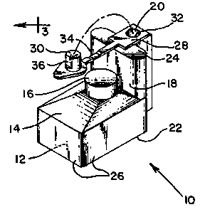
; Figure 1 is an isometric view o~ a preerrcd
embodiment of an apparatus constructed according ~o ~he
principles o~ the present i.nvention.
Figure 2 is a top plan vi.ew Or an alternative
preferred embodiment of the present inven~ion, comprising a
two-dimensional array of devices as depicted i.n Figure 1.
Figure 3 represents a cross section o~ the appara-
tus shown in Figure 2, on t.he line designa~ed 3-3.
As noted above, current devices and ~ecllniques
for detecting gas production are accompanied l~y numerous
disadvantages. Presently avail.able me~hods arc Lediolls,
ti.me-consuming, and/or reql-ire a cer~ain level o~ skil.l to
per~orm eeproducibly. In addi~ion, some methods of gas
detection do not maximi.7.e gas co~1ec-tion, since only a
portion of the gas produced ~y a given microbial culture is
'. ..: ~
`
., . -
:,'. ' . :
- '''`. . - .~ :
,, 1313114
collected ~or de~ec~lon. Various devices employed Lor gas
co.l.lecLi.on and detec~ion are fragile, cumbersome, and/or
spat.iall.y inconvenient. Further, the devices presently
used for gas co'.l.lection and detection are susceptible to
spi.llage of biohaæardous materials contained wi.thi.n ~he
devices. ln addition, because ~ests utiliæing gas col.lcc-
ti.on, such as the MPN technique, are o~en done on a large
scale, the disadvantages noted above become ampliied.
: The present invention describes a simple,
: 10 improved device for entrapment and visualization of gas
produced by microorganisms within a liquid growth medium.
: The present device provides a more efficient means of gas
collection and is well su.ited to the multip~le unit testing
needed or estimation by the MPN procedure of the number of
lS microorganisms present in numerous test samples.
Referring to Figure 1., a preferred embodiment of
the gas collection device 10 o~ the presen~ i.nventi.on i.s
~: ~ shown. The gas collection device 10 may be constructed
primarily from transparen~ materials, such as plastic or
glass- The base of the gas collection device may be con-
structed from nontransparent materials to better visualize
turbidi.ty and gas bubbles. E'urther, the gas collectior
: device must be constructed with gas barrier material.s of
sufficient thickness so as to signi~ican~ly reduce 1~2 migra-
: 25 tion through the barrier material. Preferab'ly, ~he gas
barrier ma~erial is polystyrene, po]yvi.nylidene chloride,
acrylonitrile-butadiene-styrene copolymer, styrene-acrylo-
nitriIe polymer, polyacrylonitrile, or pol.yvinyl chloride.
This device comprises a primary chamber 12 for accommodat-
ing a volume of growth medium. 'l~he base 22 or ~he primarychamber 12 is substantially rectangular, with opposi~e
walls 26 bei.ng substantiall.y mutual.ly para.l.lel, and adja-
cent walls 26 being substantially mu~ually perpendicu~ar.
.~ The primary chamber 12 has a top surface l~ that defines a
35 dome 16. The dome 16 is adapted to collect gas produced by
microorganisms contained wi.thin the primary chamber 12.
1`he dome 16 extends above the top surface l4, and the
` `:
l2 13131 14
highest interior point o th~ dome l~ e~-ab.l.i.shes a fJ.uid
leve]. The l-op surface 14 i9 s]anted upwardly toward the
dome 16 to faci.1.;ta~e entrapment of gas w;th.in the dome i6.
A fi.ll sect.i.on 1.~ is pos.it;oned adjacent to one
5 end oC the primary chamber 12, anc1 comlnunicateY w.ith the
primary chambcr 1.2. The r i.l.L se~t.ion l8 extends ~bove th~
flu.id leve.l establishe~i by the dome l6. ln acldit.i~n, l.i1e
fi'l~1 sect.ion 18 is spaced apart from the dome ~.6 o~ the top
surface 14. The ~ill section l8 and the primary chamb~r 1.2
share a contiguous base 22 that i.s subsLan~ia.1.ly perpendicu-
].ar to the walls 26 of the primary chamber 12. An inlet
surface 24 defines the upper boundary o~ the fill section
18, and further defines an opening 20 for the introduction
of a test sample (not shown) into 'the primary chamber 12.
In a preferred embodiment, the inlet surface 2~ is substan-
tially parallel ~o the base 22.
The openings 20 of the device :l.0 may be sel.ec-
tively closed by means of a removable closure 28. The
remova~le cl.osure comprises a plug 30 that is coup.l.e-l to a
pl.asti.c insert 32 by means of a flexible hinge 34. The
outer surface of the plastic insert 32 Fri.ctiona.l.1.y engages
the i.nner surface of the opening 20 t.o provide a first seal
therebetween. When the associated plug 30 is ful.l.y
depressed into the opening 20, a second sea~. is creatcd
between the outer periphery o.f the plug 30 and the i.nner
surface o~ the plastic insert 32. In a prc~erred embodi-
ment, the first and second seals created are hermetic sea].s.
'I'he outer surface of the plug 30 defines a r:lat sur~ace 36
that provides for selective venting of the dcvice 10
thrc~ugh partial insertion of the p.lug 30 into the plastic
i.nsert 32. I-~artial insertion of the plug 30 creates a gap
beLw~en the flat surface 36 and the i.nner surface of the
plas~ic insert 32 to a.l..low- pressure eq~a].i~.ation bel;ween
the interior of the device lO and the atmosphel-e.
In an alternative pre~erred embodimcnt, shown in
F`igures 2 and 3, an apparatus lO0 comprises an array of`
substantially identi.cal, adjacent units of a gas collecti.on
.
' ': '
.
- ;
13 1313114
dev.ice l0. In a prc~erred embodiment, the apparatus 100
contains three rows and three columns of adjacent units of
the gas coll.ection de~ice ]0, with the fil]. sections .ll~
disposed prox;mal to one end of the apparal;us l0n. lt
would be obvious to one skil1ed in the art ~hat ~rrays
comprising other comb;.nati.ons ~ rows and col.umns Or aclja-
cent units ~ould be employed. F`or examp.le, a three-by-
three, a three- by-~ive, a five-by-five or a one--by-fi-~e
array may be preferred for some applications.
All devices within the apparatus 100 pre~erably
share a contiguous base 122, and all adjacent primary cham-
bers 112 share at least one contiguous wall 126. Wjthin
the apparatus 100, opposite walls 126 of the primary cham-
bers 112 are substantial.l.y mutually-parallel, and adjacent
lS walls 126 are substantially mutually perpendicular.
Each primary c.hamber 112 has a top su~face 114
that de~ines a dome 116. rhe dome 116 extends above the
top surface 114, and the highest interior point of the dome
116 establishes a fluid level. Gas produced by micro-
organisms contained within the primary chamber 112 collectsin the dome 1.16. The top surface 114 is slanted upwardly
to faci.lit.ate the entrapment of gas within the dome ~16.
Each device of the apparat.us l00 has a fill
section 118 that is disposed adjacent to and communi.cates
with one end of a pfimary chambcr 1].2. ~.l.ong one axis oC
an apparatus 100, ~.ill sect.ions 118 wouJ.d be disposcd
adjacent to each other. ~long the perpendi.cular ax:is of an
- appara~us 100, lill secti.ons 1l8 would be disposed adjacerlt
to, but would not communicate with, a primafy chamber l.l2.
Each fill section 118 extends above the fluid
level establ.ished by a dome 11.6, and i.s spaced apart rrom
dome 11.6. Fach ~i.l.l section .l.18 is con~.inuous with an
inlet surface 124 that is substantiall.y paral1e~l. to the
:: base 122. Each inlet sur~ace 124 furthef delines an open-
ing 120 for the introduction oE test samples (not shown)
into he primary chambe~f 112.
1'1 13131 14
I~`i.gure 3 depi.c~s tll~ internal gconlelry Or ~he
apparatus 100 of li`.igure 2, wh.ich compr.ises an array of
devices, each of wh.i.ch has structu~e anal.ogous to ~he
device 10 shown in Figurc 1.
The fi.l.. l section 18 def.ines an ;.nLermediate
volume 138 and a distal volume 1~0. 'l'he intermediate
volume 138 is selccted to accommodate a vo.lume of flu.id,
such that no part of the total vo.lume of fluid added to the
apparatus 100 can 5pill through the opening 120 as the
1,0 apparatus 100 is tilted 70-120 from a horizontal refer-
ence plane to purge gas from the dome 116. The dista].
volume 140 is designed to minimize the volume of fluid
contained within the fill section 118 when the device is
horizontally disposed. ~ ;
The interior edge 142 of the dome 116 defines a
plane which intersects the plane of the base 122, such that
the portion of the edge 142 adjacent to the fil.l section
118 is elevated at a greater distance from the.base 122 --~
than that portion o~ the edge 142 that .is distal to the
fill section 118. Th;.s, arrangement provides t.hat a pre-
determined maximum volume of gas produced by microorganisms
within thc culture medium can be trapped within the dome
116, and that any additional volume of gas wil.L be conven-
;ently released i.nto the fill section 118. Due to the
ana],ogous i.nternal structure of the preferred embodiment
device 10 of Figure 1 and each device of ~he preferred
embodiment appara~us 100 o[ I~igure 3, there exists
comparab].e functionalily between these embodimerlts.
The openings ].20 of the apparatus 100 may be
selectively closed by means of a removable closure 1.28.
1'he removabl.e cl.osure 128 comprjses ~hrce p.lugs 130, each
coup,Lcd to a plastic insert 132 by means of a flexible
hinge 13~. The outer surface of the plastic insert 132
frictionally engages the inner surfacc of an opening 120 to
provide a first seal therebetween. Whcn fully depressed
into the opening 120, a second seal is created between thc
ou,ter periphery of ~he plug 130 and.the inner surface of
.
. .... ;: - .: ,
. '. ' ~
.. : - :,
,
: : ......... .
.... ..
13131 14
.l.5
.he plasti~ ins~r~ :l32. ln a pr~~rr~d embodimel~t, the
~irst and second seals cr~at~d are hermetic seals. 'I'he
outer surface o~ each plug 130 defines a flat surface .l.36
tha~ provi.des for selective venting o~ a device by
partially inserting the plug l.~0 in~o the pi.asti.c inser~
]32 that is ~rictlonally engaged wi~hin ~he opening 120,
thus creating a gap to allow pressure ~qualization between
the interior o.f thc device and the atmospher~.
The device and ap~aratus described above o~rer
1.0 several advantages over gas co].lection devices pr~v.iousJy
avai.lable. Individual gas collection devices can be easi.ly
adapted into various arrays oE chambers, facilitating
multiple testing of numerous samples. While mu.Ltiple test-
ing with vertical tubes may lead to tipping and potenti.al
spilling ol' biohazardous materials, the major plane of the
; apparatus of the present i:nvention is horizontal, offering
stability and sa~ety. Fur~her, the gas cvl.lection devi.ces,
: ~ either singly or in an array, are self-supporti.n~, and do
not require the use of accessory racks, as wi~h ver~.i.cal
tubes. The elimination of ~he need for racks makes the
~device, either s.ingly or in an array, spatiall.y more effi-
cient. Further, the apparatus can be stacked vertica.l.ly
~or storage, incubation, and sterili~ation aL~er use,
lead;.ng to spatial efficiency.
: 25 ~nother advantage of the apparatus is s.imul~aneous
ai.r evacuation rom the gas col.lection area. Current:Ly
avai.lable gas collection devices in which air was present
or introduced into the gas colJ.ectiorl compar~mellt cluri.ng
~preparation or inoculatiorl would generally be discarded
(:~urham tubes) or evacuated individuall-y (J-tubes).
: : ~ Apparati. of the present invent.ion racilitate simultarleous
air evacuation through the simpJ.e process of tilting ~he
apparatus.
'I`he removable closure of the device may utiJ.i~.e
~5 an i.ndivi.dual hinged pJ.ug, providing ease o~ opening and
closing the entry to the devi.ce. 'l`he hinge ensures that
plugs of the removable closures are not dropped, contami-
..- :
. '' ~
13131 14
I.6
nat~d, or mlsplaccd ~ker r~moval from the plas~ic inscr-s.
The two-position insertion o~ ~he plug a.l.lows ~he dcv.icc to
be either hermetically sealed or ventable. Coupling o~
removable closures in groups of ~hree racili.tates rapid
remova.l. and i.nsertion of pl.ugs in~o ~he apparatus.
A further advantage o~ the devica or appar~Lus Or
the pr~sent invention is disposabi.lity. l)urham ~ub~ and
J-tube devices are steri:l.i.zed aC~er use, c.l.eanqd, and
reus~d. These steps are cumbersom~, ~ime-consumi.ng, and
]o are accompanied by breakage. ~`he device and apparatus
described above provide ease in hand].ing, because ~.he
dev.ice or apparatus is merely disposed of after use. The
device and apparatus are made of inexpensive, transparen~
materials, and are suited to large-s-c'ale production.
15The device of the present invention further pro-
vides superior gas collection. I'he dome of the chamber
collects gas from approximate:l.y 75'3 o the~ volume of the
culture med.ium contained wi.thin the device. Im~ro~ed co~
lection of gas imparts more sensitivity to gas detection
through use of the present inventi.on as compared to the
~ conventional. r-urham- or J-tube devices. In addition, if
; ~ concentrated cult.ure medium is uti.lized, the equili.bration
of the medium after inoculation of the sample is rapidly
achieved through use o~ the present device. In contrast,
use of concentrated medium with convent:ional. devices is
problematic, because the concentrated medium equilibrates
slowly by di.ffusion aEter inoculation.
: The device and apparatus described above are use-
ful for the collection and detection oE gas ~roduced by
mi.croorganisms in test samples. ln an alternate prererrecl
embodiment, he device may contain a steri.le cullure medium
in a concentrated form. 'I'he culture medium should contain
0.01~ to 1.0~ of a wetting agent or surfactal)t. I'rerel-
ably, the surfactant is a nonionic or anionic sur~a-tant.
Most preferably, the surfactant is al.kylphenyl polye~her
: alcohol (i.e., Triton X-100~ or Tri.ton X-102~), silicon
glycol surfactant (i.e., ~ow X2-5211 or L~cw X2-5212~),
, . ..~ .
: ~ -, :
. . .
17
sod.i.um heptadecyl su~ate (Niacet~), nonion.ic secondary
alcohol ethoxylate (Tergi~ol~), polyal.kalene oxide-modiEied
dimethyl. po~ysiloxanes (S;lwet~), or sodium dioctyl su].fo-
succinate (~erosol~). ']`h.is cul.tur.~e medium is appropr.ial:ely
pr~par~d so that the additi.on o~ an i.noculum ~o the cul.ture
med.ium woul.d result in a d~sir~d flnal concent~a~ion of
culture medium. For instance, MPN tes~ing Or samples to
determine the number of coliform bacteria conta.ined wi~hin
each of the samples would involve thc addition o~ an
inoculum to sterile growth medium in the device to ~ield a
vo].ume greater than the volume defined by the primary
chamber, the top surface, and the dome of the device. The
culture medium is placed in the device by means of the
opening in the ill section of the device. After addition
of an inoculum to the device, any air remaining in the dome
of the top surface is easily and rapidly evacuated by
tilting the device such that the dome is comp.lete:ly filled
with growth medium. When the device is returned to the
unti].ted position, the enti.re enclosure, as de~ined by the
primary chamber, the top surrace and the dome, is free o~
entrapped air. lf air is subsequer,tly introduced into the
dome dur.ing further manipulations the device may be ti.lted
at any time to once again remove entrapped ai.r.
1'he device and apparatus provide a s;ngl.e-chamber
format ~or performing presump-ive and conL`irmatory stages
of coliform MrN tests. For instance, cultures that produce
gas after 24 or 48 hours may be confirmed as positive by
supplementing the culture wi.th a concen~raLed so.l.utiorl o[
bril.liant green bile (sG~) med.i.um to an appropria~e ri.nal
concentration. 'l`he device or apparatus is i.l.~ed severa.l
times to remove entrapped air and equilibrate the 13Gl3
medium. A successful single-chamber ~ormat, as described
above, requires proper equ.ili.bration of supplemented
medium, which cannot be rapidly achieved wiLh J-tube or
l~urham tube devi.ces, which depend on dirrusion as noted
above.
~ :;
.... . ~
`` 13131 14
1~
Ihe presence of aerogcnic mic.roorganisms .in a
plurality cE samples may be del.ec~ed l:hrough i.noculati.on o~
individual. devices of Lhe apparatus of thc prcsen~ .inven-
tion. lur~her, ~he apparatus may be u3ed Cor :stiln~ion Or
s numbers of mi.croorgan.isms wi~h;n tcsl. sampl.e.~, for exanl~c,
by appropriately d;l.uti.ng test samp.les Col Mll~ ~esting.
I;`ither three-by-three, three-by-five, five-by-five, or one-
by-fiv~ arrays may be ~mployed .in a preLerred embod;ment,
the choice o~ apparatus emp~oyed to be d~termined by the
.0 number of diluLions and the number of r~pli.cat.es to be
testcd. For MPN testing, tri.pl.i.cate samples from 10-fol.d
dilu~ions may be added to each row of three dev.ices through
the opening in each fill section. Inoculated apparati may
then be stacked for spa~iall.~ ef~icient incubation.
In addit.ion, the dev.ice and the apparatus of the
present invention may be employed for the detection of
mi.crobial gas production and the detect.ion of microbia.lly
produced f~uorescence or co]or resulting from the hydroly-
sis of f~uorogenic or chromogeni.c substraLes in the growth
medi.um. ~or instance, microbial enzyme rcactions may
provi.de a hi.ghly specific, rapi.d and sensitive assay ~or
detection of specific strains of microorgarlislns.
Examp.Les of suitable fluorogenic substl-ates which
may be utili.zed within the present inventior) are l..isted in
Tabl.e .1.
3q
~5
.. .....
.
'''-,``
:, ' ' ` ` : ' ,
1 31 31 1 4
,.~
']'~
~luoro~enic subs~rates
l;'luorescei.n diacetate
~-Methylumbelliferyl ace~ate
4-MethylumbelliferyJ. case.in
4-Methylumbellieryl-Q-I,-arabinopyranoside
4-Methylumbelliferyl-B-t)-fucopyranoside
4-Methy.lumbell.if~ryl.-Q-l,-fucopyranoside
4-Methylumbe~ eryl-B-L-~ucopyranoside
4-Methylumbelliferyl-Q-D-ga~actopyranoside
4-Methylumbelliferyl-a-D-galactopyranoside
4-Methylumbelliferyl-Q-D-glucopyranoside
4-Methylumbelliferyl-B-D-glucopyranosi.de
4-Methylumbelliferyl-B-D-glucuronide
4-Methylumbel~.iferyl nonanoate
4-Methylumbe]liferyl oleate
9-Methy]umbelli.f~ryl phosphate
bLs(4-Methylumbelli.eeyl)phosphate ---
4-Methylumbell.iferyl pyrophosphate diester
4-Methylumbelliferyl-B-D-xylopyranoside
A list of suitable chromogen;c subslrat.es ~or
use within the present invention is contained in 'I`ab.le 2.
TA~L.F. 2
_ _ _ _ _ _ _
Chromogenic subst at~s
o-Nitrophenyl-B-D-gal.actopyranoside
p-Nitrophenyl-B-D-galactopyranoside
o-Nitrophenyl-B-D-glucopyranoside
p-Ni~rophenyl-Q-D-glucopyranoside
p-Nitrophenyl-B-D-glucopyranoside
:~ p-Nitrophenyl-B-D-glucuronide
p-Nitrophenyl phosphate
~ ~ o-NiLrophenyl-B-D-xylopyranoside
: 35 p-Nitrophenyl-Q-D-xyl.opyranoside
p-Nitrophenyl-R-D-xylopyranoside
Phenolphthalein-B-l~-9lucuronide
.
:: '
: `
.
-` 13~31 ~
More specifical.~y, approximately 97% of 1:. co].i
strains tested produce the enzyme B-glucuroni.dase, while
other genera of ~nte_o act~rl.aceae general.l.y do not synthe-
size this enzyme. Ori.ginal.ly, ~-glucuronidase was detectecl
through use o~ chromogenic substrates, but recen~.l.y a mor~
sensitive assay for ~-glucuronidase has been developed,
ut.ilizing the fluorogenic substra~e 4-methylumhel.li~eryl-
R-V-glucuronide (MU~). B-g].ucuronidase cleaves the non-
fluoresccnt compound MUC, producing 4-methylumbelliferone,
: : a highly fluorescent compound detected under long wave
ultraviolet light. One advantage of MUG testi.ng for
~ E. coli is that anaerogenic strains of~ E. co.li may be
:: ~ detected. Further, MUG detection of-E~ col.i may be used in
; ~ 15 combination wi.th the presumptive coliform MPN test to estab-
: lish the presence and/or estimate the number of E. coli in
~: a test sample. The use of ~UG in MUN coliLorm media ~hus
offers a rapid test for E. coli that can be performed with
samples containing mixed popula.tions of microorganisms.
20A significant advantage of the present device i.s
amenabi].ity to incorporati.on of colorimetric or fluorescent
: detection of specific strains of microoorganisms into
~ : methods for improved gas visualization. For example, the
:~ 1uorogenic substate MUG can be added to the Ml'N co].i~orm
: 25 medium to specifi.cally detect the presence of E~. co i in a
:test sample. The apparatus permits simultaneous detection
: ~ of gas and colored or fluorescent products resul~ing from
hydrolysis`of chromogenic or ~luorogenic substrates without
;~ ~further manipulation. ]:n contrast, individual. tubes util-
i~ed for MPN testing usua].ly must be l-eld in a vert;.cal
.posi.ti.on for observation of co.lor or fluorescence. Obsorva-
: tion of fluorescence is difficult because the tubes must be
: ~ positioned essentially perpendicular to ~he ul~ravi.olet
light source. Further, the apparatus of the present inven-
tion is designed to be adaptable to automated surface scan-
ning fl.uorometers and colori.meters. 'I.`he combination Or the
;advantageous design features of the present invention with
~ .
:: :
-'. .': :. .. .. .
. ^ ..,
1 31 31 1 4
~ I
i.mpro~d de-ecti.on Or col.orcd or. fluore3~ent en~ymic prod-
uct-.s offers an lmproved, rapi.d, and easy m~thod ror detec-
~ion and es-imation o~ a speci.fic group and a spec;fic
genus within a heterog~neous popu.1ation of m.icroorgani.sms.
To summar.iæe the examp:les whi.ch follow, I`xam~l.c 1
describes a method for de~.ecti.on Or gas produced by a
mi.crobial i.solate ten~ati.vel.y identi.~ied AS one capab1c o~
gas production. ~xampJe 1l reJates to the detection oC
coli~orm bacteria i.n a water sampl.e by the presence-absence
technique, using the device o t.he present inventic-n.
Example I~I describes a method for esLimat.ic-n of numbers of
. _ .
col;.form bacteria in food or water samples wit:h the MPN
techni.que, utilizi.ng the apparatus of the present inventi.on.
Example IV relates to the estimatiori of numbers of co~.iform
bacteria, as well as t.o the specific detection and estima-
tion of E. coli in food samples, ~h~ough use of the appara-
tus of the present i.nvention in conjunction with a specific
metabolic assay. ---
The following examples are offered by ~lay o~
i.llustration and not by way oE limitation.
EXAMI'I.E I
r)etecti~n of Mi.crobial Gas rroduction by an Tso.l.a~ed S~rain
A bacterial isolate tcntativel.y .idcnti~i.ed by
cultural characteristi.cs as capable oC gas producti.on
(e.g., E. coli) i.s inocula-ed into appropri.a-e growtl1
medium contained within the dev.i.se. l3liefl.y, ll ml Or
purple brol:h (I~IFCO) containing 0.5~ lactosc is sterileJy
pipetted into the gas col.lection devi.ce. ~he device is
: tilted to remove residual lubbles from the gas collec~i.on
dome. A si.ngle col.ony l.entatively identi~ied as ~. co.li
(possessing a green metallic sheen on l,osin methylene bJ.ue
: 35 agar) is picked and suspcnded in phosphate burrered sa1ine
(PBS; pl~ 7.2). A loopfuJ. of the suspension is ~hen trans-
ferred to the purple broth within the device, and the
.
.
22
cul~ure i.ncubat~d at 37C rOI^ 2~ hour~. Ilaclcria.l isol~Les
cap~hl.e oC me~aboll~.ing l~ctosc, wi~h pro~uc~ion Or a~id
and ga~ ~nd produc~s, wi.l.l d~mons-rat~ a ~olor ~hange fron
purple ~o yellow ;.n the growth medium l.hat is indi.cat.ivr of
acid producLion. ln add.ition he gaseous cnd products,
hydrogen and carbon dioxide, wi.l.l. co.l.Lect uncler ~he dome of
the apparatus, permitting ready v.isua.l;7Jat.ion.
~XA~
Detec~ion of Coliform Bacteria in a ~rinking Wa-er Sample
by the E'resence-Absence Technique
The device of the present invention may be used to
assay water samples ~or the presence of coliform bacteria,
according to the presence-absence technique described i.n
Standard Methods for the r~,xamination of Wa-er & Wastewater
_ _ _ _ _ . _ _ _ _ . . . _ _ . . . _ . . _ _ _ _ . . _ . _ _ _ _ . . _ _ . _ _ _ . _ . . _ .
(L7th ed., American r!ublic llealth Association, 198.1.).
13rief].y, ~00 ml of dri.nkillg water are aseptica.ll.y added to
50 ml of triple strength Clark's medium in the device. Tl-le
device is tilted to remove residual bubbl.es rrom the gas
coll.ection dome, subsequently incubated a~ 35C, and
.inspected for the prcsence of gas and ac;d al 2~ and ~8
hours.
Presumpt;.vel.y positive cul~ures are thell sub-
cultured to conirm ~he presence of coli.forms. A ~oopful.
of ce].l suspension i.s transferred from each yas-positive
cul.ture to an evacuat.ed dev;ce cont.aining .1.1. ml of bril-
I.iant green lactose bile medium. 'I'he subcul.tul-es are
incuba~ed at 35C and exami.ned at 2~ and ~ hours ror the
presence Or gas.
. . ': ' ,
.
`' ` , ; :.- ~
-
2~ 13131 14
I.XAMPI,~ Tll
l~.stimation of Numbers of Coliform Uacteria in Food and
Water Sampl.es by the MrN 'I'echnique
F~ood or water sampJes may ~e assayed ~r coli~orm
levels in Lhe apparatus, according to me~hods descrlbed in
Compendium of Methods for the Microbioloqlcal ~xamination
of Foods (2nd ed~, American Public Health Associ.at.ion,
1984) and Sta_d_r.d Methods for the ~'.xamina~ion of Water _ nd
Wastewater (17~h ed., American Public Health Association,
1981). For MPN testing of water samples, 1.0 ml aliquots
of 10X concentrated lauryl tryptose broth were added to
devices of an apparatus. Replicate 10 ml test samples were
then aseptically added to each o~ five devices. The
lS apparatus was ti].ted to remove residual gas bubbles from
the gas collect.ion dome and incubated at 35C. At ~4 and
48 hours, the domes of the devices were examined for lhe
presence of gas.
Presumptively positive cultures were then sub-
cultured to confirm he presence of coliforms. Briefly, aloopfu]. of cell suspension was transferred from each
gas-positive culture to an evacuated device containing l.L-
ml of bril].i.ant green lactose bile medium. The inoculated
subcultures were incubated at 35C and examined at 24 and
48 hours for the presence of gas.
Food samples were diluted 10-fold prior to MPN
assay. One part (by mass) of solid food was added to 9
parts (by volume) of sterile diluent bufer ~13utterfie.ld's
Phosphate), and the mixture was blended for 2 minutes. Two
additional serial 10-fold dilutions were made from the 1:10
diluted food sample. Ten ml of each 10-fold dilution were
added to each of thrce separate devices of an apparatus,
each containing ].0 ml of 10X concentrated laury.L ~ryptose
broth. The apparatus was lhen tilted to evacua~e gas from
the collection dome. The cultures were i.ncubated at 35C,
and examined for the production of gas at 2~ and ~ hours.
A loopful of cell suspension was then transferred from each
-~
- :
-
-`-` 13131 14
2~
ga~-posltive culture to an cva~uaLed device conta.ini.ng .1.1.
m.l. o~ br;lliant green la(tose bi.l.e medium ~o contiL-m the
- presencc oF coli~orms. ~rhe ;noculated sul~cul.~ure~ we~e
incubatcd at 35OC ancl examincd at 2~ and ~ hours ~or ~he
presence of gas. '~'he number o deviees ~hat contain gas at
each di.l.ution is used to ~stimate thc number Or bact~ria
eon~ained within the orig.inal sample through use of a
s~andard statist.i.cal (Ml'N) probabil.ity tab.le.
.L0 . EXAM~LE'_tV
-Detection of E. coli and Esti.mation of Numbers of Coli.~orms
and E. col.i. in Food Samples by the MPN Technique
., .
... The presence of F.'. c-oli in food samples may be
speci.fical].y detected and estimated in an Ml'N assay by
adding 4-methylumbelliferyl-B-D-glucuronide (MUG) to lauryl
tryptose broth. l~rie~1y, food samplcs were diluted 10-foJd
(1 part mass of so~ food to 9 parts voMIme steri.l.e
di.luent buffer), and the mixture blended for ~ minutes.
Two additional serial l.0-fold di.luti.ons were made. Ten ml
aliquots rrom each 10-fc)ld di~uti.cn were added to each of 3
separate devices of an apparatus, each containing 1.0 m.l. of
lOX conccntrated laury~ tryptose broth, suppl.emcntecl with
50 ug/ml MUG. The apparatus was tilted to remove resid-lal
bubbLes from the gas collection dome. 'I`he cul.tures were
incuba~ed at 35C, and inspected for gas production at 2~
and ~8 hours. :I:n addition, ~he apparati werc exami.rled at
2~ hours under long-wave u:ltravi.olet illuminatioll for
f.Luorescence resulti.ng rrom B-glucuronidase c]cavage of ~lUG.
Inoculated cultures were coml~ared to an uninoGulated devicc
to determine positive ~luorescence. I)~vices e~hibi~.ing
fluorescence with or without gas are considercd to be
posit.ive ror the presence f !` cc 1 i . Gas ancl rluorescence
at the various dilutions arc used to cstimate thc num~cr o~
coliforms (gas) and E. coli (f~uoresccnce) lhrouyh use of a
standard stati.sti.cal (MPN) probabi1ity table.
,,,
.
"
~ :`
~5 1313114
I`rom ~he foregoing ;t wi.l.l bc apprec;.atod thaL,
all.hough specifi.c embodimcnts o~ ~hc inventioll have been
described herein for purposes of illus-ration, various
modifi.cati.olls may be made wi.~hout deviati.ng from ~h~ spirit
and scope Or ~he invention. Ac.~ordingly, the inven~:ion i5
not limited except as by Lhe appended claillls.
]O