Note : Les descriptions sont présentées dans la langue officielle dans laquelle elles ont été soumises.
131 16~8~
FLUID-CARRYING CONPONENTS OF APPARATUS FOR
AUTOMATIC CONTROL OF INTRAOCULAR PRESSURE
Backaround of the Invention
1. Field of the Invention
The present invention relates to ophthalmic
microsurgical instruments and, more particularly, to such
surgical instrumentation which automatically controls
internal ocular globe fluid pressure during ophthalmic
surgical procedures and the like.
2. General BaGkground
A large number of microsurgical procedures inside the
eye are performed through "closed systems" which maintain
the integrity and internal pressure of the ocular globe
while microsurgical instruments are used to penetrate the
eye through one or more small incisions. Exemplary
functions performed by these instruments are:
!,''~i
~ 3 ~ ?~
fragmentation - the cutting and separation of ocular
tissue, such as the lens in cataract surgery or fibrous
and membrane-like growths inside the vitreous (e.g.,
vitrectomy, membranectomy);
emulsification - the mechanical digestion of tissue
(usually the lens~ by means of ultrasound in order to
facilitate its removal through small incisions;
irrigation (infusion) - the introduction of a saline
solution into the operating field by means of gravity or
positive pressure; and
aspiration (suction) - the removal of fluid and/or
entrained tissue fragments by means of vacuum.
The surgeon combines irrigation and aspiration to
transport tissue fragments away from the operating field.
He or she also uses these functions to maintain
intraocular pressure during the surgical procedure.
Control of pressure in irrigation and aspiration is
extremely important. If aspiration suction is too strong
(due to excessive vacuum), it may damage endothelial cells
during anterior chamber surgery or may result in retinal
detachment in vitrectomy procedures. Too high an
irrigation pressure or excessive variations in the
pressure or flow rate of the irrigation fluid may
traumatize ocular tissue.
Instruments for ophthalmic microsurgery made in
accordance with prior art are based on the premise that
the important parameters in the different surgical
procedures are the static levels of intraocular pressure
and aspiration vacuum. Static intraocular pressure is
controlled by the height (hydrostatic head) of the
infusion bottle that contains the saline solution used in
ophthalmic surgery. Prior-art instruments provide for
raising and lowering of the bottle at the surgeon's
command using either manual or mechanical means.
Likewise, aspiration vacuum can be controlled by the
surgeon either presetting or continuously varying (via
foot-pedal control) the pumping rate in the aspiration
03090l~ 131ll
3 1 3 ~ ~ 7 ~ ~ ,
line (see for example Douvas: U.S. patent 4,158,707). In
systems where measurement of intraocular pressure is
attempted, a pressure sensor is typically placed (at some
distance from the ocular globe as taught by Bittner U.S.
S patent 3,572,319) and manifested by curre~t commercial
instruments.
In February 1986 the inventors of the subject
invention published the results of original research
(Archives of Ophthalmology, Vol. 104, pp. 269-272) in
which they demonstrated on the basis of theory as well as
experimental data that the standard surgical maneuvers
involved in common ophthalmic procedures (cataract
surgery, vitrectomy) produce sudden, large changes in
intraocular pressure. These pressure changes are due to
perturbations in the rate of fluid flow into or out of the
eye associated with enlargement or closing of incisions;
the removal of tissue and vitreous humor; and the cutting
action of surgical instruments inside the eye. Such
sudden pressure changes include "spikes" with peak
intensities as high as 160 mm Hg and rapid periodic
fluctuations with fre~uencies as high as 300 cycles per
minute. These dynamic changes in intraocular pressure
cannot be controlled by manipulation of the infusion
bottle height, nor can they be measured at remote
locations, such as the console and the fluid line (where
the pressure sensors are located in current, commercial
instruments) due to rapid attenuation of the pressure
disturbances, as they travel along the fluid conduit.
The research findings prompted the subject invention
by the same inventors of the invention described in U.S.
patent 4,548,205, which teaches the incorporativn of
pressure sensor/transducers into various types of infusion
or mechanical cutting tips for use inside the eye, so as
to provide signals for feedback control of irrigation or
aspiration during ophthalmic procedures.
03090/1/l-1-13/l1
-4- 1 3~ ~ ~ n
SummarY of the Present Invention
The subject invention is directed to improvements in
the fluid-carrying components of the apparatus described
in U.S. patent 4,548,205, which enhance the safety of the
apparatus and increase the 6peed of its response to sudden
and/or rapid periodic changes in intraocular pressure.
The apparatus of U.S. patent 4,548,205 operates to
sense intraocular pressure exerted on the tip of a
microsurgical instrument or local suction forces on the
tissue removed through aspiration. An electrical signal
generated in response to relative pressure changes is used
to automatically regulate aspiration vacuum level or
irrigation flow rate within acceptable ranges for
providing an extra measure of safety to those surgical
procedures.
The surgical instrument includes a needle-like
instrument with a pressure transducer mounted so that,
when the instrument penetrates the ocular globe, the
transducer lies either immediately outside the globe or
inside the globe, where it can communicate directly with
the fluid therein. The instrument measures the pressure
of the ocular fluid surrounding the instrument relative to
ambient atmospheric pressure or local suction forces in
the instrument opening exerted on diseased tissue as the
tissue is aspirated.
The surgical instrument utilizes a miniature pressure
sensor located adjacent to a thin, flexible diaphragm.
The diaphragm can be constructed from natural rubber or
other suitable elastomer and serves as a barrier between
the fluid, the pressure of which is to be measured, and
some appropriate reference environment. The diaphragm is
connected to the transducer and operates to transmit
forces to the transducer as a result of pressure
differences between these two environments causing the
diaphragm to move.
The transducer is a suitable, miniaturized pressure
transducer with appropriate sensitivity and stability. ~n
03090/1/1-1-13/11
_5_ ~ 3~ ~ r~ ?
electric signal is generated by the transducer, which is
transmitted to an instrument console where it is amplified
and displayed. The signal can be used to activate known
feed-back control circuits to operate a valve for
regulating or limiting suction vacuum or irrigation fluid
thr~ugh the same or another instrument.
One improvement over the teachings of U.S. patent
4,548,205 includes a closed loop through which a pump can
continuously circulate a saline solution compatible with
intraocular fluid. The closed loop system is also
equipped with a device to selectively divert saline
solution from the closed loop to a transfer conduit which
is in communication with the ocular globe. When the
transducer detects pressure fluctuation in the Pye outside
a predetermined range, the signals generated by the
transducer, which are received by a microprocessor
controller, cause the diverter to either increase or
decrease the amount of fluid diverted from the closed loop
to the transfer conduit in communication with the ocular
globe thereby causing fluid to be supplied to or removed
from the eye.
Another improvement is the use of a damping device to
attenuate rapid changes in intraocular pressure. The
damping device can be in the form of a hollow chamber,
capable of holding fluid at a positive pressure, connected
by a way of a relief tube to the transfer conduit, which
is in communication with the ocular globe. Sudden
increases in ocular pressure cause fluid to be expelled
through the relief tube into the damping chamber. Sudden
decreases in ocular pressure cause fluid to be drawn from
the damping chamber through the relief tube, into the eye.
When the damping device is used in conjunction with the
pressure feedback control system, both devices are capable
of reacting to changes in pressure in the frequency range
of .5 to 10 cycles per second.
Accordingly, it is an object of this invention to
provide an ophthalmic surgical instrument which accurately
03090/1/1-1-13/11
6 1 3 ~
and safely measures the pressures exerted by ocular fluids
or tissues at the site of microsurgical activity and to
maintain intraocular pressure within safe levels.
Another object of the invention is to provide an
accurate pressure signal to feedbac~ control circuits which
automatically regulate and/or limit suction vacuum or
regulate the flow and pressure of the irrigation fluid in
response to sensed intraocular pressure.
The instrument which is the subject of the present
invention provides a number of controls during anterior
chamber or cataract surgery such as, for example:
1. control of anterior chamber depth (space between
cornea and iris);
2. better regulation of bleeding by precise
tamponade;
3. accurate measurement of intraocular pressure
through a second site during wound closure;
4. better control of suture tension during wound
closure to avoid astigmatism; and
5. better approximation of physiological intraocular
pressure after wound closure.
Controls afforded by the invention during vitreous
surgery include:
l. measurement and control of aspiration forces
applied to the diseased tissue at the instant of excision
and limitation of these forces to avoid retinal detachment;
2. regulation of vitreous pressure from a second site
in order to control bleeding during surgery; and
3. better approximation of physiological intraocular
pressure after wound closure.
6a 1 3 ~
In accordance with one aspect of the invention there
is provided an apparatus for controlling fluid pressure in
an ocular globe, comprising: (a) a feed circuit loop ln
which fluid can be continuously circulated; (b) transfer
conduit means connected to the conduit loop and adapted to
communicate with the interior of the ocular globe; (c) pump
means for circulating fluid through the feed conduit loop;
(d) a fluid reservoir communicating with the conduit loop
for supplying fluid to the conduit loop; (e) pressure
sensing means adapted to communicate with fluid in the
ocular globe for generating signals in response to changes
in intraocular pressure; (f) regulating means for regulating
the volume of fluid that can circulate through at l~ast one
portion of the conduit loop; and (g) control means for
receiving signals from the pressure sensing means and
controlling the amount of fluid that can flow through the
regulating means in response to fluctuations in intraocular
pressure.
In accordance with another aspect of the invention
there is provided an apparatus for controlling fluid
pressure in an ocular globe, comprising: (a) a transfer
conduit means adapted to communicate with the interior of
the ocular globe; and (b) damping means communicating with
the transfer conduit means for attenuating transient and
rapid periodic disturbances in intraocular pressure.
Brief Description of the Drawinas
For a better understanding of the nature and objects
of the present invention, reference should be had to the
following detailed description, taken in conjunction with
the accompanying drawings, in which:
'~
_7_ ~ ?~
Figure 1 is a schematic section view illustrating a
"closed system" surgical procedure in the eye;
Figure 2 is a sectional view of the tip of a
microsurgical instrument for performing vitreous surgery
which is known in the prior art;
Figure 3 is a sectional view of one embodiment of the
invention where a pressure transducer is mounted to
provide communication between the interior of the ocular
globe and an internal conduit of the instrument of the
type shown in Fig. 2;
Figure 4 is another embodiment of the invention in
which the transducer communicates directly with the
interior of the ocular globe;
Figure S is a sectional view looking along lines 5-5
of Fig. 3;
Figure 6 is a sectional view looking along lines 6-6
of Fig. 4;
Figure 7 is a sectional view of another embodiment of
the instrument similar to those of Figs. 3-6, in which the
transducer is located outside the eye but adjacent to an
opening that communicates with the interior of the eye
when the instrument penetxates it;
Figure 8 represents a schematic view of an embodiment
of a cloæed loop system where the pressure transducer is
mounted on a surgical instrument responsible for
irrigation/aspiration as shown in Figs. 3-7;
Figures 9, 9A and 9B represent a second embodiment of
a closed loop system where a flow restrictor is used;
Figure 10 represents a third embodiment of a closed
loop system;
Figure 11 is a schematic view of a damping chamber
that can be incorporated into the systems of Figs. 8, 9 or
10;
Figure 12 is another embodiment of a damping chamber
shown in use on a patient;
Figure 13 is a sectional view of a third embodiment
of a damping chamber;
03090/1/1-1-13/11
-8- 1 3 3. ~
Figures 14 and 14A are sectional views of other
embodiments of a damping chamber.
Detailed Description of Preferrecl Embodiments
Fig. 1 illustr~tes an ocular globe or eye 12 which
includes a lens 13, cornea 14, anterior chamber 15, iris
16, ciliary body 17, vitreous body 18, optic nerve 20,
retina 21, cilera 22 and choroid 23. An instrument 25,
the tip of which is shown in greater detail in Fig. 2, is
a surgical needle 0.4 to 1.00 mm in outside diameter
formed of stainless steel which is attached to a handpiece
(not shown) for manipulation by the surgeon. The
handpiece can be connected through flexible plastic tubing
(not shown) to both a saline solution reservoir fox
irrigation (not shown) and a pumping system for aspiration
(not shown). The details of elements not shown are known
to those with ordinary skill in the art and need not be
described in detail in order to practice the invention.
The instrument 25 is known as an
irrigation/aspiration/cutting tip and is shown in Fig. 1
as being inserted in the vitreous 18. Section is used to
aspiration diseased tissue 30 into a side opening 31 of
the instrument 25. As shown best in Fig. 2, the tissue is
cut by a curved micro guillotine blade 32 which is
actuated by the surgeon and slidable in the instrument 25.
A saline solution or the like is discharged through
outlets 33, 34, and infuses the operation site. The
infusion, in c~mbination with controlled section through
the opening 31, helps to draw the tissue fragments 30 into
the instrument 2S for removal after they are cut by the
blade 32. Arrow 36 in Fig. 2 illustrates both the
discharge of the saline solution and suction action
mentioned above.
The conventional instrument shown in Figs. 1 and 2,
however, has no provision for accurately measuring the
local section force used to draw the diseased tissue 30
into the instrument 25 priox to cutting. ~ince the tissue
removed by the vitrectomy procedures is usually located in
03090/1/1-1-13/ll
9 1~
the immediate vicinity of the retina 21, the danger of an
inadvertent damage of the retina 21 or other healthy
tissue by excessive suction force during vitrectomy is
considerable.
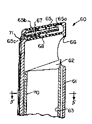
The embodiment of the invention illustrated in Figs.
3 and 5 solves this problem by enabling the suction force
to be monitored constantly. An instrument similar to the
one in Figs. 1 and 2 has been modified to measure pressure
differences between the external and internal forces of
its irrigation/aspiration/cutting tip. The modified
instrument is referred to generally by reference numeral
60 and includes an outer elongated housing 61 which
surrounds an inner concentric guillotine 70 which carries
a cutting blade 62 that cooperates with an opening 66 for
surgically removing tissue fragments as described above.
An inner bore or channel 63 operates to convey fluid
and/or tissue. Only the tip of such an instrument is
shown in Fig. 3 and additional features such as the
discharge outlets 33, 34, shown in Fig. 2 were omitted to
simplify the description.
A pressure transducer 65 is mounted in a chamber 65a
located near aspiration inlet 66, the chamber 65a being
bounded by two parallel diaphragms 67, 68, formed of the
silicon rubber inserts that are about 1 mm in diameter.
The diaphragms 67, 68, are connected to the instrument 60
by means of an epoxy resin. The transducer 65 is
preferably mounted at the outer end 61a of the tip of the
housing 61.
Pressure transducer 65 is a piezo~electric or
photo-electric device known to the art which is capable of
measuring intraocular pressure with the required
sensitivity (plus or minus 1 mm Hg), stability and
linearity. Other types of transducers, such as sensors
operating in conjunction with fiber-optic light guides
which transmit signals in the form of variations in light
intensity caused by pressure differences moving a
reflective surface, can also be used in con~unction with
03090/1/1-1-13/11
1 o 1 3 ~ ~ 7 ~ D, 3
the invention without substantially altering the size,
shape or function of the instrument. An electrical signal
generated by the transducer 65 is carried through wire
leads 61 to a monitor console which is known in the art
and contains a suitable power supply as well as the
necessary electrical circuits for conditioning, amplifying
the displaying the pressure measurement.
The piezo-electric elements 65b are attached to a
cantilever beam and a rigid base 65c, which is anchored to
the wall of the instrument. Wire leads 71, which carry
electrical signals from the transducer 65, are connected
to the exterior surface of the instrument 60 so as to
avoid interference with the action of the guillotine
cutter 70. ~he leads 71 are bonded to the instrument 60
so that they are part of its smooth outer surface.
The vitrectomy suction instrument 60 significantly
enhances safety through sensitivity to suction force and
consequently intraocular pressure during surgery. As the
surgeon aspirates strands of diseased tissue into the
opening 66, the local pressure difference measured between
diaphragms 67, 68, by the transducer 65 results in a
relative pressure reading that reflects the forces exerted
on the tissue strands as they enter the aspiration inlet
66. These forces fluctuation continuously because of
differences in the viscoelastic properties of the
manipulated tissue and the viscosity of the surrounding
vitreous. The force level at any given time can fall in a
range that departs considerably from the average force and
the pressure in the vacuum line can be adjusted to
accommodate these fluctuating force levels. By using the
transducer 65, a signal can be generated to activate
momentarily a vacuum relief valve in a known way (not
shown) when the local pressure exceeds preset levels to
adjust the suction when the force level falls outside the
permissible range. Thus, the instrument 60 operates to
reduce considerably the danger of damage to healthy tissue
03090/1/1-1-13/11
by preventing excessive instantaneous peaks in the local
suction forces.
Referring to Figs. 4 and 6, another embodiment of the
invention is illustrated, this one being directed to a
surgical instrument which can measure intraocular
pressures while performing an irrigation or aspiration
procedure. The instrument is generally designated by
reference numeral 40 and is an elongated body 41 formed of
surgical grade stainless steel with an outside diameter of
approximately 1 mm. The body ~1 is divided through
substantially its entire length into two parallel channels
42, 43, that are separated by an internal wall 49.
Channel 43 is an irrigation/aspiration channel which is
connected to a handpiece (not shown) to either a vacuum
system (not shown) or a saline supply reservoir. The
channel 43 has an outlet 44 located near the apex 45 of
the tip of the instrument 40.
A transducer 50 is mounted in the portion of the
channel 42 adjacent to the tip of the instrument 40, the
channel 42 being vented to the atmosphere at a suitable
site away from the operating field. The transducer 50 is
of the type described above where the embodiment of Figs.
3 and 5 and is connected to the instrument 40 through a
base 55a. At the tip of the instrument 40, the transducer
42 terminates at a window 46 which is located adjacent to
the outlet 44. The window 46 is approximately 1 mm in
diameter and is fitted with a diaphragm 47 formed of
silicon rubber. The diaphragm 47 is connected to a window
46 by means of epoxy resin. Wire leads designated by
reference numeral 52 carry electrical signals generated by
the transducer 50 to suitable instrumentation (such as
that described below) for translating the signals into
useful information for monitorins and regulating
intraocular pressure.
The intraocular pressure probe 40 is suitable for
measurement and control of intraocular pressure during
closed system procedures in the anterior chamber 15 as
03090/1/1-1-1~/11
~ 3 ~ ? `~
-12-
well as in the vitreous chamber 18. The instrument 40 can
be inser~ed at a site separate from the operating incision
and remain in place throughout the entire procedure,
providing the surgeon an independent source of determining
and/or controlling intraocular pressure for providing
information used in tamponade, suture tension controls and
final approximation of physiological pressure at the end
of wound closure.
One disadvantage of placing the transducer in the
portion of the probe that penetrates the eye, as done in
instruments 40 and 60 (see Figs. 4 and 3, respectively),
is that this configuration requires the probe to have a
larger diameter than would be otherwise necessary. This
problem can be eliminated without effecting the accuracy
or speed of the device by relocating the pressure
sensitive diaphragm and the transducer outside the eye but
in a position where significant signal can be generated in
response to changes in intraocular pressure.
Fig. 7 illustrates one such alternative embodiment of
the invention. The instrument, generally designated by
reference numeral 95, includes an elongated needle section
96 with an opening 97 which can be inserted into the
ocular globe. The opposite end of the needle section 96
opens into a chamber 98 which is designed to remain
outside the ocular globe. A transducer 99 is mounted in
the chamber 98 opposite the opening 97. ~lthough the
transducer 99 is not located inside the ocular globe, it
position adjacent to the opening into the globe supplies a
pressure reading nearly as accurate as one obtained
through internal placement.
The transducer 99 can be of the type described above
for the embodiments illustrated in Figs. 3-6, or refused
silicon-type such as ~ntran Model No. EPIL-F080-55
manufactured by Antran Devices, Inc., Fairfield, NJ, which
is separated from the chamber 98 by a diaphragm 100 formed
of paraline, or the like. Wire leads 102 carry electrical
signals generated by the transducer 99 to external
03090/1/1-1-13/11
-13- ~ 3 ~ ~ r~ ,J,~
instrumentation that is described in detail below. The
chamber 98 is eguipped with an input opening 103 that can
be connected to a flexible plastic tubing 104 for
supplying fluid in appropriate clmounts to the ocular
globe.
The instruments shown in Figs. 3-5 and 7 can be
incorporated into any number of systems for controlling
pressure within the ocular globe 12. For example, signals
generated by the transducer can be used to control the
suction level through the same probe on which the
transducer is located (Figs. 1 and 2) or a second probe
when the surgical procedure requires fluid to be
circulated through the eye. For other surgical
procedures, pressure in the eye can be maintained within
the predetermined range through a single probe.
The instruments described above can be used in a
system of the type shown in Figs. 8, 9 or 10 where a
pressure level within a predetermined range is maintained
and controlled more accurately than in any other known
system. This is accomplished through the use of a closed
feed loop through which saline solution is continuously
circulated. This closed circuit feed loop is connected to
a conduit that is in turn connected to the eye so that
reaction to a change in pressure detected by the
transducer, will act to supply or withdraw fluid from the
eye as required, thereby controlling intraocular pressure.
Referring to Fig. 8, an instrument I of the type
shown in Figs. 3-6 or 7 penetrates the ocular globe 12 and
is connected to a fluid conduit 104. A flow loop 110 is
connected to the conduit 104 through a flow splitter
connection 109. When the system is operating, a
peristaltic pump 107 continuously circulates saline
solution through the loop 110 in the direction of arrows
111. A reservoir of saline solution 108 is connected to
the loop 110 for supplying additional solution when
needed. A pressure relief value 112 can be provided at
03090/1/1-1-13/11
-14- ~3~$~
the splitter connection 109, but it is not considered
necessary for successful operation of ~he circuit.
If the instrument 100 detects a pressure change in
the ocular globe 12, a signal is transmitted through a
S line 102 to a monitor/console 105 of a type known in the
art, which contains a suitable power supply as well as the
necessary electrical circuits for conditioning, amplifying
and displaying the pressure measurements. The signal is
in turn transmitted to a microprocessor controller 106 of
a type known in the art, which is operatively connected to
the pump 107.
The microprocessor controller is programmed to allow
the pump 107 to circulate fluid through the loop 110 at a
predetermined flow rate when signals received from the
transducer indicate that the pressure of intraocular fluid
is within a preset range. This flow rate will operate to
maintain a p~edetermined a pressure when a pressure drop
is detected by the instrument I; the resulting signal to
the microprocessor controller operates to speed up the
pump a predetermined amount for infusing additional saline
solution into the eye.
Conversely, if a pressure increase is detected, the
pump speed is reduced. The use of a flow splitter in
relatively close proximity to the instrument 100 (for
example, by resting it on the forehead of the patient) and
the continuously circulating saline solution in the loop
110 provide for a much more rapid response t.o pressure
changes in the eye than if a long fluid column were used
or if a pump had to be activated in response to each
pressure change.
Fig. 9 illustrates an alternative embodiment of the
closed-loop circuit. A saline solution is continuously
circulated in the feed conduit loop 120 by a variable
speed peristaltic pump 122 in the direction of arrow 123.
A fixed flow restrictor 124 is located within the conduit
loop 120. A suitable reservoir 126, in which saline
03090/1/1-1-13/11
~ 3 ~ d~ g c~
-15-
solution is stored, supplies additional saline solution to
the conduit loop 120 as needed.
When an instrument 128 of the type shown in Figs. 3-6
or 7 detects a pressure change in the ocular globe 130, a
signal is transmitted through a line 132 to a
monitor/console 134 of a type known in the art, which
contains a suitable power supply as well as the necessary
electrical circuits for conditioning, amplifying and
displaying the pressure measurements. The signal is in
turn transmitted to a microprocessor controller 136 of a
type known in the art, which is operatively connected to
the pump 122.
The microprocessor controller is programmed to allow
the pump 122 to circulate fluid through the conduit loop
120 at a predetermined flow rate when signals received
from the transducer indicate that the pressure of the
intraocular fluid is within a preset range. This flow
rate operates to maintain a predetermined pressure level
within the ocular globe 130. However, if a pressure drop
is detected by the instrument 120, the resulting pressure
signal to the microprocessor controller operates to speed
up the pump 122 and raise the pressure of the circulating
saline solution. The fixed flow restrictor 124 in turn
causes an increased amount of the circulating saline
solution to be diverted into the conduit 138, resulting in
additional saline solution to be infused into the eye 130.
Conversely, if an increase in intraocular pressure is
detected, the pump speed is reduced which operates to
decrease the back pressure and reduce flow through the
restrictor 124. Less fluid is diverted into the conduit
138, which reduces pressure in the conduit 138 and allows
a net outflow of fluid from the eye 130.
Figs 9A and 9B show the use of a fixed flow
restrictor at other locations in the conduit loop 120. In
both Figs. the restrictor is located between the conduit
138 and rese~voir 126, with the Figs. showing different
configurations of the conduit loop 120.
03090/111-1-13/11
-16- 13~
Another embodiment of the invention is shown in Fig.
10, where an instrument 128 of the type shown in Figs. 3-6
or 7 is in turn connected to an ocular globe 130. When
the system is operating, a peristaltic pump 140
continuously circulates saline solution through a feed
conduit loop 142 in the direction of the arrows 144. A
suitable reservoir 146 containing saline solution is
connected to the loop 140 for supplying additional
solution when needed.
When the instrument 128 detects a pressure change in
the eye 148, a signal is transmitted through a line 150 to
a monitor/console 152, similar to the monitor/console 134
described above in conjunction with Fig. 9. ~he signal is
in turn transmitted to a microprocessor controller 154 of
a type known in the art, which is operatively connected to
a stepper motor 156 mounted on a base 158. The conduit
loop 140 is formed of a flexible tubing so that a rotating
eccentric cam 160 mounted on the stepper motor 156 can
control the flow of saline solution through the tubing by
alternately pinching and releasing a pinching force on the
line depending on the position of the cam 160.
The microprocessor controller 154 is programmed to
allow the pump 142 to circulate fluid through the loop 140
at a predetermined flow rate when signals received from
the transducer in the instrument 128 indicate that the
pressure of the intraocular fluid is within a preset
range. This flow rate operates to maintain a
predetermined pressure level within the ocular globe 148.
If a pressure drop is detected, the resulting signal
operates to activate the stepper motor 156, which rotates
the cam to pinch the flexible feed conduit tubing against
the base 158, creating a back flow pressure which diverts
additional fluid to conduit 162 and infuses additional
saline solution into the eye 148. Conversely, if a
pressure increase is de~ected, the stepper motor 156
rotates the cam 160 to a position that enlarges the
opening in the flexible, tubing permitting flow to
03090t~ 13/11
-17- ~ r~ ~ ~
increase within the feed circuit loop 140 and consequently
lower the intraocular pressure.
Incorporation of a closed loop in the infusion fluid
conduit, as exemplified by the embodiments of Figs. 8-10,
S increases significantly the response speed of the fluid
delivery system to changes in intraocular prèssure, as
compared to a simple infusion conduit from the pump to the
eye. In addition, conduit loops of the types described
provide an important safety element in the event of pump
stoppage, due to equipment malfunction. The system can
revert to passive, gravity flow from the infusion bottle
through the return portion of the loop of the eye, thus
bypassing a stalled pump or other malfunction.
It has been found that the effectiveness of the
pressure-activated, feedback-controlled system in
responding to rapid fluctuations in intraocular pressure
reaches an upper limit at fluctuation frequencies of ca.
200 per minute. Pressure changes in this frequency range
can be effectively attenuated by the use of damping
devices, described in greater detail below, which are
mounted in series or parallel with the fluid infusion
conduit in close proximity to the infusion cannula. In
general, these damping devices utilize the elasticity of
thin membranes or the damping properties of air or other
~5 gases, confine~ into a small chamber.
one embodiment of such a damping chamber is shown in
Figs. 11 and 12, where a damping or compliance chamber 120
is connected to a fluid conduit 104, either downstream
from a splitter connection (Fig. ll) or at the
splitter connection 109 (Fig. 12). The compliance chamber
120 operates to accommodate sudden changes in pressure in
the ocular globe 12 caused by surgical manipulations such
as pressing on the globe, pulling on the ocular muscles or
tightening of stitches where pressure is raised or
starting or enlarging an incision where pressure is
lowered. Such pressure fluctuations tend to be very
rapid, on the order of 10-2-10-l per second. The normal
03090/1/1-1-13/11
- 13 ~
-18-
response time of the systems, shown in Figs. 8-10 might
not be fast enough to react to many such pressure
fluctuations because of inertial and frictional forçes in
the equipment and associated flow lines.
In order to provide a quic~er response time to these
sudden fluctuations, the compliance chamber 120 is
included in the flow line leading to the eye, in close
proximity to eye. Preferably, the compliance chamber 120
is located from 6-10 cm. from the tip of needle section
96a.
The compliance chamber 120 in Fig. 11 is formed as a
small, spherical chamber that is 4-8 cm. in diameter with
highly elastic walls. The compliance chamber 120 can be
completely filled with the fluid F flowing through the
15 flow lines 102, 104 (Fig. 12). However, the reaction time
to intraocular pressure changes can be increased by
initially filling the chamber 120 with air or other gas G,
as shown in Fig. 11, for more rapidly accommGdating
pressure changes because of the greater compressibility of
the gas G.
As shown in Fig. 11, the compliance chamber 120 can
be formed as part of or connected to the conduit 104,
downstream from the splitter connection 109. In such
case, the conduit 104 can be formed separately from the
25 conduit 102, with individual needle sections 96, 96a,
respectively, connected to the flow line 104 and
instrument I as described above. Alternatively, as shown
in Fig. 11, the compliance chamber 120 can be connected to
the loop 110 at the flow splitter connection 109.
Figs. 13, 14 and 14a show additional embodiments of
these damping devices in the form of small thin walled air
chambers, a few cubic centimeters in volume, constxucted
from metal, rigid plastic, or other suitable material. In
Fig. 13 the damping chamber 200 is attached to an infusion
35 conduit 202, close to the i~flow cannula 204. Fig. 14
shows a damping chamber built into the hollow handle of an
irrigation handpiece ~0~.
03090/1/1-1-13111
--19--
Sudden increases in intraocular pressure cause a
small amount of fluid to be expelled from the ocular globe
210 and bac~flow into an open tube 212 inside the damping
chamber, thus relieving pressure in the eye and
compressing the air in the damping chamber. The opposite
happens with sudden reductions in intraocular pressure,
which cause the fluid in tube 212 to flow forward into the
eye 210. In Fig. 14A, aside from the different
configuration of the tube 212, an aspiration line 218
leading to a vacuum pump (not shown) is shown in the
handpiece 206.
The damping characteristics of these air chambers may
be fine-tuned to the si~e and elasticity of the particular
ocular globe involved in a given procedure by varying the
air-chamber volume. This can be done by the surgeon at
the start of the procedure, by holding the chamber 200 or
206 upside-down, thus allowing a variable amount of
irrigation fluid 216 to enter the chamber and remain there
during the procedure. Similar tuning takes place
automatically during the procedure in the event of
deliberate increase in the pressure setting, e.g., when
the surgeon uses tamponade to stop bleeding; the increase
in infusion line pressure forces infusion fluid into the
air chamber, thus decreasing its volume until equilibrium
is reestablished at the high pressure level.
These damping devices are optimally used in
conjunction with a feedback-controlled infusion system of
the type described above in conjunction with Figs. 8-10,
whereby the two systems act in concert. However, the
damping devices may be used alone, if desired, there~y
providing only attenuation of transient and rapid periodic
pressure changes, without overall pressure control.
The automatic maintenance and control of intraocular
pressure, achieved by the foregoing invention has
significant therapeutic potential in reducing edema in the
retina after vitrectomy, decreasing intraocular
inflammation after irrigation/aspiration procedures and
03090/1/1-1-13/11
-20- ~ J,g~
minimizing postoperative astigmatism incurred during wound
closure in cataract surgery.
The inventions embodied in the instruments and
apparatus above are useful in constantly monitoring and
controlling both intraocular fluid pressure and suction
forces during ophthalmic surgery. By allow the surgeon
the benefit of this type of equipment, much of the
guesswork of maintaining optimum intraocular pre~sure
during surgery is removed, resulting in safer, more
accurate surgical procedures. Moreover, the control
systems can automatically regulate intraocular pressure
according to a predetermined set of instructions more
rapidly and accurately than before.
Although different embodiments of the invention may
vary in detail, they are still intended to be within the
scope of the inventive concept described above. The
details described in the foregoing preferred embodiments
are intended to illustrative and not limiting in any
sense.
03090/l/1-1-13/11