Note : Les descriptions sont présentées dans la langue officielle dans laquelle elles ont été soumises.
2~ %40
This invention relates to apparatus and a method
for processing and analyzing blood serum and more
particularly to a system that automatically processes whole
blood specimens to separate and withdraw the blood serum
from the red blood cells and that also removes from further
processing those specimens which are defective before the
blood serum is withdrawn.
The process of separating the red blood cells
from the blood serum of a whole blood specimen by
centrifuge and then removing the blood serum is conducted
on a large scale in hospitals and laboratories. This
process is usually conducted manually by a technician. In
the process, a stopper sealed test tube, containing a whole
blood specimen and a separating gel, is centrifuged so that
its contents are separated into three layers, a top layer
containing the serum, a middle layer containing the
separating gel, and a bottom layer containing the red blood
cells. After the test tube is centrifuged, the technician
must examine the blood specimen to determine whether it is
defective. If the sample is not defective, the technician
. . ,
, ~ :
:.
2~ 4~)
then inserts a needle through the rubber stopper, eyes the
placement of the needle in order to insure the needle does
not contact the separating gel, and withdraws a sample of
the blood serum from the top layer and places the sample
into a cup.
Efficiency, accuracy, and maintaining the
integrity of the blood specimen are essential to this
process. More important is the safety of the technician
while completing the process. By fully automating this
process, these factors are greatly enhanced. The danger
to the technician of being exposed to any transmitted
diseases in the specimens during this process is
eliminated.
U.S. Patent Nos. 4,713,974 and 4,478,095 disclose
devices for automatically piercing container lids and
withdrawing samples. Neither of these patents disclose any
means for sensing an appropriate level inside the test tube
for positioning the tip of the sampling needle. Also, these
patents do not disclose devices for use with centrifuged
blood samples and do not disclose any means for
automatically detecting defective samples.
U.S. Patent Nos. 4,120,662 and 4,311,484 both
- disclose blood sample processing systems for delivering
blood from closed vacutainers to a Coulter Counter. These
systems are not suitable for use with centrifuged blood
samples in that tubes are sampled in an approximately
horizontal position and are agitated prior to sampling.
2~ 4~)
U.S. Patent No. 4,326,851 discloses a level
sensor for use with a fluid transfer mechanism for
determining when the bottom tip of a fluid aspirating probe
touches the top surface of a sample fluid. This device
cannot be used with blood samples in conventional test
tubes. In addition, the patent discloses no method or
apparatus for automatically sensing whether the sample is
defective.
This invention is accordinyly directed toward
apparatus and a method for automatically centrifuging
blood specimens and separating gels in stopper sealed test
tubes, determining whether the centrifuged specimens are
defective, and removing and then dispensing blood serum
samples from only those sealed test tubes in which the
specimens are not defective.
The method of the present invention includes
centrifuging a test tube containing a whole blood specimen
and separator wax, moving the test tube into an optical
sensing unit, and analyzing the electrical signals
generated by the sensing unit to evaluate the success of
the separation and to determine the position of the
boundary surface between the separator wax and the blood
serum.
In the preferred embodiment, the optical sensing
unit includes a vertical cavity operative to receive a test
tube. The cavity includes a light source disposed on one
2~ 40
__4
of its sides that emits a light beam that extends generally
normally to the longitudinal axis of a test tube placed in
the cavity. The cavity also includes at least one
photosensor disposed on its opposite side and positioned
to receive the transmitted portion of the light beam. The
photosensor generates electrical signals proportional to
the amplitude of the transmitted beam. The apparatus of
the present invention further includes means for receiving
these electrical signals and analyzing them to evaluate the
success of the separation, and to determine the position
of the boundary surface between the separator wax and the
blood serum along the longitudinal axis of the tube.
In an alternative embodiment, the vertical
cavity of the optical sensing unit includes a vertical
array of photosensors disposed on one side of the cavity
and a corresponding vertical array of light sources
disposed on the opposite side of the cavity. Each of the
plurality of light beams emitted from the light sources
extend generally normally to the longitudinal axis of a
test tube placed in the cavity. The photosensors each
receive the light beams transmitted from their
corresponding light sources and generate electrical signals
proportional to the amplitude of the transmitted beams.
The apparatus of the present invention also
includes an input station adapted to hold test tubes each
containing whole blood specimens and separator wax, and a
centrifuge for separating the whole blood into serum and
red cells separated by a layer of separator wax.
2~ 4~)
__5
The apparatus further includes a needle
apparatus, responsive to the means for determining the
position of the boundary surface between the separator wax
and the blood serum, connected to suction means for
insertion into a test tube and for drawing a blood serum
sample from the tube. The needle apparatus is also
connected to means for dispensing the serum that was drawn
out of test tubes by the suction means.
The apparatus also includes a pair of output
stations for receiving both successfully and unsuccessfully
separated test tubes, and a robotic arm for moving the test
tubes from station to station, under the control of the
means for evaluating the electrical signals generated by
the photosensor(s).
The preferred embodiment also includes a feeder
station adapted to dispense empty containers that are
operative to hold blood serum samples. The robotic arm is
also adapted, under the control of the means for evaluating
the electrical signals generated by the photosensor(s), to
remove an empty container from the feeder station and to
move it to a position underneath the needle apparatus.
The present invention makes the process of
centrifuging and analyzing blood specimens efficient and
accurate. It also eliminates the danger of a technician
being exposed to any transmitted disease, such as AIDS,
during the process.
.qs
2~Q~4~)
----6
Other objectives, advantages, and applications
of the present invention will be made apparent by the
following detailed description of the preferred embodiment
of the invention. The description makes reference to the
accompanying drawings in which:
Figure 1 is a perspective view of the preferred
embodiment of the automatic blood serum processor and
analyzer;
Figure 2 is a cross-sectional view of a test
tube, containing a centrifuged blood specimen, of the type
used in the present invention;
Figure 3 is a cross-sectional view of the optical
sensor of the preferred embodiment of the present
invention;
Figure 4 is a side view of the
aspirator/dispenser needle unit of the preferred embodiment
of the present invention:
Figure 5 is a cross-sectional view of an optical
sensor of an alternative embodiment of the present
invention; and
Figure 6 is a general flow diagram of the
algorithm for evaluating the output of the optical sensor
of the preferred embodiment of the present invention.
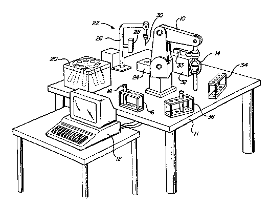
Referring to Figure l, the preferred embodiment
of the present invention is supported on a work station
11. In Figure 1, a robotic arm, generally indicated at 10,
2~ 4`~:)
__7
is positioned so that it can axially rotate about a
vertical axis to reach any of six different stations. The
robotic arm is connected to a control computer 12. In the
preferred embodiment, the robot is a five axis articulated
arm. Such robotic arms are well known to the art. The
~Q~4~)
--8
¦robotic arm 10 includes a multi-purpose gripper 14 of
'conventional construction.
The first station constitutes a rack 16 for
holding test tubes 18 of conventional construction. The
rack may be either manually loaded by an operator, or
automatically loaded. In the preferred embodiment each
test tube is bar coded for identification purposes. The
bar code is read either by the robot 10 or by equipment at
the hospital or clinic where the blood was drawn. The
second station is a centrifuge 20. The centrifuge is used
to centrifuge whole blood specimens along with separating
~'gels in stopper sealed test tubes by rotating the tubes
!inclined with respect to a vertical axis about that axis
so that the contents of the test tubes are separated, as
indicated in Figure 2, into a top layer 40 containing the
blood serum, a middle layer 42 containing the separating
gel, and a bottom layer 44 containing the red blood cells.
The separator gel (or wax) has a density half-way between
the densities of the serum and the red blood cells. As
;,20 indicated in Figure 2, after centrifuging the separator gel
may not lie in a horizontal plane normal to the
longitudinal axis of the test tube, but rather at an angle
from the horizontal plane. The orientation of the middle
layer 42 is determined by the type of centrifuge used.
Centrifuges are well known to the art.
The third station is a serum sensing and
i~ aspirator/dispenser unit, generally indicated at 22. This
., .
station includes an optical blood serum sensing unit 24,
2~ 4~1
and an aspirator/dispenser unit 26. The station 22 also
includes a bar code reader 28 of conventional construction.
Both the sensor and the aspirator/dispenser units are
connected to the control computer 12. The optical sensor
24 receives test tubes containing centrifuged blood
specimens and outputs signals to the computer 12 so that
the computer may determine whether the specimen is
defective, and, if it is not, an appropriate level in the
test tube to position the tip of an aspirator needle for
removing a blood serum sample. The aspirator/dispenser
unit 26 functions, under the control of computer 12, to
lower a sampling needle 30 to puncture the stopper seal of
a test tube held in the optical sensor 24, for withdrawing
samples from the test tube. Automatic aspirator/dispensers
are well known to the art.
The fourth station is a serum cup feeder 32 that
stores empty cups 33 for holding blood serum samples
dispensed by the aspirator/dispenser unit 26. The serum
sample cup is preferably formed of plastic. The fifth
station is an output rack 34 for holding cups containing
blood serum samples along with their corresponding test
tubes. Finally, the sixth station is a rejection unit 36
for receiving those test tubes which are determined
defective by the computer 12.
The preferred embodiment of the present invention
operates, under the control of computer 12, as follows:
First, the robotic arm 10 loads test tubes, each
containing whole blood specimens and separating gel, into
2~ 4~
----10
the centrifuge 20 one by one from the input rack 16. The
centrifuge 20 is then activated. After the centrifuging
process is completed, the robotic arm 10 removes the
centrifuged test tubes, one by one, from the centrifuge 20
and places them into the optical sensor 24.
If the signals from the optical sensor 24
indicate that the centrifuging results in lipemic (white),
hymolized (red) or otherwise unsuccessful specimen, then
the robotic arm 10 removes the test tube from the sensor
24 and places it in the rejection unit 36.
If the signals from the optical sensor 24
indicate the specimen is not defective, the sampling needle
of the aspirator/dispenser unit 26 is lowered to
puncture the stopper seal of the test tube and to the level
in the test tube previously determined by the analysis of
the output signals from the optical sensor 24. A sample
of the blood serum is drawn from the test tube through the
needle 30 by the aspirator unit. In the preferred
embodiment of the invention, approximately 1.5 milliliters
of blood serum is withdrawn.
At the same time that the blood serum is being
withdrawn, the robotic arm 10 removes an empty serum cup
from the serum cup feeder 32, using the gripper 14. After
the blood serum sample is withdrawn from the test tube, the
aspirator/dispenser unit 26 lifts the sampling needle 30
out of the test tube into a stow position. The robotic arm
then moves the empty serum cup into a position
underneath the sampling needle 30. The
. ' '' '
'.' ~
.,. ,,: ~ , . .
2(~ 4~)
----11
asp:irator/dispenser unit 26 then dispenses the blood serum
sample through the sampling needle 30 and into the cup that
is supported by the robotic arm lO.
After the cup receives the blood serum sample,
the robotic arm places the cup on top of the stopper seal
of the test tube resting in the optical sensor 24. The
robotic arm lO then removes the test tube, along with the
serum sample cup, from the optical sensor 24, positions it
by the bar code reader 28 for identification purposes, and
places the test tube along with the serum sample cup in the
output rack 34. In the preferred embodiment of the
invention, the serum sample cup is constructed so as to
snugly fit on top of a stopper.
The above process is repeated until all
centrifuged test tubes are examined. The system may then
load a new batch of test tubes into the centrifuge.
Figure 3 is a cross-sectional view of the optical
sensor 24 of the preferred embodiment of the present
invention. The sensor 24 is located in a vertical cavity,
generally indicated at 50, of a housing 52. The vertical
cavity 50 has an opening 54, and is adapted to receive a
test tube of conventional construction. The sensor 24 is
connected to a power source via a power cord 55.
A light source 56 is disposed on one side of the
cavity 50, near the opening 54. The beam emitted by the
light source 56 extends generally normally to the
longitudinal axis of a test tube placed in the cavity 50.
In the preferred embodiment, the light beam is a pulsed
,
Z~?~Q~4~)
--12
infrared rectangular sliver of light extending across the
diameter of the cavity 50. The beam is pulsed at a high
frequency to avoid ambient noise.
A sensor 58 is disposed on the opposite side of
cavity 50 from the light source 56 so as to receive the
transmitted portion of the light beam emitted from the
light source. In the preferred embodiment, the sensor is
a horizontal array of photosensors adapted to receive the
entire sliver of light when the cavity is empty. Optical
filters that only transmit light having the frequency of
light source 56 are positioned in front of the photosensors
in order to avoid noise. The contents of a test tube
displaced between the light source 56 and the sensor 58
will partially occlude the light beam from the sensors.
The sen~ors have an analog output proportional to the
portion of the beam that is occluded. The sensor 58 is
connected to the control computer 12 via a connection line
in order to provide it with the outputs of the
photosensors. In the preferred embodiment, the output from
photosensors is passed through an analog to digital
converter before being received by the computer 12.
The control computer 12 processes the signals
received from the optical sensor 24 in order to determine
the success of the centrifuge separation and the level of
the separator wax (the middle layer) in the centrifuged
specimen. In the preferred embodiment of the present
invention, the robotic arm 10 is controlled to lower the
centrifuged test tube to be analyzed down through the
", ' ', . '' .
.
2~ 4~)
--13
opening 54 and into the cavity 50 of the optical sensor 24.
Whi.le the robotic arm lo is moving the tube down into the
cavity 50, the computer 12 receives the output signals from
the sensor 58. If a successfully centrifuged blood
specimen, as indicated in Figure 2, is being moved into the
cavity 50, first the light beam is occluded to a relatively
high degree by the bottom layer 44 of red blood cells, then
to a lesser degree by the middle layer 42 of separator wax,
and then to an even lesser degree by the top layer 40 of
blood serum.
If the output signals from the optical sensor 24
do not indicate these three layers, then the robotic arm
removes the test tube from the optical sensor and places
it in the rejection unit 36. In the case where the
centrifuging is successful, knowledge of the position of
the test tube relative the optical sensor at the point in
which the sensor 58 signals indicate a transition between
the separator wax and the blood serum allows the computer
12 to determine a level, spaced above the separator wax,
for positioning the sampling needle 30 of the
aspirator/dispenser unit 26.
Figure 6 is a general flow diagram for the
algorithm used by the computer 12 in evaluating the outputs
from the optical sensor 24 in the preferred embodiment of
the invention. For the purposes of illustration,
well-known housekeeping functions, such as error checking
features, have been omitted from the flow diagram of Figure
6.
., .
-I : .
2~ 4~
--14
Th~ algori~hm make~ use oP t~e ~ollowing
va~iable~:
DEFECT : boolean ~nri~ble ~or lndicntlng
whet~er the peclmen i8 de~ective;
~AST: lnd1cateG tîle Whlch portlon of the
centri'ug~d blood ~;~mple Wa~ last ln
between ~he ~enSor and ~le 1~ ght
source -- X (for l~lti~lization), Rs
(20r re~ blood cell~), SEP (for
s~p~r~ion wax)~ and BS (~or bloo~
serum):
TRA~SITIol~ : boolean, set to ~rue w~len ~ensor
first detects blood serum;
OCCL : var~ahle rOr readlng ln amow~t o~
occluslon sen~ed by ~en~or sn;
RBMIN~ RBMAX: cozlstants repreqentlng the
minimum and mAxlmum val~e~
o~ 0~CL that would indlcate
red blood ~ells;
S~PMII~, ~EP~AX : constant~ ~epre~erlting th~
minimum ~nd m~ximum valu~s
of OCCI, th~t woul~ indlcat~
separator wax)
8SMlt~ BSr~x : cvnstant~ represent~ng the
minimUm and maximum value~
o~ OCCL that would lndicate
blood nerUm?
CU~ me ~ype a5 LAST, rOr ~torl~lg t~e .
curront portion o~ the ccnt~ifuged
blood eerum be~ween the sensor and
the llght ~ourceJ
PO~:ITION, Z : for storlng the vertical posltlon
~o of the robot gripper;
FACTO~ conitant;
REFEI~EI~CE : vertical po~it lol1 of rc~]~ot grlpper
when holding a t:e.~t tube t;lla t i8
fully placed ln the opti~al
~en~orl and
SE~SO~.Z : vertical po~ition vf the ~ensor 58 .
~0
~ir~t,, the alyorlthm lnltializes DEFECT to fal~e,
LAST to X, and TRAI~SITI0~ to fal~e. I~ext, at the ~tep
.
:
,
.
2~ 4~)
--15
indi~ated ~t 1~0, i~ is cl~ecked whe~hcr ~EFECT i~ f~l~e,
the robot arm is movillg ~he tube in~o the ~n~or, and
TRA~lsITIo~ i~ falQe. I~ one o~ the abo~ cond~tlons i~ not
true, then the algor~thm goes to the ~tep ~ndicated a~ 114.
Otherwise, the algorithm cont1nue~ ~t ~top 102.
At 102, the output ~rom ~ensor 5~ i~ rcad lnto
the variable OCCL. Then, i~ 18 c~ecked whether the valuc
of OCCL is ln the range that lndlcates th~ ~ensors ar~
detecting red blood cells~ is I~Ot, th~ ~lgor1thm
lo goes to ~he step indicated at 10~ t~e senGor ls
detecting red blood cel1s, cu~ s~t to ~ and lt 1~
checked whether the la~t sensor read ~ndlc~ted separat.or
wax or blood serum. If not, the a1gorlthm 3klps ~ the
step ~ndicated at 11~. If the varlable L~ST le set to
15 either SEP or BS, thetl t~le algorithm c~oes to the step
indicAted at 108.
At 104, l'c is checl~.ed whetl~er the varial~le OCCL
is set to a value lndi~ative o~ ~epnrator wax. I~ not, the
algorithm continues at the step indicated at 106. I~ so,
CURR is set to SEP, and 1~ ls checked whether LAST ls ~et
to X (just initialized) or BS (the last ~en~or read
indicated blood Qerum). If LAST is not ~et to ~ither o~
~hese values, the algorltllm continues at th~ ~'cep lndlcated
at 110. ~ l` 1s set to elt~er X or I3S, th~ Algorlthm
2s goes to s~cp 108 .
At 106, ~t is ~lecked w~letller oCCL 1~ ee~ to
v~lue indi~ative of th~ 6~nsorQ detectlng blood. ~erum. I~
not, the alc~orithm sklps to ~tep 108. I~ ~o, CU~R i3 ~et
2~ 4~
--16
to ~S, an~ i~ is checked whether LAST 1~ S~e ~o ~leher RB
or X. If no~, ~he algori~hm skips to st~p 110. I~,
however, LAST ls set to either R~ or X, the algorlthm skips
to eitep l~R.
At 6tep 108, ~EFECT i6 6et to true. Nex~, the
alyorithm continue~ at the ~tep indicated at 100.
At ~teU 110, lt 18 checked whether ~oth CURR iB
set to ~S and L~ST i~ q~t to SEP. If not, tl-e alg~ritllm
oontinue~ ~t the qte~ indlcated at 112. I~ 60, PO~ITION.2
1s set to the vertloal pO~tiOI~ o~ the ro~ot gripper and
T~At~SITION is set to true. ~he algo~lt~m t~en contlnue~
' at ~tcp 112.
At 112, h~ST is set to CU M, and the ~lgorlthm
jump~ back to ~cp 1~0.
A~ 1~4, lt ls cheeked wll~ler eltl~er DEFE~T 15
true, or transi~lon i~ ~al6e. I~ so, t~e cen~rlfuged ~lood
specimen is defective and a routlne for controlllnq the
rohotic arln to di6pose o~ the defectlve te_t tube i~
called. If both ~EFECT is falYe and TRANSI~I~N i6 true,
then POSITION.~ is ~et to FA~TOR + ~F~RENC~ - (POS~TION. Z
- SENSOR, 2) . Tllis i~ the de~lred ~ertlcal pu~ltion for
positioning ~ samp1ing needle ln the te6t tu~e ~or
witl~ wi~ lvod ~;erum. Next, a ~outln~ 1~ call~d to
contro1 the ~spirat~r/di~~en~e~ unlt to withdr~w the blood
serllm.
....
~ he descrlptlon of the above a1~vrl~llm le no~
i~tended to 1lmlt the pre~ent lnvention. Many ~ eren~
al~orithms m~y bo im~1emen~ed fcr t~le purpo~eY ~f ~he
.
. .
.' ' ,
. ~ .
Z~ 4~
--17
in~entlon. In alternatlv~ en~odlme~ts, the sen~or and
analyzlng al~orithm may ~ further adapted to sen6e
dl~colorizations in the ~peci~en that would indic~te an
un~uccessful centri~uglng.
Figure 5 1~ a cross-~ectional v~ew oP an optical
sensor unit that may be utlllzsd in an alternati~e
embodiment of the presen~ i.nvent~os~. A houslng 80 c~ntalns
a vertical cavlty, generally lnd~cated at 82. ~ha cav~ty
82 ha~ an opening 84 and iR adapted to receiv~ a t~t ~uhe
I0 of conventional constr~tion. The hou~rl~ 8~ i~ connected
to a power source via a po-"er cord 85
A vertical array of ll~ht so~rces, indlc~ed at
8G, is di~po~ed on one side o~ ttle cavity 82 ~nd ex~end~
~rom the top o~ the cavlty, ne~r th~ opening 84, to th~
15 ~ottom. Each light source emits a light bea~ th~ ext~nds
~enerally normally to the longituainal ~xis o~ a test tube
pl~ed in the cavity ~2.
A cor~e6ponding vert~cal array oP photo~en~or6,
indicated at S8, is disposed on tho opposlte 61de of cavity
82 from the vertical array o~ llgl~t sources ~6. Eanh of
the photosensors ~s adap~ed ~o recelve the llght be~m
t~ansmltted from it~ correspondlng llght ~o~rce and then
generate signal~ proportlonal to the ~mplltude of the
transmitted beam. ~he slgnals outputted by the ~en.~or~ ar~
provided to the computer 12 via connectlon llne 90.
In thi~ embodiment, t~le computer ~2 An~lyZe~ the
sign~ls generated ~y each photo6en~0r after ~ te~t t~be ~9
placed into the cavity B~ by the robo~ic arm 10. I~ thç
2~ 4~)
6i~rlals do not lndlca~e that the blood ~peclmen is
~e~aIated into three di.~erent l~ycr~, then th~ aample 18
defective and ~he robotlc arm 10 1~ controlled to remove
the test tube rrom ~he sen~or 24 and place it ln th~
S re~ection unit 3~. Ir the ces~tri~uge w~s ~ucce~ul, the
computer 12 determine~ ~ level ln the ~est tube to position
the sampling n~edle by lo~atil~ the !owest pho~o~n~or in
the array 88 tha~ ~ geneLating ~lgnals ind~catlve o~ the
~lood serum l~yer.
10Figure 4 ls a side ~lew o~ the
~spir~tor/dispenser nee~le unlt of ~he preferred e~hodiment
of ~he present invent10ll. A vertlcully oriented ~ampling
needle 70 15 ~onnected to an extendible vertlc~l nr~ 71
that ls ~upport~d on an overhead arm 73 by a vcrtical po~t
1575. The nee~le 70 i6 ~onnected to an ~pirator~di~pen~er
7~ of conventional con~truction by a tubing GB. In ~e
pre~erred embv~imellt, tl~e nëedle 70 is po~itioned directly
above the cavity 50 o~ t}le optical sen~or 24 ~o th~t when
: lowered, ~he needle 70 may pun~ture the ~topper ~eal o~ a
~0 test tube resting ln tlle cavi~y through ~ts center.
The extendible vertical arm 71 ls conne~ed to
a motor 74 di~po~ on the over~ead ~rm 73, th~ control~
the verti~d' posi~loll of tll~ needle 70, and m~a~f lower the
needle into a stopper seal~d test tube that lg ponlt~oncd
ln the ~ptic~ n~or 24. Tl~e overhead ~L~ 73 i~ also
capable o~ ~loviny laterally to en~ure proper posltionin~
o~ the 6ampling needle 70 ill relatlon to a ~est t~e b~low
it. The m~tor 74 and the a~plrator/dl~pen~er 72 are
.
2~ 4~)
----19
connlectsd to the ~ompu~er l~ ViA co~t-ect~ on 1 ino~ 77 and
76, respectively. The motor 74 and ~xtendible vert~al ~rm
71 ~re of conventional Con~trUction and controlled by lnput
si~nals transmit~.ed by the computer 12 through t.he
connection line 77. In an alternative ~mbodlment, the
overhead arm ~3 may be ~x~endible so that the hor~æontal
positlon of the needle may alqo be ad~usted.
The preferred embodlm~nt Or the pre~ent ~nvent~on
also includes a bar code reader 78. The re~der ~8 1~
posi~ioned so that the robotlc ~rm lO may place a bar coded
test tube in front of it. ~I~e bar code re~d~r 78 ~8
connec~ed to the computer 1~ by connection l~ne 79 so that
th~ computer ¢ont~ol~ when the r~a~er ~ 5 ac~,lvated. rrhe
co~lputer 12 also recei-~es the ln~rma~ioll ob~lned ~y ~he
a~tivated reader 7~ via th~ conl~ection llne ~9. In the
preferred embodimen~, the bar coded test tube~ contalning
whole blood 6pecimens and qeparator gel are ln~t~ally
lo~ded into the input rack 16 in ~ predef~ned orl~ntatlon,
so that the robotic ar~ 10 may properly place the prooes~ed
~bes in front of t~e bar code reader 78. Al~ernat~ve
embodimen~s may not include a ~ar code reader and thereore
may r~ot require the test tube~ to be inltiAlly l~Aded ~n~o
rack 16 in pr~d~termlned orlcntatlon~.
The above de~crip~ion i~ not lntended to limlt
the present invention. I~ lc und~r~tood thAt lt la
possible to make modi~`ications and variatlo)-c ln light o~
the aho~e ~eachings without departing from the prescnt
inYent ion.