Note : Les descriptions sont présentées dans la langue officielle dans laquelle elles ont été soumises.
20(~9~91.
IMPLANT
FIELD OF THE INVENTION
This invention relates to a soft tissue implant
and more particularly to a meniscus cartilage replace-
ment for a patient.
BACKGROUND OF THE INVENTION
Wall U.S. Patent No. 4 502 161 discloses a
meniscus cartilage replacement for a patient which
consists of a woven fiber sheet coated with a resilient
material with a lateral extension of the sheet extend-
ing outside the joint for anchoring to the side of the
tibia with a screw. However, the wall replacement is
thin and flat ttwo dimensional) and hence is non-
anatomical in shape.
The following several patents are several of the
references in the aforementioned Wall patent.
Kenny U.S. Patent No. 4 344 193 discloses a
meniscus cartilage replacement of three dimensional
shape. However, the Kenny replacement consists simply
of a non-reinforced molded silicone rubber member.
Although other possible ways are mentioned briefly in
passing, the Kenny drawings show sutures and increased
20039~31
thickness en~s as ways to hold the replacement in place
in the joint, the increased thickness ends being
discussed in detail.
Stubstad U.S. Patent No. 3 879 767 discloses an
artificial implant but formed as a spinal disc replace-
ment.
Homsy U.S. Patent Nos. 3 971 670 and 4 127 902
merely disclose artificial tension members which may be
led through holes in bone and stapled for use as
replacement tendons and ligaments. No cartilage
replacement is shown.
In so far as I am aware a fully satisfactory
meniscus cartilage replacement has not been achieved in
the prior art.
Accordingly, the objects and purposes of the
invention include provision of a soft tissue implant in
the form of a meniscus cartilage replacement for a
patient, wh~ch combines an anatomical shape with woven
and felted fiber interior reinforcement for strength
and durability, in which coated top and bottom surfaces
are capable of sliding with respect to adjacent tissues
of the patient in a manner to simulate a natural
meniscus cartilage, in which a convex, exterior edge is
capable of receiving natural fibrous tissue ingrowth of
the patient to, in time, naturally anchor the implant
in the joint of the patient and in which, optionally,
the implant can be positively anchored to adjacent bone
while awaiting such natural fibrous tissue ingrowth.
Other objects and purposes of the invention will
be apparent to persons acquainted with apparatuses of
the general type upon reading the following specifica-
tion and inspecting the accompanying drawings.
SUMMARY OF THE INVENTION
The objects and purposes of the invention are met
~()()39~1.
by providing a soft tissue replacement implant, such
as a meniscus cartilage replacement, for a patient,
which comprises appropriately shaped top and bottom
layers sandwiching therebetween at least one inter-
mediate felted layer, and a resilient bonding material
coating the layers and holding same in a laminated
condition. The intermediate layer(s) is cut narrower
than the top and bottom layers and the layers have a
common side edge. The top layer being contoured, to
provide a wedge shaped cross section and a contoured
three dimensional shape. A fabric member is bonded to
the thickened edge of the resulting laminant and is
porous to invite ingrowth of patient tissue to anchor
the implant eventually in place. In addition, a method
of making the implant involves coating of layers with a
resilient bonding material, applying the layers one
atop the next, and curing the resilient bonding
material after each successive layer is applied.
BRIEF DESCRIPTION OF THE DRAWINGS
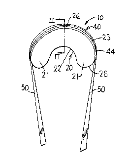
Figure 1 is a pictorial view taken from the top
and convex, exterior perimeter edge of a meniscus
cartilage implant embodying the invention;
Figure 2 is an enlarged cross sectional view
substantially taken on the line II-II of Figure l;
Figure 3 is an exploded view of woven and felted
components of the Figure 1 implant;
Figure 4A is a top view of a fragment of woven
fabric superimposed by the outline of one of the Figure
3 components;
Figure 4B is an edge view of the Figure 4A sheet;
Figure 5A is a top view of a sheet of felted
material superimposed by the outline of a corresponding
Figure 3 component;
Figure 5B is an edge view of the Figure 5A felted
sheet;
20~39~1.
Figure 6 is a fragment of the Figure 2 cross
sectional view but in an unfinished state;
Figure 7 shows in dotted lines a top view of the
top woven layer component of the Figure 1 implant in a
flat state, such component as shown in solid line being
distorted into a three dimensional bowl segment shape;
Figure 8 is a top view of a tube of the Figure 1
implant;
Figure 9 is a fragmentary pictorial view of an end
portion of the Figure 8 tube prior to trimming;
Figure 10 is a top view of an optional elongate
tape of the Figure 1 implant;
Figure 11 is a schematic pictorial view showing a
Figure 1 implant installed as a replacement for the
natural medial meniscus cartilage in the knee joint of
a patient.
DETAILED DESCRIPTION
Figure 1 shows a soft tissue implant 10 embodying
the invention.
Although the present invention in its broader
aspects is applicable to implants in other portions of
the body of a human (or other mammal) patient, for
convenience of illustration of a preferred embodiment,
the particular implant 10 here shown is adapted for
replacement of a meniscus cartilage in a human knee.
The present invention is readily applicable to both the
lateral and medial meniscus cartilages, but for
convenient illustration, the embodiment shown is a
replacement for the medial meniscus cartilage.
Thus in Figure 11, an implant 10 embodying the
invention is shown installed atop the tibia 11 and
below the corresponding condyle of the femur 14 of a
patient.
The medial meniscus cartilage implant 10 is
anatomically shaped, namely three dimensionally shaped
Z0039~1
like the natural medial meniscus cartilage of the
patient.
More particularly then, the implant 10 is a
generally C-shaped (or kidney bean shaped) implant as
seen in Figure 1 and is of wedge shaped central cross
section, as shown in Figure 2.
The implant 10 comprises a body 20 having spaced
apart ends. The body 20 has a perimeter edge 22, 23
comprising a concave perimeter edge 22 and a convex
perimeter edge 23 (Figures 1 and 6). The convex
perimeter edge 23 defines the ends 21 of the generally
C-shaped body 20. The concave and convex perimeter
edges are oppositely facing and spaced across the width
of the generally C-shaped body 20. The body 20 has a
flat bottom face 24 (Figure 6) and a sloped, preferably
slightly concavely curved top surface 25. The central
portion of the convex perimeter edge 23 is much thicker
than the concave perimeter edge 22. For example, the
central portion of the convex perimeter edge 23 may be
about one-quarter inch high, whereas the concave
perimeter edge is preferably a feather edge. The
convex perimeter edge 23, at least in the central
portion thereof, upstands substantially perpendicular
from the bottom surface 24 of the body 20. The convex
perimeter edge 23 tapers from the thick central portion
26 thereof toward the ends 21 of the body 20 (as can be
generally seen in Figure 11), so that the convex
perimeter edge 23 tapers substantially to a feather
edge in the central portion 26 of the ends 21.
The body 20 (Figure 6) is a multi-layer laminate.
In the preferred embodiment shown, such laminate
comprises a woven cloth bottom layer 30 (Figures 3 and
4), and in successively stacked relation thereatop, at
least a first felt intermediate layer 31, preferably a
second felt intermediate layer 32 and a top cloth layer
~0(~39~1.
33. More than two felted layers normally will not be
needed. A resilient bonding material 34 covers the
bottom and top faces 24 and 25 of the body 20 and
quantities of the bonding material 34 are interposed
between and coat the opposed surfaces of the layers
30-33 within the laminate to bond the layers 30-33
together and to help provide the tapered cross section
above discussed.
As seen in Figures 3 and 6, the layers 31-33 are
of varying width. The bottom woven layer 30 is of
substantial width, as measured between its convex and
concave perimeter edges, and defines the shape, in
plan, of the implant 10. The first intermediate felt
layer 31 is of less width than bottom layer 30 and the
second intermediate layer 32 is of lesser width than
the first intermediate layer 31. Whereas the ends of
the bottom woven layer 30 are semicircular, the ends of
the intermediate felt layers 31 and 32 are generally
much narrower and, in the embodiment shown, are
pointed. The top woven layer 33 is generally similar
in shape and size to the bottom layer 30 but may be
slightly narrower in width.
As seen in Figure 6 (and in broken line in Figure
3), the layers 30-32 stacked one atop the other with
the central portions of their convex perimeter edges
vertically stacked and their ends and concave perimeter
edges stepped progressively inboard. Due at least in
part to its slope, the top cloth layer 33 preferably
has its ends and concave perimeter edge slightly
stepped inward from the corresponding edges of the
bottom cloth layer 30, again as indicated in Figures 3
and 6.
This varying width of the layers and stepping of
the ends and concave perimeter edges of the layers,
along with the initial flowability of the resilient
20039~1
bonding material 34, determines the wedge shaped cross
section of the implant lO.
A porous tube 40 (Figure 9), preferably of knitted
fiber, may be of any desired hollow cross section, for
example circular cross section. However, in the
preferred embodiment shown, the tube 40 is of generally
rectangular cross section, having four evenly circum-
ferentially spaced crimped corners 41 integrally
connecting the edges of four side walls 42. The tube
40 is however soft and pliable, and thus is readily
deformable in shape. The porous material of the tube
permits fibrous tissue grown by the patient to enter
the adjacent open mesh of the side wall 42 and crimp
corners 41 for interlocking the tube with the adjacent
tissue of the patient in a manner more fully discussed
hereafter.
Resilient bonding material 43 fixes one side wall
42' (Figures 2, 8 and 9) of the tube 40 to the rela-
tively thick central portion of the convex perimeter
edge 23 of the body 20. The tube 40 follows the convex
perimeter edge 23 through about 180 to 200 of arc and
is located symmetrically with respect thereto. The
ends of the tube 40 are preferably trimmed at an angle,
as indicated at 44 (Figures l and 8), so that the angle
cut open ends 44 of the tube 40 lie substantially
tangentially with respect to the curved, convex
perimeter edge 23 of the body as the latter approaches
the ends 21 of the body. In this way, the ends of the
tube 40 blend smoothly into the shape of the body near
the ends 21 thereof.
Preferably a high tensile strength tape 50
~Figures 1, 2 and 10) of woven fibers extends loosely
through the tube 40 and has end portions extending
considerably beyond the tube 40 and body 20 for
purposes appearing hereafter.
20039'~
While other materials may be employed, in the
preferred embodiment shown, the following materials
were found satisfactory.
Thus, the woven bottom and top cloth layers 30 and
33 were cut from commercially obtained sheets of woven
polyester (e.g. Dacron TM) cloth. The woven Dacron
cloth is relatively thin, having a thickness ap-
proximately comparable to writing paper. The woven
polyester fabric used in one unit made according to the
invention was of a type already made for implantation
in the cardiac field, e.g. for peri-cardium patches.
Also, the felt layers 31 and 32 were of felt-like
material of matted polyester (e.g. Dacron TM or Teflon
TM) material which is very soft and fluffy and whose
surface has a fuzzy, fleece-like texture. The felt
layers 31 and 32 are several times thicker than the
woven fabric layers 30 and 33. In one unit constructed
according to the invention, the felted layers, prior to
coating, were of thickness approximately equal to or
somewhat exceeding 1/16".
The above-mentioned woven and felted fabrics (at
30-33) are for example available from Meadox, located
at Oakland, New Jersey, under the respective model
nos. 019254 and 019304, 019306, 019314, 019316, 019324,
and 019326.
Also, the tube 40 was knitted in a continuous
length tubular configuration from polyester (e.g.
Dacron TM) fiber of approximately one-quarter inch
diameter. A suitable tube is available from Meadox
located at Oakland, New Jersey under model no. 130-10.
Also, the tape 50 was of high tensile strength,
woven polyester ~e.g. Dacron TM! fiber. In the
embodiment shown, the tape was about one-eighth inch
wide and had a tensile load rating of about 150 pounds.
20039~1.
Suitable tape can be obtained from Meadox located at
Oakland, New Jersey under model no. 130-20.
Also, the resilient bonding material employed was
a polyurethane liquid used as a coating to bond to the
above-discussed components (as detailed further
hereafter), the coated members then being subjected to
a curing step to remove the curing agent (dimethura-
cedimide) by subjecting the coated member to a special
environment of controlled temperature and humidity in a
conventional manner. Polyurethane bonding material
marketed under the trademark Surethane, available from
Cardiac Control Systems located at Palm Coast, Florida
has been found suitable.
The cured polyurethane forms a smooth layer which
tends to reject patient fiber ingrowth and tends to be,
when coated with body liquids present in joints,
slippery and of low friction, to simulate the similar
characteristics of the natural meniscus cartilage.
Although dimensions may be varied at will to suit
the needs of a particular patient cartilage to be
replaced, in one particular medial meniscus cartilage
constructed according to the invention, the length of
the body 20 (measured horizontally in Figure 1) was
about one and three-quarter inches, the maximum width
thereof (measured along the vertical cutting line II-II
in Figure 1) was about one-half inch and the shape was
generally that shown in Figure 1, the thickness of the
bcdy 20 at its convex perimeter edge 23 maximum
thickness being about 3/16".
A favored method of manufacturing an implant 10
according to Figure 1 is as follows.
The flat, generally C-shaped layers 30-33 of woven
fabric and matted material are cut to desired size and
shape (depending on the size range and configuration of
the type of natural cartilage to be replaced).
Z0039~1
-- 10 --
In making one unit, the woven bottom layer 30 was
coated at least once (preferably twice) with the
resilient bonding material 34, being cured after each
coating. Unless otherwise stated hereafter, the coated
layer (and the partial laminate formed as hereafter
described) is laid flat during curing since it tends
after curing to return resiliently to the shape (bent
or flat) in which is maintained during curing. Curing
was carried out by a conventional polyurethane curing
method, in a conventional polyurethane curing chamber
in which temperature and humidity are conventionally
controlled.
Thereafter the top face of the bottom layer 30 was
coated once again with the resilient bonding material
and the first felt layer 31 was placed thereon in the
manner generally indicated in dotted lines in the
bottom portion of Figure 3, namely with the central
convex edges of the layers vertically aligned. The
resulting initial laminant 30, 31 was then cured.
Thereafter a coating of the resilient bonding
material was applied atop the felted layer 31 and
the coated surface of the underlying layer 30. The
felted intermediate layer 32 was then placed upon
the coated layer 31. The resulting partial laminant
30-32 was again subjected to curing of the resilient
bonding material.
Thereafter, a coating of resilient bonding
material was applied to both sides of the top woven
fabric layer 33 and the top woven layer 33 was held in
a three dimensional semi-circle shape, much like the
shape of a segment of a rounded bowl, namely with the
concave perimeter edge 60 substantially in one plane
and the central portion 61 of the convex perimeter edge
62 spaced above the plane of the edge 60 by about the
20~399~
-- 11 --
desired thickness of the central portion 26 of the
convex perimeter edge 23 of the body 20 to be formed.
This was done by pulling the ends of the coated top
layer 33 from their normal planar position indicated at
63' in dotted lines in Figure 7, to a more closely
laterally spaced position indicated in solid lines at
63 in Figure 7 and fixing, by means of pins 64 or the
like, such ends 63 to a rigid substrate, such as a
styrofoam plank and the result was subjected to curing.
After curing the layer 33 tends to hold its thus dis-
torted shape even when the pins 64 are removed and the
layer 33 is removed from its substrate 65.
Preferably the layer 33 was given a second coating
of resilient bonding material and again cured. During
this second cure, the now double coated layer 33 may be
once again temporarily secured by the pins 64 to the
substrate 65 in its solid line position shown in Figure
7 so that it more rigidly is fixed in its distorted
bowl segment shaped configuration.
Thereafter, a further coating of resilient bonding
material 34 was placed atop the upper felted layer
32 and covered the exposed edges of the coated layers
31 and 30. The distorted, three dimensional bowl
segment shaped layer 33 was then placed upon the coated
underlying layers 30-32, and subjected to another
curing step. This produced the generally wedge cross
section laminated body 20 of Figure 6.
The coating penetrates only partway through the
thickness of the felt layer so that a central thickness
of the felt layer remains fluffy and pliable and
substantially free of the resilient bonding material in
the finished implant, such that the finished implant is
pliable.
Thereafter, a further layer of resilient bonding
material was applied to the convex perimeter edge 23 of
~()()39~1
- 12 -
the body 20 and the long side 42' of the end trimmed
(at 44) tube 40 was placed thereagainst. The resulting
structure was subjected to curing to firmly bond the
tube 40 to the body 20 in the manner illustrated in
Figures 1 and 2. It will be understood that the
resilient bonding material 34 interpenetrates the
openings in the knit side wall 42' of the tube 40, so
that upon curing of the resilient bonding material to
the usual resilient rubbery mass, the filaments of
bonding material interpenetrating the openings of the
knitted tube wall 42' firmly interlock together the
tube 40 and body 20.
Optionally, before the tube 40 is subjected to
contact with the resilient bonding material on the body
20, the tape 50 may be inserted through the trimmed
tube 40 to extend beyond the ends thereof in the manner
shown in Figure 1. Upon completion of the above
described bonding of the tube 40 to the body 20 and
curing of the intervening resilient bonding material
34, the tape 50 is thus caused to stay in place along
the convex perimeter edge 23. As a practical matter,
the resilient bonding material 34, prior to curing, may
extend far enough through the opposed tube wall 42' to
contact parts of the tape 50 and thereby endwise fix
the tape 50 to the body 20. However, such endwise
fixing is not essential and it suffices that the tube
40 alone be bonded to the body 20, with the tape 50
free to run longitudinally in the tube 40.
OPERATION
~s seen in Figure 11, the implant 10 here shown is
insertable in place of the natural cartilage (here the
medial meniscus cartilage) of the patient, to seat upon
the top of the tibia 11 and assist in supporting the
overlying condyle 13 of the femur. In time, natural
fi~rous tissue growth will enter the openings in the
zo~9~.
- 13 -
knitted fabric of the tube 40, such as the top and
bottom surface thereof and particularly the lateral
outboard facing surface 42 " thereof, to firmly hold
the implant 10 in place in the joint, while yet
permitting a natural deqree of sliding movement of the
implant 10 with respect to the tibia 11 and condyle 13
during normal flexing of the joint.
Optionally, to help anchor the implant 10 in place
during ~.ealing and fibrous tissue ingrowth, the surgeon
may elect to utilize the exposed ends of the tape 50.
This can be done, as shown in Figure 11, by boring
angled holes 70 downward through the top of the tibia
11, to emerge at the sides thereof. The exposed ends
of the tape 50 can then be extended down through such
holes 70 and be secured, as by conventional surgical
staples 71, to the side of the tibia 11, thereby
limiting relative movement between the implant 10 and
the opposed tibia 11 and condyle 13. If the surgeon
decides he does not need to use the exposed ends of the
tape 50 for anchoring purposes, he can simply trim same
off where they emerge from the ends of the tube 40.
In use, the liquid normally present in the joint
of the patient will coat the polyurethane coated and
sealed bottom and top faces 24 and 25 of the implant 10
just as it would the corresponding bottom and top faces
of a natural medial meniscus cartilage similarly
located. The implant 10 will thus interact with the
relatively moving tibia 11 and condyle 13 during
patient movement of the joint, such as would a natural
medial meniscus cartilage.
It will be understood that while the invention has
been above disclosed, for illustrative purposes and by
way of convenient example, in connection with a
replacement for a medial meniscus cartilage in a
patient, it is contemplated that the invention will
20039~11
also be applicable to other, more or less similar,
cartilage replacement situations.
Although a particular preferred embodiment of the
invention has been disclosed in detail for illustrative
purposes, it will be recognized that variations or
modifications of the disclosed apparatus, including the
rearrangement of parts, lie within the scope of the
present invention.