Note : Les descriptions sont présentées dans la langue officielle dans laquelle elles ont été soumises.
--1--
MEA8~REMENT OF CARDIAC PERFORMANCE
Field of the Invention
The present invention relates to cardiac monitors
generally and more particularly to cardiac monitors which
measure left ventricular performance.
Baokqround of the Invention
Various cardiac monitors are known in the art. the
known monitors typically utilize measurements taken
invasively using cardiac catheterization or noninvasively.
The prior art is summarized in an article entitled "Method
for Noninvasive Measurement of Central Aortic Systolic
Pressure," by A. Marmor, et al., Clinical Cardioloqv,
1987, 10:215, and the references cited therein.
8ummarv of the Invention
The present invention seeks to provide an improved
cardiac monitor and method for cardiac monitoring.
There is thus provided in accordance with a preferred
embodiment of the present invention a method for reliably
measuring cardiac performance under resting and/or
exercise stress conditions to enable measurement of the
cardiac power index including the steps of:
measuring the left ventricular pressure;
measuring the left ventricular volume;
determining the product of the left ventricular pressure
and the left ventricular volume as a function of time;
--2--
1 determining the time derivative of the product; and
determining the slope of the time derivative, as it
rises thereby to provide an indication of the cardiac
power index,
characterized in that the step of measuring the
left ventricular pressure includes the step of:
measuring the arrival times of cardiac pressure
pulses at a given site at a plurality of pressure values,
especially z set of optimized pressure values.
Further in accordance with an embodiment of the
present invention the method is further characterized in
that the step of measuring the left ventricular pressure
also comprises the step of employing an optimization
algorithm which concentrates the largest number of
pressure measurements in the interval during the early
ejection phase.
Additionally in accordance with a preferred
embodiment of the present invention, the method is
additionally characterized in that the step of measuring
~0 the left ventricular pressure also comprises the step of
measuring the arrival times of cardiac pressure pulses at
a given site during the time period during which the left
ventricular pressure rises from 100% to 125% of the
end-diastolic value.
The method may also comprise the step of displaying
real-time electrocardiogram and blood pressure wave forms
on a continuously updated basis.
There is also provide a method for reliably measuring
cardiac performance under resting and/or exercise stress
conditions to enable measurement of the cardiac powsr
index including the steps of:
measuring the left ventricular pressure and the left
ventricular volume;
determining the product of the l~ft ventricular
pressure and the left ventricular volume as a function of
time:
determining the time derivative of said product; and
.,
J 2 i ~
--3--
1 determining the slope of the time derivative, as it
rises thereby to provide an indication of the cardiac
power index,
characterized in that it also includes the step
of displaying real-time electrocardiogram and blood
pressure wave forms on a continuously updated basis.
In accordance with a preferred emhodiment of the
invention, the method is also characterized in that it
includes the steps of displaying, simultaneously and
together with said electrocardiogram and pressure
wave forms, the calculated delayed left ventricle pressure
values and the calculated corresponding left ventricular
volumetric values.
Additionally in accordance with a preferred
embodiment of the invention, the method is further
characterized in that it comprises the step of measuring
during one or more cardiac cycles, the arrival time for
the given occlusive pressure, and storage of the measured
times for each pressure.
Further in accordance with an embodiment of the
present invention, the step of measuring the time of
arrival includes the step of rejecting time values having
unacceptable variance.
Additionally in accordance with a preferred
embodiment of the invention, the step of measuring the
time of arrival also includes the step of statistical
averaging of several acceptable sample points to reduce
the effects of beat-to-beat variance, artifactual signals
and noise.
Further in accordance with an embodiment of the
invention, the step of measuring left ventricular volume
includes the steps of taking least one measurement within
15 msec of QRS.
Additionally in accordance with an embodiment of the
invention, the step of measuring left ventricular volume
includes the steps of carrying out multiple volume
measurements within 40 msec of each other.
~; 2i~
1Further in accordance with an embodiment of the
invention, the method is further characterized by the
steps of measuring the systolic and diastolic blood
pressure.
SIn accordance with a preferred embodiment of the
invention, there is also provided the step of calculating
the cardiac power index as the slope of the best least
squares regression fit to an entire set of instantaneous
power values up to a maximum power point, excluding points
10whose values lie outside the range of variance that is
commensurate with the other points.
Another preferred embodiment of the inventive method
relates to a method of measurement of the left ventricular
pressure as a function of time, i.e., according to this
15embodiment not the cardiac power index based on the
product of pressure and volume as a function of time is
ascertained, rather the arrival times of cardiac pressure
pulses at a given site at a plurality of pressure values,
especially a set of optimized pressure values, are
20measured, and indices from said arrival times at sa~d
plurality of pressure values are derived, including but
not limited to the time derivative of the pressure. These
indices can be taken or evaluated for the characterization
of the cardiac performance.
25The measured arrival times are preferably used for
fitting a curve, said curve estimating the time varying
wave form of the left ventricular pressure. The slope of
the curve is calculated and defines one of the preferred
indices.
30An especially preferred embodiment of the inventive
method resides in measuring the arrival times by
measurement of Doppler signals of blood flow at the given
site. For this a specific Doppler ultrasound sensor and
processor are used which are described below.
35The inventive method has the advantage that cardiac
performance can be reliably measured under exercise stress
' conditions of the patient. This is especially achieved
h
-5
1 by the Doppler blood flow measuring method used together
with a very specific processing of the received Doppler
signals which results in a clear and noise-free
characterization of the cardiac performance, i.e.,
pressure and volume-time or pressure-time curves.
Additionally in accordance with an embodlment of the
invention, there is provided an apparatus for reliably
measuring cardiac performance under resting and/or
exercise stress conditions to enable measurement of the
cardiac power index comprising:
apparatus for measuring the left ventricular
pressure;
apparatus for measuring the left ventricular volume;
apparatus for determining the product of the left
ventricular pressure and the left ventricular volume as
a function of time;
apparatus for determining the time derivative of said
product; and
apparatus for determining the slope of the time
derivative, as it rises thereby to provide an indication
of the cardiac power index,
characterized in that the apparatus for
measuring the left ventricular pressure comprises
apparatus for measuring the arrival times of cardiac
pressure pulses at a given site at a plurality of pressure
values, especially a set of optimized pressure values.
Further in accordance with an embodiment of the
invention, the apparatus for measuring the left
ventricular pressure also comprises apparatus for
employing an optimization algorithm which concentrates the
largest number of pressure measurements in the interval
during the early ejection phase.
Additionally in accordance with an embodiment of the
invention, the apparatus is additionally characterized in
that the apparatus for measuring the left ventricular pressure
also comprises means for measuring the arrival times of cardiac
pressure pulses at a given site during the time
2~2~
--6--
1 period during which the left ventricular pressure rises
from 100% to 125% of the end-diastolic value.
Additionally in accordance with an embodiment of the
present invention, there is also provided apparatus for
displaying real-time electrocardiogram and blood pressure
wave forms on a continuously updated basis.
Further in accordance with an embodiment of the
present invention, there is provided apparatus for
reliably measuring cardiac performance under resting
and/or exercise stress conditions to enable measurement
of the cardiac power index comprising:
apparatus for measuring the left ventricular pressure
and the left ventricular volume;
apparatus for determining the product of the left
ventricular pressure and the left ventricular volume as
a function of time;
apparat~s for determining the time derivative of said
product; and
apparatus for de~ermining the slope of the time
derivative, as it rises thereby to provide an indication
of the cardiac power index,
characterized in that it also includes apparatus
for displaying real-time electrocardiogram and blood
pressure wave forms on a continuously updated basis.
Additionally in accordance with a preferred
embodiment of the present invention, the apparatus is also
characterized in that it includes the apparatus for
displaying, simultaneously and together with said
electrocardiogram and brachial pressure wave forms, the
calculated delayed left ventricle pressure values and the
calculated corresponding left ventricular volumetric
values.
Additionally in accordance with a preferred
embodiment of the present invention, the apparatus is
further characterized in that it comprises apparatus for
measuring during one or more cardiac cycles, the arrival
2~ iisJi~
--7--
1time for the given occlusive pressure, and storage of the
measured times for each pressure.
Further in accordance with a preferred e~bodiment of
the present invention, the apparatus for measuring the
5time of arrival includes apparatus for rejecting time
values lying outside the range of variance of the other
values.
Further in accordance with an embodiment of the
present invention, the apparatusfor measuring the time of
10arrival also includes apparatus for statistical averaging
of several acceptable sample points to reduce the effects
of beat-to-beat variance, artifactual signals and noise.
Additionally in accordance with a preferred
embodiment of the present invention, the apparatus of
15measuring left ventricular volume includes apparatus for
taking at least one measurement within 15 msec of QRS.
Further in accordance with a preferred embodiment of
the present invention, the apparatus for measuring left
ventricular volume includes the apparatus for carrying out
20multiple volume measurements within 40 msec of each other.
Additionally in accordance with a preferred
embodiment of the present invention, there is also
provided apparatus for measuring the systolic and
diastolic blood pressure.
25Furthermore, the invention concerns an apparatus for
carrying out the method according to one of the claims 14
or 15.
Additionally in accordance with a preferred
embodiment of the present invention, there is also
30provided a pulse wave sensor and/or pulse wave processor
with reduced motion artifact~effects.
Further in accordance with a preferred embodiment of
the invention, the apparatus for detecting the arrival of
the cardiac pressure waves at a given site, preferably at
35the brachial artery site, is a Doppler - ultrasound arterial
wall motion sensor.
j~ J ~
1 According to an especially preferred embodiment of
the inventive apparatus, the means for detecting the
arrival of the cardiac pressure waves at a given site,
preferably at the brachial artery site, is a Doppler
ultrasound blood flow sensor. the sensor itself and a
corresponding processing unit combined therewith allow the
rejection of motion artifact effects.
The Doppler ultrasound sensor (transducer) is
advantageously held by an armband mount comprising an
adjustable transducer mount fixed to an adjustable
attachment strap. The Doppler ultrasound sensor
(transducer) is preferably formed as a flat package with
Doppler crystals mounted so as to provide fixed angle of
illumination, typically 30 to horizontal.
Said pulse wave processor preferably contains a
high-pass filter separating the high frequencies from the
audio signal and an RMS-amplitude-to-DC converter
measuring the power of the high frequency spectrum by
converting the total RMS (root mean square) into a
proportional DC voltage.
f.
1rief Descripti~n of the Drawin~s
The present invention will be understood and
appreciated more fully from the following detailed
description, taken in conjunction with the drawings in
5which:
FIG. 1 is a functional block diagram of the cardiac
power index monitor (CPIM) constructed and operative in
accordance with a preferred embodiment of the present
invention;
10FIG. 2 illustrates a system implementation based on
the embodiment of FIG. l;
FIGS. 3A, 3B and 3C illustrate the derivation of
points on a pressure-time curve using a cuff, an ECG, and
a distal pulse wave form sensor;
15FIGs. 4A, 4~ and 4C are a collection of idealized
graphs of ECG, brachial arterial pressure and brachial
arterial wall motion as a function of time, which are
useful in understanding the operation of the apparatus of
FIG. 1;
20FIG. 5 illustrates one possible version of a cuff
pressure control algorithm for optimal decrementing of
cuff pressure;
FIGS. 6A, 6B and 6C illustrate the acquisition and
synchronization of composite volume and pressure curves,
25and the calculation of the resulting cardiac power curve,
from which the cardiac power index (CPI) is derived;
FIG. 7 is a flow chart describing the operation of
the apparatus shown in FIGS. 1-6:
FIG. 8 shows a specific embodiment of a pulse wave
30form sensor together with holding means;
FIG. 9 shown another embodiment of the holding means
for the pulse wave form sensor;
FIG. 10 is a block diagram of a processor for the
pulse wave form sensor;
35FIG. 11 is an exact circuit of the processor
according to FIG. 10;
~ ~J U ~ IJ f-~ ~ 3
--10--
l FIG. 12 is a block diagram of a cuff pressure control
unit; and
FIG. 13 is an exact circuit of the cuff pressure
control unit according to FIG. 12.
s~
1 Detailed Description of the Pre~ent Invention
In an article entitled, "Noninvasive Assessment of
Myocardial Performance," by A. Marmor, et al.,
published in the Journal of Nuclear Medicine, vol. 30,
No. 10, Oct. 1989, and incorporated herein by reference
as Annex A, the author defines a measure of cardiac
performance known as the ejection rate of change of power,
which is referred to herein as the cardiac power index or
CPI. CPI represents the rate at which cardiac power
changes during the period of ejection of blood from the
heart, known as early systole, and is estimated from the
cardiac power curve., The cardiac power curve is obtained
by taking the time derivative of the product of the
cardiac left ventricular pressure and volume during the
early part of systole.
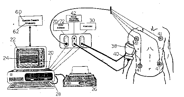
Reference is now made to FIG. 1 which illustrates,
in block diagram form, a cardiac power index monitor,
constructed and operative in accordance with the present
invention. Reference is also made of FIG. 2, which
illustrates a system implementation based on the
embodiment of FIG. 1. the cardiac monitor, denoted by
reference numeral 10, comprises a microcomputer 20, which
is preferably IBM-PC compatible. the microcomputer 20
preferable controls all monitor functions and drives a
physiological data display 22, such as an EGA graphics
video monitor, and a cardiac power index (CPI) display 24,
which may be provided by the same apparatus used for
display 22. The microcomputer 20 also stores data in and
retrieves data from a mass storage device 28, preferably
a hard disk drive with at least 10 mbytes, and drives a
hard copy device 26, preferably an Epson compatible
dot-matrix printer.
The monitor of FIG. 1 also comprises noninvasive
blood pressure measurement (NIBP)/cuff pressure controller
(CPC) apparatus 30, such as a Bosch EBM 502 D, for
measuring the brachial arterial pressure and heart rate,
and which operates a sphygmomanometric cuff 38. Cuff 38
P ~
-12-
1 is preferably a wrap-around type such as that used in the
PediSphyg system by CAS Medical, Inc. of Branford,
Connecticut, U.S.A., or a Bosch cuff. The cuff pressure
controller incorporates appropriate interface and con~rol
circuitry and software to enable the operation of
apparatus 30 in the mode of pressure control of cuff 38
instead of its conventional mode of operation for blood
pressure measurement. A block diagram of the controller
is shown in FIG. 12.
The monitor 10 also includes an ecg monitor 70 and
an R-wave detector and trigger generator 72, both
typically contained in standard ecg monitor systems such
as a Mennen Horizon 2000 patient monitor.
Also included in ~onitor 10 is a pulse wave form
sensor 40, namely, a Doppler ultrasound wall motion and
blood flow detection sensor, such as MedaSonics model 94G,
attached to the same arm as the cuff 38, and approximately
1-3 cm distal to lt. A pulse wave form processor 42
(shown in FIG. 10), preferably an analog and/or digital
circuit whose input is the wave form from sensor 40,
provides an analog output which is preferably proportional
to the blood flow.
Alternatively, the output may be proportional to the
wall motion or the velocity of wall motion. In either
implementation, high-pass filters eliminate most of the
influence of motion artifacts from the output signal to
the A/D converter 44, whose digital data output is read
by microcomputer 20.
A gamma camera 60, which may be a commercial
field-of-view gamma camera, such as an Elscint Model APEX
and its associated CPU 62, receives a gating R-wave
trigger either from an ECG monitor 70 or from its own
internal ECG monitor. In response thereto, camera 60
records a plurality of frames of several milliseconds'
duration at intervals of typically 25-40 milliseconds
throughout each cardiac cycle, averaging together the
frames from many (typically 300) cycles to obtain the
h ~, t~ `3
--13--
1 averaged volumetric frame values along the time curve
through the cardiac cycle.
A gamma camera CPU 62 communicates the resulting data
values to microcomputer 20 via a digital link, preferably
RS232 or Centronics parallel, or alternatively via disk
transfer.
As illustrated in FIG. 2, cuff 38 is attached
preferably above an elbow, and is controlled by
microcomputer 20 via cuff pressure controller 30. An
R-wave detector and trigger generator 72 senses the sharp
spike-like wave of the ECG, known as the QRS complex, and
provides a digital trigger pulse corresponding to the
occurrence of the R-wave (the center of the QRS spike).
It is proposed in the article by A. Marmor, et al.,
~5 of Annex A to measure a cardiac power curve and from it
to calculate a cardiac power index. Cardiac power is
defined as the time derivative of the product of cardiac
volume and cardiac (or aortic) pressure with time. the
cardiac power index is defined as the slope of the portion
of the power versus time curve from onset of systole to
the moment of maximal power.
Determination of the cardiac power curve and cardiac
power index (CPI) using the cardiac monitor 10 is
described hereinbelow.
E~timation of ~eft V~ntricular Pressure
occlusion of brachial flow during most of the cardiac
cycle creates a standing fluid column between the aorta
and the brachial artery, such that the rising intra-aortic
pressure wave form is transmitted to the brachial artery
with minimal distortion. Accordingly, the pressure values
obtained at the brachial artery very closely represent
those in the left ventricle.
In order to enable later combination with left
ventricular volume measurements made at the heart, the
brachial pressure values must be shifted in time to
account for the propagation of the cardiac pressure wave
-14-
1 from the heart to the brachial artery. The post-QRS time
required for a cardiac pressure wave to travel from the
heart to the brachial artery measurement si~e is known
herein as the propagation time, as is discussed ~elow in
conjunction with FIG. 5. The propagation time for a given
patient during the examination period is presumed constant
under all conditions of heart activity.
The operation of the cardiac monitor 10, including
the calculation of the CPI, is described in the flow chart
of FIG. 7. Patient preparations for gamma camera
ventriculography are completed, and 3-4 ECG electrodes 41
are attached in standard thoracic montage, for input to
ECG apparatus 70. While the patient is at rest, cuff 38
is applied just above an elbow, and the pulse wave form
sensor40is attached 1-3 cm distal to the cuff on the same
arm. The pulse wave form signal is acquired by
microcomputer 20 from apparatus 42 and displayed together
with the ECG, on the physiological data display 22, where
the c~ality of both ECG and pulse wave form signals are
used as visual feedback to verify proper signal
acquisition or to guide any required adjustment.
FIG.S 3A, 3B and 3C illustrate the technique by which
the sample points on the composite pressure-time curve are
determined, through the relationship between brachial
arterial pressure, cuff pressure, the ECG QRS complex, and
the detection of a pulse wave form distal to the cuff.
Two simplified cardiac cycles are shown with
representative parameter values in FIG.S 3A-3C. In the
first cardiac cycle, systolic pressure is 110 and cuff
pressure is set to 100 Torr, while in the second cycle,
systolic pressure is 115 and cuff pressure isset to 90 Torr.
Shown in Fig- 3A are the brachial pressure wave form, the cuff
pressure, and the ECG wave form, indicating the relative
timing of the QRS complex o each cardiac cycle and the
resulting brachial pressure wave form.
Point Al of cardiac cycle 1 occurs at the first
instance during the cycle when brachial pressure exceeds
i'~J~ J
-15-
1 cuff pressure. i~eferring to Fig. 3B, which depicts the pulse
wave form produced by pulse wave form processor ~2, it is
noted that tha pulse wave form abruptly rises at point Bl,
whose occurrence coincides in time with point Al of FIG.
3A, as the blood pressure wave passes the cuff, i.e.,
breaks through, and causes arterial wall motion that is
sensed by device 42.
The time delay from the QRS complex to the beginning
of the abrupt rise of the pulse wave form, labeled Tl and
having a value of 220 msec in FIG, 3B represents the time,
after the QRS complex, when brachial arterial pressure
reached 100 Torr. In FIG. 3C, which represents the
composite pressure-time curve, point C1 has a pressure
value of 100 Torr and a time of 220 msec, in accordance
with the pressure and time values of points Al and Bl
above. It is noted that the time scale of FIG. 2C is in
msec, whereas that of both FIGS. 3A and 3B is in seconds.
In similar fashion, in cardiac cycle 2, where
systolic pressure is shown as 115 Torr and cuff pressure
is shown as 90 Torr, points A2 and B2 correspond to the
time when the blood pressure wave breaks through the cuff,
which occurs at 180 msec after the Q~S of cardiac cycle
2. In FIG. 3C, point C2 is shown at a pressure of 90 Torr
and a time of 180 msec, in accordance with the pressure
and time values of points A2 and B2 above. In actual
implementation, each point on the composite pressure-time
curve is determined by averaging together the delay times
measured for a given cuff pressure maintained over a
plurality of cardiac cycles.
While the patient is still in resting position, the
operator causes the cardiac monitor 10 to commence
measurement initialization. During initialization, prior
to application of any pressure on cuff 38, the arterial
pressure propagation time from heart to brachial artery
iS estimated, and the pulse wave form is characterized.
Cardiac monitor 20 is operated to measure the maximum
and minimum pulse wave form values. Pulse waveform values
-16-
1 MAX~MP and MINAMP are the respective average maximum and
minimum values of the pulse wave form output of detector
42 during a plurality of cardiac cycles, preferably 10.
MAXAMP is preferably obtained by averaging together the
maximum a~plitude value of the output of detector 42 from
the aforementioned plurality of cardiac cycles, while
MINAMP is preferably obtained by averaging together the
minimum amplitude value of the output of detector 42 from
each of the aforementioned plurality of cardiac cycles.
FIGURES 4A, 4B and 4C illustrate a method for
calculating the propagation time, which is also used for
calculating the breakthrough time referred to below and
in Procedure ARRIVAL of FIG. 7. FIGURES 4A, 4B and 4C,
respectively, show the ECG wave form, brachial arterial
pressure wave form, and pulse wave form for two idealized
cardiac cycles. The propagation time is calculated by
first detecting the steep upswing of the pulse wave form
shown in 4C.
A regression line, labeled S1 in the first cycle and
S2 in the second cycle, is fitted to the early portion of
the upswing, preferably to the samples from the first 30
milliseconds of the upswing. A second regression line,
labeled D1 in the first cycle and D2 in the second cycle,
is fitted to the last portion of the wave form prior to
the upswing, preferably to the samples during the last 30
milliseconds prior to the upswing. The time interval T1,
from the R-wave of QRS 1 until the intersection point Bl
between lines S1 and Dl~ is the arrival time of the pulse
wave of cardiac cycle 1 at the pulse wave form sensor 40.
Similarly, the time interval T2, from the R-wave of QRS
2 until point B2 is the arrival time of the pulse wave of
cardiac cycle 2 at sensor 40. When determining
propagation time, the above arrival times are preferably
averaged together from a plurality of cardiac cycles,
preferably 10 cycles.
The operator then causes the apparatus 30 to obtain
the diastolic and systolic pressure values, and the heart
~3
-17-
1 rate, via microcomputer 20. A cuff pressure control
algorithm, one embodiment of which illustrated in FIG. 5,
uses the measured diastolic and systolic pressure values,
and selects the pressures to which the cuff is to be
inflated.
In a particularly important characteristic of the
present invention, the series of pressure values to be
implemented by the cuff 38 are defined such that the
largest number of pressure measurements are concentrated
during the early ejection phase, typically defined as the
phase between 100-125% of the end-diastolic pressure. An
example optimization algorithm for defining the pressure
values is illustrated in FIG. 5, wherein the pressures P0
through P9 are set as follows:
for DP - Systolic pressure - Diastolic pressure
P0 - 1.25 Systolic
Pl - Systolic pressure
P2 - Systolic - 0.25 DP
P3 - Systolic - 0.50 DP
P4 - Systolic - 0.65 DP
P5 - Systolic - 0.75 DP
P6 - Systolic - 0.85 DP
P7 - Systolic - 0.90 DP
P8 - Systolic - 0.95 DP
P9 - Diastolic pressure
The number of points, and their precise dependence
on systolic and dia~tolic pressure, may vary from the
foregoing, so long as there are a plurality of points in
the pressure ran~e from the end-diastolic point to midway
up the systolic rise, i.e., from diastolic pressure to
(systolic - 0.5 DP). In response to an operator
instruction to monitor lOj cuff 38 is inflated to pressure
P0, and the pulse detector output used to verify occlusion
of flow by the cuff.
-18-
1 The threshold for confirmation of occlusion is when
the output amplitude of pulse wave form detector 42 is
less than a fraction of the difference between
aforementioned MAXAMP and MINAMP, preferably 0.05
(MAXAMP-MINAMP). If the original cuff pressure P0 does
not reduce the output of detector 42 per above, the value
of P0 is increased, preferably by 10% of its previous
value, and the confirmation procedure repeated. The above
is repeated until occlusion is confirmed or until P0
reaches a maximum of 150% of systolic pressure. once
occlusion is confirmed, the detected pulse wave form
values are averaged together over a plurality of cardiac
. cycles, typically 10, to obtain an average baseline value
AI~P.
The operator then operates monitor 10 to commence the
measurement of the pressure-time curve. Cuff pressure is
reduced to value Pl, intended to allow breakthrough only
near the systolic peak. Microcomputer 20 analyses the
pressure wave form signal in real time during the current
cardiac cycle to determine if and when breakthrough
occurs. Breakthrough is typically defined as thepoint
when the wave form value first rises significantly above
the baseline, which in the preferred embodiment is defined
as a rise of more than three standard deviations above the
aforementioned baseline average value AMP.
If and when breakthrough is detected, the method
described above in determining propagation time is used
to estimate the breakthrough time. The above procedure
is repeated during at least 2, typically 5-10, cardiac
cycles for the same cuff pressure setting, providing at
least 2, typically 5-10, estimates of the breakthrough
- time for the pressure, from which mean and variance are
calculated for said breakthrough time. Before proceeding
to a new cuff pressure value, the set of breakthrough time
estimates is reviewed, and outlying values (typically
those lying more than three standard deviations from the
mean) are excluded from the set, and a new final mean
-19-
1 value calculated. The final mean value is the one stored
in the pressure-volume curve for the cuff pressure value
used.
Once the final pressure-time point has been determined
for a given cuff value, the cuff pressure is then reduced
to the next value determined in the cuff pressure control
algorithm, until the last value has been completed.
It will be appreciated from a consideration of FIG.
3 that at low pressures, such as those close to the
diastolic pressure, the above-mentioned method may be
unreliable as the required standing column of blood is
not well established prior to the onset of systole.
Hence, the pressure-time value for onset of systole is
taken to be the most recently measured diastolic pressure
value and its time is taken to be the aforementioned
propagation time determined when the patient was at rest.
The set of pressure values thus obtained is
interpolated typically by a piecewise polynomial curve fit
by least squares minimization to provide estimated
pressure values at any desired time point during the
systolic portion of the cardiac cycle. The pressure curve
as shown in FIG. 6B, which typically comprises an average
of pressure values recorded over a multiplicity of cardiac
cycles as described hereinabove, is then shifted by the
amount of the propagation delay, thereby producing an
estimated left ventricular pressure curve.
Left Ventricular Volume Determination
Reference is now made again to FIG. 1~ As noted
above, in the preferred embodiment, the invention
additionally comprises a field-of-view gamma camera 60,
such one commercially available from Elscint of Haifa,
Israel, and its associated CPU 62. The gamma camera 60
and CPU 62 measure the volume of the left ventricle using
gated radionuclide ventriculography according to the count
rate method as described in "Left Ventricular
Pressure-Volume Diagrams and End-systolic Pressure-Volume
- ~o -
1 Relations in Human Beings," by McKay, R.G., et al., and
published in Journal of the American Colleqe of
Cardioloqy, vol. 3, 19~4.
In accordance with a preferred embodiment of the
invention, the R-wave detector 72 detects the R-wave of
the ECG signal. Alternatively, if gamma camera 72
incorporates an ECG apparatus and associated QRS or R-wave
detector, the QRS or R-wave is detected by the detector
of the gamma camera.
A predefined amount of time later, typically 10-20
msec, the gamma camera 60 counts the number of gamma rays
coming from the left ventricle during a predefined time
frame, typically 5-10 msec. The gamma camera 60 repeats
the measurement every typically 20-50 ms, producing
sampled points on a curve of the left ventricular volume
with time. The volume curve thus produced is typically
synchronized to the QRS complex via the R-wave detector,
and is illustrated in FIG. 6A.
Typically, the volume curve will have only a few
points and, thus, it is typically interpolated by least
squares piecewise polynomial curve-fitting methods. Thus,
an interpolated volume curve, illustrated in FIG. 6A, is
calculated which has data at the same time points as the
pressuxe curve calculated in accordance with the method
described hereinabove. The cardiac power curve can thus
be calculated from the volume curve and the pressure
curve, as illustrated in FIGS. 6A, 6B and 6C.
Calculation of Cardiac Power Curve and CPI
At a plurality of points throughout systole, typically
32 points, the product of the corresponding pressure and
volume values is calculated. The time derivative of the
product is typically estimated using a second order
central difference methodj to produce corresponding points
on a cardiac power curve, illustrated in FIG. 6C. In the
preferred embodiment, the CPI is calculated from the
cardiac power curve values as follows:
-21-
1A linear regression line is fitted to the points of
said power curve between the start of systole and up to
and including its maximum value. Any data points whose
value lies more than two standard deviations from the
5linear regression line are excluded. After having
excluded the outlying points, a new regression line is
calculated, and its slope is used as the final CPI value.
The entire sequence of operation of monitor 10, as
described above, is summarized in FIG. 7.
10FIG. 8 shows a pulse wave form sensor 40 together
with its mounting means. The sensor 40 is a Doppler
ultrasound arterial blood flow sensor and comprises a
Doppler ultrasound transducer 80 which is formed as a flat
package. This enables a stable, compact mounting on the
15patient's arm. The Doppler crystals are mounted so as to
provide a fixed angle of illumination, typically 30- to
the horizontal.
The transducer is held by a transducer mount 81 which
is adjustably supported in a bracket 85, the two legs of
20which serve for the attachment of a strap 83 which is put
around the arm of a patient. The strap 83 can be fastened
around the arm in a tight manner by an adhesive-free
connection of its ends, for instance by means of Velcro
material. At its inner side, the strap has a plurality
25of pieces 84 of a compressible material which serve for
the absorption of shocks and movements.
After initial attachment of the transducer in
approximate location, a fine adjustment of transducer
position is made by an adjustment means including a screw
30shaft 82 extending through corresponding bores in the
bracket 85 and the transducer mount 8i and through two
retaining rings 87 on both sides of the bracket. The
screw shaft can be manually operated by a ~nob 86 at its
one end. By turning the knob 86, the mount 81 and then
35the transducer 80 is moved transversely with respect to
the arm of the patient.
~332 ~3
-22-
1 This embodiment allows a reliable attachment to the
arm without adhesives and maintenance of adequate pressure
of transducer against the desired skin location.
Another embodiment of the mounting means for the
transducer is shown in FIG. 9. The transducer 200 is
identically shaped as in FIG. 8. It is also held by a
transducer mount 201 having the shape of an inverted U.
According to this embodiment, the mount can be moved
vertically in the drawing so that the pressure with which
the transducer is pressed against the arm can be adjusted.
This is realized by means of an adjustment screw 203 which
can be manually turned (at 204) and which extends through
a screw bore in a bracket 202. Accordingly, by turning
the screw, the distance between the mount 201 and the
bracket 202 is varied and the transducer package is thus
pressed against the arm.
As in the embodiment of FIG. 8, the two legs of the
bracket 202 serve for the attachment of a strap 205 which
can be put around a patient's arm. The strap can be
fastened in a tight manner by means of a similar
connection as shown in FIG. 8.
It is now referred to an embodiment of a pulse wave
form processor 42 of which a block diagram is shown in
FIG. 10. The processor has the following components:
120 BIDIRECTIONAL DOPPLER PROBE, model MEDASONICS P 94-A,
is a 5 MHz Doppler blood flow transducer connected
to the driving circuit.
121 PHASE SHIFT BOARD, MEDASONICS p.n. 109-0051-010,
separates the sounds of the advancing blood flow,
providing two high level audio outputs.
122 AUDIO BAND PASS, passes the frequencies between 70
Hz and 15,000 Hz, suppressing noise, especially the
50/60 Hz "hum".
2~ f'
-23-
1 123 POWER AMPLIFIER provides the speaker drive and volume
control from the front panel.
124 HIGH-PASS FILTER separates the high frequencies from
the audio signal. The blood break-through generates
high frequencies (beyond 1400 Hz). This filter also
attenuates the sound generated by the receding flow
which has lower frequencies.
125 RMS to DC CONVERSION measures the power of the high
frequency spectrum by converting the total RMS (root
mean square) into a proportional DC voltage.
126 PROGRAMMABLE GAIN CONTROLLER, allows amplification
of the RMS value under computer control. Three bits
set eight levels of gain. The processed Doppler
signal is available at the BNC output connector.
127 ISOLATION BUFFER, transfers the processed Doppler
signal to the A/D converter which is isolated, according to
patient safety standards.
According to this embodiment, the processor provides
an analog output which is preferably proportional to the
total rapid blood flow, i.e., the portion of the blood
flow detected by sensor 40 which is flowing with
significant velocity. The processor produces an output
to an A/D converter which is proportional to the root mean
square (RMS) amplitude of the Doppler audio shift
frequencies above the smaller of 300 Hz or a frequency
equal to the multiple of the Doppler carrier frequency and
the factor 6 x 10 5.
FIGURE 11 shows an exact circuit of the processor
according to FIG. 10.
FIGURE 12 is a block diagram of a cuff
pressure control unit, i.e., of the pump controller 36
-24-
1 shown in FIG. 1. An exact circuit of this unit is shown
in FIG. 13.
The cuff pressure controller has the following
components:
101 PARALLEL INTERFACE, configured as an 8-bit parallel
port, D-15 connector, receives the commands from the
PC (Dell Computer). The available commands are:
- INFLATE
- STOP
- SLOW DEFLATION OF GIVEN RATE
- FAST DEFLATION
102 8 BIT LATCH stores the received command, controlled
by STROBE pulse.
103 DIGITAL TO ANALOG CONVERTER uses the six most
significant bits to generate 64 voltage steps(2.56 V
full scale, 40 mv per bit).
104 VOLTAGE CONTROLLED CURRENT SOURCE converts the
constant voltage into constant current, according to:
current = input voltage/20k ohm
which means 2 microamp per bit (126 microamp max).
105 CAPACIIOR DISCHARGER is a circuit capable of
discharging a 1000 ~f capacitor, with constant
current provided by block 104, in a floating mode
(none of the terminals connected to the ground).
Due to the constant current discharge the voltage
across the capacitor falls with a constant rate given
by:
dv = l/c time current
which gives a min of 2 mv/sec and a max of 126
3s mv/sec.
" . ~
-25-
1 106 COMMAND DECODER receives the two least significant
bits of the received byte, decoding the four basic
commands: inflate, stop, quic~ deflate and deflation
of given rate.
107 CHARGE/DISCHARGE SWITCH connects the low leakage
capacitor (used as sample & hold) to the charge or
discharge circuit. The analog switch is DPDT type.
108 L0W LEAKAGE CAPACITOR, 1000 ~f, is used as a voltage
memory. The voltage across the capacitor follows the
actual cuff pressure value. Discharging it with a
constant current generates a linear decreasing
voltage.
109 CAPACITOR CHARGER & COMPARATOR, determines the
voltage across the capacitor to follow the actual
cuff pressure value. The value is received from the
Bosch unit as 1 volt per 100 mm Hg pressure.
110 QUICK RELEASE CIRCUIT is a driver for the quick
release valve of the Bosch unit. Quick deflation
occurs upon receiving the corresponding command or
when the pressure reaches the maximum allowed value
(300 mm Hg).
111 OVER PRESSURE PROTECTION is an emergency circuit
which completely deflates the cuff at 300 mm Hg
pressure. This factory value can be changed by use
of an internal potentiometer.
h 1~ 2 ~ f.~
--26--
1 112 VOLTAGE COMPARATOR is the feedback loop controlling
the Bosch's deflation valve. During the slow
deflation, the capacitor is discharged with a
proqrammed constant current. The voltage across the
capacitor is a linear descending ramp. The
comparator compares this voltage with the actual
pressure value. The amplified error value drives the
deflation valve. As a result the pressure decreases
at the programmed rate.
113 OFFSET CORRECTION, allows the calibration of analog
pressure value against a standard manometer.
The cuff pressure controller has the following principle of
operation: -
Upon receiving (through the parallel port) the
command INFLATE the pump is energized and inflates the
cuff until the STOP command is received. During the
inflation the capacitor is accurately charged to a voltage
value equal to the actual pressure.
The SLOW DEFLATE command contains six bits which
finally are converted into a constant current. This
current discharges the capacitor generating an internal
built-in linear voltage ramp. The comparator compares
this voltage to the pressure value amplifying the
difference. The error voltage drives the deflation valve
forcing the pressure to follow the ramp. With the
described values the minimum deflation rate is 0.2 mm Hg
per sec and the maximum 12.6 mm Hg per sec.
The QUICK DEFLATION command deflates the cuff
immediately.
The STOP command freezes the cuff pressure to the
last value.
It will be appreciated by persons skilled in the art
that the present invention is not limited to what has been
particularly shown and described hereinabove. Rather the
scope of the present invention is defined only by the
claims which follow.
-
,
--2 7 -- t `
~nn ~ ~ /4
__ _ ~ __ _
Radionuclide Ventriculography and Central
Aorta Pressure Change in Noninvasive
Assessment of Myocardial Performance
Alon Marmor. Tali Shanr, Izhar Ben Shlomo, Rafael Beyar, Alex Frenkel,
and Dov Front
Depanmen~ o~Cardiology, Rebecca SieD ~ospital. Safed: Depanmen( of l~luclear .1Sedicine,
Rambam llospi~a/, Haifa; Depanment of Biomedical Engineenng and Department of
Cardiology, Rambam Medical Cemer: Technlon-llT, Far,~lly orl~edicine, Hai~a, Israel
Systolic pressure-volume diagrams were obtained noninvasively by measunng the systo~ic
eentral aonic pressure with a new devic~ and by combining the pressure measurements. thus
obtained, with a~solute volume measurements obtained by radionuclide ventncu~ography
during ejection. By dividing the peak power by the time elapsed from the beginning ot ejection
to the peak power point, the ejectbn rate of change of power (ERCP) was calculated. The
ability of this index to assess left ventncular function at rest and exercise was evaluated in
ten healthy subjects. ERCP proved to be more sensitive than global left ventricular ejection
f raebon increasing tivefold ~rom rest to exercise compared with only 2û% increase in global
ejeetion fraction. ERCP inr,reased dramatically postexercise from 3411 ~ 2173 to 18 162
14 633 gm/secZ, median 12 750, 95% confidence interval 970û-29 600. in healthy, whil~ in
pabents it increased twofold from 2637 + 824 to 5û62 ' 1897 gm/sec2, median 4û70. 95%
confidence interval 2800-7030, p < 0.001. ERCP had an excellent discriminative power in
differentbting healthy subjects from patients. having 100% sensitivity, 9û% specifidty. 95%
aeeuraey, 95% positive prr,~dictive value, and 90% negative predicbve value. Thus, this
noninvasive index seems to have a more comprehensive ability to evaluate changes in le~'
ventrleulat ~unction and shows a promising potential lor elinieal applications.
J Nucl ~led 30:1657-1665.1989
Maior efforts were made in the last decades to powerandtheejectionr,lleofchangeofpower(ERCP)
develop a reliable index to evaluate left ventricular to evaJuate left ventricular perforrnance. They demon-
perfor,mance. These effons included the development strated the superiority of their inde~t jD animal studies
of invasive indices obtained during cardiac catheteri- and in catheterization studies in patients. The invasive
zation such as ma~dmal left ventricular DP/DT. as well nature of this method prevented it from becoming an
as noninvasive indices such ns left ventricular ejection everyday clinically accepted diagoostic tool.
fraction at rest and during exercise. In recent studies In the present study an attempt was made to generate
(1-3) pressure volume diagrams were generated for noniovasively the power indices described by Stein and
diagnostic purposes for the evaluation of left ventric- Sabbah and to nssess their ability to evaluate leR ven-
ular performance using radionuclide ventriculography tricular function in healthy subjects under varying con-
for volume measurements and left ventricul~r intrlc:~v- ditions. The noninvasive measurementswere made pos-
itary pressure recordings. In spite of their invasive na- sible by a new instrument allowing noninvasive meas-
ture, these studies demonstrate the clinical importance urement of the central aorliC pressure (6). This method
of pressure-volume loops and provide a new insight was found to yield a good correlation with the ascending
into the leh ventticular fuDction. Stein ~nd Sabbah aor~ic pressure wave as measured by a Millar tipped
(4,5) in a series of studies used left ventrtcular systolie manometer in p~tients during heart eatheterization. By
combining this method with radionuclide measurement
Received Dec.8,1988; rev~sion ~ccepted May 9,1989. of leh ventricular absolute volume, pressure-volume
For reprinu contacc A. Mar~nor, MD, Dept. or Cardiology, curves were generated and the leR ventricular systolic
RebeccasierrHosp~ srael l3loo~ work and power were calculated:.The mean ejection
Volume 3û Number 10 October 1989 1 657
~ _
- 2 8 - ~ ~ h ~
rate of ch tnge of power (ERCP) in early systole waS mean power, peak power. and the rate of change of power
calculaled from the power-time curve. We attempted were compared with the change in global LVEF.
to deterrnine v~hether this index would be more sensi- The De~ice
tive than other ejection indices (ejection fraction. mean The components and the theoretic pnnciple of the device
power, tnd p~: tk power) in assessing leh ventricular and its elemems were reponed in a previous work (6) and are
function tt rest and Ltfter exercise. brieflv summanzed here. The device is composed of four
elements:
(a) a sundard sphygmomanometric cuff with an internal
MFIl~ODS transducer measuring ~he imracuff pressure. The cuff is pro-
vjded with an automDtic dellecting device controlled by a
Patient Populsltion microprocessor allowing gradual and constant delllation;
The study was performed in nine male patients 3 to 6 mo (b) an addiuonal narrow sphygmomanometer cuff con
aher an acute myocardial infarction (Ml), mean age 56 + 5 nected to a high sensitivity pressure transducer placed 1-3 cm
yr (patients) and ten healthy subjects (male, mean age 56 + below the occlusive cuff:
12 yr). The patients were included if they had a documemed (c) a sundard ECG monitoring svstem: and
Ml by elevation of total CK above 120 IU/I (>90 IU normal (d) all three elements are connected through an analog to
value), and CK-MB fraction above 4~O. and had a normal or digital convener to a censral processinr unit ICPU) consisting
near normal ejection fraction (>44%). There were six padents of an INTEL 8088 micro-processor. The output is displayed
with an inferior Ml and three patients wjth a non-Q wave Ml. on a monitor screen (Fig. I ).
mean total CPK being 655 + 160 IU with 6 + 2L/-o CK-MB. The method is based on the creation of a sunding fluid
Global left ventncular ejection fraction ~LVEF) measured by column from the aona to the occluded brtchial anery during
radionuclide ven~riculogrtpny was 57.11 1 9.3%. ranging theentireprrJcessofpressuremeasurements.Bvapplyingan
between 44% to 73%. No regional wall abnormalities were occlusive pressure on the brachial anerv dunng systole using
detected in any of the patients. an intlauble cuff. 3 temporarv sunding IIuid column is created
The nommal group included ten subjects wjth no symptoms in which the nsing intraaonic pressure is transmined to the
of angina pectoris. Mean global LVEF in the healthy subjects periphery with minimaJ distonion. The time intervals needed
was 61.1 ~: 7%. rangjng from Sl% to 75%, p = N.S.. when for the aonic pressure wave to overcome a ,iven occlusive
compared to the study group, All subjects underwent supine brachial pressure applied bv the inll tuble cuff on the ann are
exercise radionuclide ventrieulography which WaS stopped equal to the time intervals needed to reach the same pressure
aher 3 min at 100 W. The increase in heart rate and blood in the central aonu plus the propag~tion tirne to the brachial
pressure are summarized in Tlble 1. None of the subjects point, which is constant in the same patient throughout the
experienced chest pain, trrhythmias. or ST-T wave chmges. measurements. Time inter als are measured from the onst
Three patients eomplained of fatigue and shonness of breath. of depolarization ~QRS comple~ servin~r IS I reference s-stem ~
Measurements were uken before (at rest~ and immediately to the detection of the pressure wave b: an extemal transducer
aher exercise. Absolute LV volumes were measured b,v the at the brachial anery level. Application ot muhiple. successive,
count rate methods ts descrioed (3). At the same time. non- occlusive pressures on Ihe brachial aQerv decreasing sequen-
invasive measurements of central aonic pressure were made tially from peak svstolic to diastolic pressures. and plotting
by the device developed by us (6) which is described brieny their values ag~unst Ihe above described time intervals results
in the following paragraphs. Pressure-volume-time systolic in the reconstruction of the central aonic pressure curve.
curves were generated and the initial systolic work and power The validity of the noninvasive method waS documented
were calculated for the rlrst half of the stroke volume, The by two different approaches (6).
rate of change of power was calculated by dividing the peak 1. The pressure values measured by the device were super.
power by the time elapscd from the beginning of ejection to imposcd on the simultaneously measured central intraaonic
the peak power point. The changcs, from rat to exercise. in pressure wava in patients undergoing cardiac catheterization
TASLE 1
Blood Pressure and Heart Rate at Rest and Postexercise in Normals and in Patients'
Rest Postexerdse
Doubb Doubb
HR beats/sec BP mm/HgproductHB beats/secBP mm/Hg product
_ . . ..
Normals 70 + 10 102 + 17 7140 121 + 5 124 + 16 15 004
nD10
PaUents 70+8103~:97210 114 1 8 128+19 14592
n-9
P value NS NS NS NS NS NS
Note: Thsre was no significant di~terer~ h the postexerdse response ot heart rate. blood preSSure and doubb product betweer
the heatthy and the patients.
1658 Marmor, Sharir, Shlomo et al The Joumal d Nudear Mediane
--29--
1 ~
~1_ ~ ~
7- f~J
~ _ __ _ _____ ~
I '~' \
,
,
FIGURE 1
Schematic representation of the nomnvasive pressure device A. Sphygmornanometer cuff with intemal transducler
measuring the inlracuft pressure. 2. Narrow cuff connected to a high sensltivity pressure transducer. ECG and computer
are also schematically represented.
using Millar micromanometers. The tvpicai invasive prcssure :n~is to cotrect for the time lag between the pressure and
time curvc was generated from _10 cycles. and the mean s.d. volume curves. The beginning of lhe imfial pressure nse was
curvcs were calcul3ted. All vrtlues measurcd by lhe dcvice l'cll aligned with the beginning of lhe decrease in left ventricular
within I s.d. ~rom lhe cenlml inlr~ortlc recordings in 1-1 om volume ( ncgalive DV/DT) when lhe inhial end diaslolic poiDl
of the 15 patients studied. signifiles the beginning of ejec~ion. In ordemo eliminate errors
2. Using linear regression analvsis. ~n e:tcellent correlation resulting Irom changes in heart rate during acquisiuon of data
of r = 0.97 was found in e tch palienl (in a mtal group of 15 lhe study was aboned and lhe palicnts were e.~cluded when
patients) between the invasive tnd noninvasive digitized pres- changes of 5 bpm or more were l'ound. The pressure curves
sure values. obtained in lhe s tme pafient for the same fime rccordcd by the device and lhc lime-acuvily curves obtained
intervals. by the 8amma camera were ploncd agunst each other and
systolic pr~ssure-volume diagrams wcre generated. As the peak
Genera~ion of S~slolic Pressure ~olume Curves and Power power occurs early in ejection (4), and as we were interes~ed
Crlculation . in dcriving indices on the initial phase of ejecuon, only the
Absolu~e venuicular volumes were dclcrmincd by Ba~ed first haJf of the ejection phase (in ~erms of suroke volume) was
radionuclide ventnculography aCCordinK lO ~he coun~ r;t~e genera~ed. namely. the portion of ~he diagram dunng the first
me~hod (3). Using red blood cells laoelcd in vivo wi~h ~0 mCi halr of the ventncular emptying~ Leh ventricuiar work at half
of techne~ium-99m (~9mTc), a standard field-of-view gamma s~roke volume, mean power, pe~k power, ;tnd ERCP were
camera was inler~aced to a dedicatcd minicomputer with a dcnved according ~o ~he following formulas:
low-encrgy, medium resolu~ion, par~llel hole collima~or. Data v_v~-"~sv
werecollec~edin45-1ehantenoroblique(LAO)positionwi~hW = 0.0136 J~ p-dv tl)a 15- caudal anguhtion. The cardlac cycle was dtvldcd ID~O _v"
20 frames. A ~otal of 5 million counts were collected. Time-
activity curves were generated for the left ventricle. Aher the p _ (2)
aquisilion of leh ventricutar volume points a smoothing ~1- T
gorithm was applied using a fast f~ourier regression analysis pp
wi~h 16 harmonics. The ejection flow was oalculated ~s IheERCP = Tl ~ (3)
first denvative of the above dcscribed Founer fit. Aner the
simultaneous acquisition of pressure and volume points thcv where W = systolic work, V0~ = the end diastolic volume in
were aligned in such a way that for every pressure poin~ a milliliters, SV = stroke volume in milliliters, p D instanta-
corresponding volume point was found from the Founer fit. neous pressure in millili~ers of mercury and 0.0136 is constant
All prtssure points were moved lehward on the htorizontal ~o e~press the work in gram'meter, P - the mean pow in
.,
Vdume 30 ~ Number 10 October 1989 1659
r~ ~) h ;.
--30--
g-m/sec. PP = Fleak power, T = time in milliseconds, Tl - of ~ spccific lest using multiplc Ih~esholds lo dislin~uish
lim~ ~o peak po-v~r in seconds, and ERCP = eJection ra~e of belween normal and abnormal ~roups. RO(: :~n~lvsis was
change Or power in g'm/sec^, done to tesl Ihe sensilivily. specificily. accuracv and prediclive
A new ind~ lomhe assessmenl of rate or chanpe of pow~r values of each or Ihc methods sludied. The Ihrcshold with Ihe
was d~veloped. In ord~r to calculal~ Ihis index. pcak pow~r highest accuracy was selcc~ed to represent the best possible
was divided by Ihe limC to peak power, namely we calculalcd Ihreshold dclincaling normal from palholo~ic~l response. The
the slopc of the line connecung the power at Ihc beginning of rollowing indices wcre dcrived ~ccording lo the following
cjeclion to the peak power (Fig. 2). The reason ror the gen~r- equalion: -
ation of this index is that it ~enectS an average eslimase of rale
of change o~ power, unaffecl~d by the vanabilily associaled senslllvlly (%) s Irue poslllve/true posltlve + ralse nceative
wilh measurements of the instantaneous power values. A x 100
possible noisiness in the instantaneous power measurements specificitv (Co) = true negati~e/lru~ ne~aDve + ralse posiuve
may result from the MUGA technique of measunng relatively x 100
few volume values. This inde~ is independent of instantaneous
prcssure and nOw variabiLity, prediaive accuracy of a posiuve test (~0) = Iruc positive/true
posiliVe + ralsc posilive x 100
Ststtisticlll Ar~lysis
The ability of Ihe subjects to perforrn and to inaease the predictive accuracy of a negative test ( 5) = Irue ne~aUve/lrue
ejection power is dependem on their physical condilion, es- negative + ralse negauve x 100.
p~cially in thc healthy subjects (sedentar,v and physically
trained subjects participaled in Ihe sludy). The populauon RESUL
thus disphyed a skeved dislribuuon. Ihe Irained subjec s TS
outstanding in Iheit perforrnance. Thaefore Ihe melhod of
calculaingconfidence imervals fora populalion median waS All subjects pcrformed supme exercise stress tests
applied rather thati a parametric approach with t-tests and starting with 75 W with an increment of '5 W ever,v 3
standard deviation. Aher calculaling a 95'0 confidence inler- min and reaching 100 W in atl subjects. glob~l ejection
val the nonparametnc rank test. Mann-Whilney. was applied. fraction increasing in the health,v subjects from 61.1
This apptoach is recommend~d in m~dicat studie5 deaLing 7% to 66 1 7%, p = N.S., tnd in the patients t'rom 57.1
wjth small and nonhomo~en~ous populalions ( 7.S). For the ~ 9,~0 to 58 1 6%. p = N.S, Pv using ~5~o conRdence
purpose of presentation and compalison ~ith ejection fraction interva~ and nonparametric r~nk test. resttng ejection
mean and stand;lrd d~viation were calculated. althou~h these fraction in healthY subjects had a medi~n value of 63,o
parameters were not used in the statistical anal~sls. as the
population is not normally dismbuled. wnh a confidence mtervat of 52-65~o. It Increased aher
Receivcr operator charaaerislic (ROC) tn3.1ysis stands lor exerQse to a medtan value of 67,b, 95 o confidence
receiver operator charactenstic curves and it is denvcd from Interval of 59-71 C~o. p < 0.03. In patients the median
a graph in which the sensitivily is ploued versus l-specificilv was 59% at rest (95æ confidence imenal ~7-685o),
while tfter e:cercise the median waS 60to (95~o confi-
~300 dence interval 48-70%), p = N.S. When the t~o groups
PE~K POYER~PP) were compared there was an overlapping in the confi-
,_ ~ ~~- dence in~en~als, rank test showing non significtnt dif-
~n ro~ /~/ --1~ , ference between the groups. The indices of m,vocardial
~ // ~ ~ performance in the control group (health,v subjects) and
a: /~/ ' in the patient group are detailed in Tables 2,3. Mean
.. // power increased in the healthy from 297 + 169 g''m/
~ 5Do ~/ ERCP-PP/TI sec to 695 + '~79 g~m/sec, while in Ihe patients from
3 // ~ 245 + 67 to 441 + 147 g m/sec. In the patients the
;~ 5~ I ~~ peak power increased only slightly from 346 + 78 to
I ~ ",~ ~ A ~ 561 + 129 g~mlsec. The ejection rate of change of
InO~ ~-.~ . . .. 130 lilO power (ERCP) increased in the healthy fivefold from
TIME (MSEC) 3411 + 2173 to 18 162 + 14633 g~m/sec' (Table 2),
t T l t median 12750 (95% confidence interval 9700-29 600)
while in the patients it increased twofold from 2637 +
FIGURE 2 824 to 5062 + 1897 g~mlsec2 (Table 3), median 4070
Schematic representation of calculation of ERCP from the (95% confidence interval 2800 7030). As shown, the
power time curve at rest and after exercise in a healthy two confidence intervals are completely different, p <
subject. A: Power-time curve at rest. P: Power-time curve O.oO I . To funher demonstrate the discriminative power
postexercise. ERCP as shown ~n the tigure is calculated Of this parameter the percent change from rest to effon
by dividing the peak power by the tlme needed to reach
peak power in early systole, and represents the slope Of was calculated: ERCP mcreased m the healthy group
the line connecting the power at the beginning ot the by 376% (95% confidence interval 304-625%) while in
ejection and peak power. the patients it increased only by 84% (95% confidence
:
1 660 Mamlor, Sharir, Shlomo et al TheJoumal d Nuclear Medidne
~ iJ h t
--31--
TABLE 2
My dial Per1onT ancc Indices at Rest and aner a 1 00W Exercise in Healthy Subjects
A. Rest
Work LVEFPower Peak power ERCP
Subject no lgml [%IIg m/secl~g-m/secl ~g-mlsec~
36 51 324 533 4517
2 65 60 462 604 4027
3 32 S3 203 354 _2360
4 95 75 733 1110 9250
34 64 293 431 3748
6 31 52 207 333 2220
7 27 64 236 330 2750
8 26 65 182 271 1936
9 20 65 145 200 1429
29 62 193 300 1875
AVG 39 61 298 447 3411
STD 22 7 169 249 2173
r~. Postexerase
Pat~ent no.
47 54 1010 1089 14918
2 88 62 1208 2072 29601
3 - 23 59 556 604 10067
4 107 ~1 1071 1402 5a417
67 670 1318 15506
6 46 64 534 759 9731
7 55 67 597 1128 14100
8 45 70 540 674 11424
9 36 70 400 619 10317
~5 77 372 604 7550
AVG 55 6~ 696 1027 18163
STD 24 6 2~8 453 14634
interval 67-21 7~o). The individual changes in ejecdon ficity was found. its poshive predicti- e v~lue being 88C~o
fraction and in the ejection rate of change of power in ~nd the neg~tive predictive value being 90~0 (Fig. j).
both groups are summnnzed in Tables 2. 3. Note that
the change in ejection fraction was relatively small in
both groups while the chnnge in ERCP waS mnrkedty DISCUSSION
higha in the normal group compared to the patient
group. ROC analysis of the studied indices showed that The function of the left ventricle ~s a pump is best
LVEF at rest had a low sensitivity and a low negntive nssessed during ejection. when simultaneous changes in
predictive value (Table 4, Fig. 3). Mean power. peak pressure and volume with respect ~o time take place.
power, and ERCP also hnd low discriminative power nt Most of the methods devised to measure left ventricular
rest, but ERCP showed m e~cellent discrtminntive function are bnsed on chnnges of one pnrameter, either
power at e~ercise with a 100% sensitivity: 90~O specific- volume (circumferentinl fiber shonening, ejection f~ae-
ity; 90% positive predictive power and 100% negative tion) or pressure (dp/dt) ns a function of time. These
predictive power (Table 5, Fig. ~). This contrasted with methods give an incomplete information. each of them
the ejection fraction which showed 675 sensitivity, 70% nssessing only one aspect of the leR ventricular pçrforrn-
speeificity with positive and nega~ive predictive powerS nnce. ignoring the simultnneous chnnges in the other
of 62% and 63%, respecdvely (Figs. 3, 4). As illustrated pn~meter. An index taking into account end-systolie
in Flgure 4, ERCP a~fter exercise separates accurately pressure-volume relationship hns been used more re-
the healthy from the patients, while ejection fraction nt cently as a relnlively load independent and sensitive
rest and nher e~ercise and ERCP at rest were unsuc- measure of ventricular contractile state ( 9~. However,
cessful in differentiating between the two groups. Even this inde~c, mensured invnsively, was shown to be rela-
when the chnnge in ejection frnction was considered tively insensitive to chnnges in the inotropie state (10).
(Flg. 5), its specifici~y reached only 60% and its posidve The only systolie inde~c bæd on nll three parameters
predictivè vatue only 40%. In contrnst. when the change (pressure, volume, and time) is the leh ventrieular
in ERCP was used. a 100g'o sensitivity and 90% speci- power. In ~hysicnl terms the power is the most impor-
.
Volume 3û Number 10 ~ October 1989 1661
TASLE 3
Myocar~ial Performance Indices at Rest and After a 100W Exercise In Patients
A. Rest
Work LVEFPower Peak power ERCP
Sub~ectno lgml [%llg m/secl Ig m/secl lg m/sec'
'l 32 49 266 429 3900
2 28 52 178 303 2c20
~3 32 47 262 3~8 3180
4 30 44 27B 360 3273
34 73 306 361 2asa
6 36 68 225 360 2118
7 16 59 91 150 a82
8 44 63 315 420 2a1s
9 3s 59 292 346 2662
AVG 32 57 246 346 263a
STD 7 9 68 78 a25
B. Postexercse
Patient no.
36 50 400 59~ 7035
2 36 60 3~8 s42 3613
3 42 4~ 410 626 6260
4 53 42 408 363 2792
46 73 575 638 5317
6 35 70 29s 514 4283
7 30 63 252 420 2800
8 72 57 720 514 4673
9 54 6s ss7 835 8789
AVG 45 59 442 516 5063
STD 12 10 148 129 1898
tant parameter which describes the function of a pump. ischemia during exercise tests. In the present studv we
The left ventricular powcr is an expression of the rate present a noninvasive method of measuring vemncular
at which the left ventricle does work, and it was used power as the product of aonic pressure and the r~le of
before to characterize the performance of the left ven- change of left ventricular oiume durin~ ejection. We
tricle in dogs ( I I ) and in man ( 1'-15 ). It was measured recently described a method to measure the ascending
invasively and calculated as the product of the aortic limb of thc aortic pressure noninvasively (6). The pres-
pressure and flow (13-15). Another invasive mcthod sure wave of the ascending aorta during e rly ejection
that was used to calculate left ventricular power was the phase represents the pressure wave of the leh ventricle
product of aortic pressure and the rate of ch tnge of left in the absence of aonic stenosis. We utilized this
ventricular volume during ejection (l6). method together with the absolu~e ventricular volume
The reason ejection power did not become popular curve obtained by radio-entriculogram to obtain ven.
as an index of leh ventricularcont}actility is its invasive tricular instantaneous power during ejection. The leh
nature and therefore the inability to use it in a clinical ventricular power increases rapidly during early ejec-
setting for the measurement of myocardial reserve or tion, reaches a peak volume and then declines, therefore
. '
TABLE 4
ROC Analysis at Rest in ~he 19 Subjects
.
LVEFMeanpower Peak power EPCP
SensiUvity 33 89- 100- 100-
Speci~aty loo 30- 30- 30.
Accu-acv - 68 58 63 63
Threshd~ 50 31 459 3957
Posi~ivepredictivev~ue 100 3s- 63' 63-
Negat~vepredictiveva~e 63 65 100- 100-
Stabsticallv sbnificant ~p ~ 0.05).
1 662 Marmor, Sharir, Shlomo et al The Journal ot Nudear Medicine
3 3 - ~ ~J t~
REST EXERCISE
,_ r~o~TIrHTS ~ o ~TIEIITS HaLTHY
e ~~ ~ sT uaUE
" ` 60
_~ . I z
~: ., ~ ~_
z 40 ~: 40
1~: z
Z o20 ~ Q 20
_ u. ~ v, FIGURE 3
Discnminative power of ejection frac-
3 _ tionat rest ana exercise.
we considered the eslrlv ejection period only (till the The noninvnsive nature of our method enables us to
volume reached half the end diastolic volume. Fig. '). use this index in ambulator,v pttients ~nd to measure
In order to nssess myocardi~l reserve we mensured the chslnges in ERCP at rest and exerc~se. in order to nssess
lefs ventriculnr power a~ rest and postexercise. myocnrdial performance and myocardi31 resene.
The inde.~c developed in the present stud,v (the slope
Ejecsion R~te of Change of Power of the line connecting the power at the be~inning of
We csllculated the mean rnte of ch3nge of power ~jection so peak power. Fig- ~) constilUtes ~n 3vernge
during enrly ejectiOn by dividing the peak power to the estim;~te of the r;tte of ch~nge of power during c y
time from beginning of ejection to peak power- Math- ejection This index is simihr to the inde~ me3sured by
ematicallY. the rstte of chsmge of power is the firsl Stein ;Ir;d Sslbbnh (4,5~1he pe3k r~te ol changc of
derivative of power ;tnd the second derivat~Ve of work power--both ~ssessing the r;tte of ch3nge of power-
with respect to time. Physiologic311Y. the r3te of ch~nge However. our indc.x represents ~n ~-crnge estim3te of
of powerduringejectionm~ansthe3cceler3tlonolwork the rate of ch3nge of power ~nd no~ ~ singlc v31ue of
generated by the left ventricle during ejectiOn- A simil,3r r3te of change of power sls Stcin ~nd S~bb3h s inde-~-
index had stlready been mestsured invasivc!y by Stcln Another difTcrcnce bctween our mclhod and the in-
and S3bb3h (~.5). It was shown to be sensttl-e to dru~- dex me3surcd by Stcin ~nd Sabbah is thc nonin-~35il~e
induced ch;tng~s of the inotroPiC st3te in dogs. while 3nd indircc~ nature of our mcthod- The nonin~35iVc
affected little by ~llter3tions in prelold or ~ftcrlo~d. in ;~ppro;~ch h3s inherent difficultics ~nd ~rrors in me~
contrast to othcr cjcction indiccs (ejectiOn fr~ction- urements ol' instantsmeous volume 3nd prcssure- The
circumferentisll fiber shonening. ventnculslr vork ~nd pressure meslsurementS were v~id~led in-~si-~ly (6)-
etc) which arc m3rliedly influenced by los~din8 The rstdionuclide technique was chosen tor olu
conditionS (~). The ejeclion r3te of change of power me$lsuremeDts because it is free of geometric ~sump-
was also shown to separ3te psltients with abnorm3l ~nd tions ;md was extensivelY validated in the p;tst ( 17-20)-
normal ventricuhr pertformance (categonzed on the
basis of the ejection frstction. me3n velocity of circum- Palien~ Selection and E.Yerr;ise Tcsffng
ferential fiber shortening and left ventricul~r end dia- The aim of this study was to assesc the usefulness of
stolic volume inde:t) with no overlap of values between power and ERCP indices in the evaluation of leR ven-
categories of p3tients (p c 0.001) (5). tricular perform3nce. P~tientc after Ml ith preserved
TAELE 5
ROC Analysis Postexercise in the Studied Population
LVEFMean power Peak powar EP~CP
( /o)~0~ (o~O) ~0/o)
Senslovity 67 67 56 too-
Speclhrity ~o 80 100- so~
Arxuracy 68 74 79 95
PosiOvepredir,~tivev~6e 62 ~s loo ga-
l,, Negaove preair,tive value 63 62 71 100
Thresho~ vaiue 63489 g~m/sec 564 g-m/sec 8970 g-m/ser,
StaOsOc~ly sigmflcanl (p < 0.05;.
.
Vdume 30 Number 10 October 1989 1663
--3 4-- ~ ~j f.; `~3 ~ ~ ~
REST EXERCISE
~RTIENTS HEP~LTHY PaTlENTS H~
I
.. ~o
~ X~O
~, T u~LUE ; ~ zoooo .
giscriminative power of ERCP at rest O . (n ~ j
and exercise. 0 0
or slightly reduced left ventricular function (as judged 1897 g~m/sec in the patient group (Tables B and 3B),
by the ejection fraction at rest) were chosen. We delib- and it differentiated the two groups ~lth 100~0 sensiliv-
erately chose these patients, who in spile of bçing aher ity. 90% specificity. 90~ accuracv and had a erv small
documented myocardial injury were diagnosed as overlap of values (Tables 5. Fig. ~). Using ~5~c conl;
healthy by LVEF at rcst~and at çxercise. failing to dence interval a clear differenlialion between ~he
differentiate between them and the heaJlhv subjects healthv subjects and the palients was obt~ined. ERCP
(Fig. 3, Tables 2A. 3A. and 4). LVEF increased from increasing in Ihe healthy bv 376 c. 95C,~o confidence
61 7%to66+6.2%inthehealthvsubjects(Table2) in~erval 30~6'5% while in the patients it increased
and from 57 l 9% to 58 9,8% ir; pa~ients. p = N.S. onlv b,v 84æ (95% confidence intenal 67- 1, 0).
(Table 3). Ejection fraction did not differentiale be- The difference in ERCP from rest to exercise also
tween healthy subjects and p~tients as shown by the separ;tted Ihe IwO groups whh hi~h sensili h- and spec-
ranktestand~he95oconfidenceinten~als. w hichwere ificily (lOO~o and 90%. respectively) (Fig. j). h in-
almost identical in the two groups at rest and after creased fivefold in the healthy group while less Ihan
exercise. In this respect. this group of pttients v.ith twofold in the patients.minimal myocardial damage served as the reference We conclude that ERCP is a useful index of m-ocar-
system when comparing left ventricular ejeaion rrac dial performance. It cm be measured nonin~ el . Ils
tion performance to ERCP performance in discrimi- physiologic meaning is the accelerttion of energy ex-
nating the healthy from the diseased. These patients pended upon the production of usetul work b- the
had myocardial damage ~documented enzvmatic in- entricle during ejection. It was shov.n to be sensitive
farction). Peak power, mean power. and ERCP did not to change in contraaile state. while relati-ely independ-
separate the two groups at rest. We concluded that the ent of loading conditions (5). It increases markedly
myocardial damage was too small to affect ejection during exercise. and has a hi~h sensitivit,V ~nd specificity
indices at basal conditions. . for detecting mvocardial reserve. In order to assess its
ERCP at the postexercise measurement was 18162 clinical importance as an indicator of leh ventricular
+ 14 633 g~m/sec- in the healthy group and 5062 I pertormance or as a detector of myocardial ischemia
EXERC I SE
a"".~r~ ms
o , . ~,' ~ oo ~ ~ -
Discrim~native power ot the change ~ ~ ~ ~ ;
in ejection tractlon and in ERCP atter ~ ~t
exerclse. 0
1664 Murnor, Shuir, Shlomo et al The Joumal ot Nucleu Medicine
~ ~. r~ j J ~ J
funher s~uclics in different groups of patients with is- K, Sag3v~a K, Compar3~i~e inlluencc o~' 1O3d ~ersus
chemic hean disease h tve to be pert^ormed. inotroplc st3tes on mde.~es o,' ~entncul~r contractlhts:
e.~penmen~al and ~heorenc~l an~lvs~s based on pres-
sure-volume rei3~ionships. Circulailon I Y87: 76:14"-
REFERE,~CES 1436.
I l. Chapman CB~ B3kcr O. ~itchell JH. Lcft ventncul~r
1. McKav RG. Spe~rs JR. Aroesty J.~/l. ct al. Instant3- runction at rest 3nd dunng e.~ercise. J Clin In~est
neous rrle3surement of left ~nd nght ventncular stroke 1959: 38:1'01.
volume and pressure-volume relationships wjth an 1~. Bunncl IL. Grant C. Greene DG, Thc me~surcment
impedancec1theter. Circula~ion 198~: 69:703-710, of le~ cn~ncular pov~er 3nd ~ cnslon in m3n.
2. M~gon3n DJ. Shafrer P, Bush CA. et ~1. Assessment Phnsiol 196': 5:115.
of lelt entncular pressure-volume rel3tions using 13. Hernandez RR, Greenfield JC Jr, .~lcCall B~V. Pres-
gated radionuclide ~ngiographv, echocardiography sure-~low studics in h~penrophic subaomc stenosis. J
andmicromanometerpressurerécordings. Circula~ion ClinIn-es~ 1964: 13:iOI,
1983: 67:844-853. 14. Greent;eld JC Jr. Harlev A. Thompson H~. et ~1.
3. MclCa~ RG. Aroes~,v IM. Heller GV. et ~1. Left en- Pressure-llow studics in rn3n dunng a~nal l;brillation.
tricular pressure--volume dia~ms and end-systolic J Clin In-es~ 1968: ~7:'411.
pressure--volume relations in human beings. J .~m 15. Snell RE. Luchsinger PC. Dctermination ot` the e!ner-
Coll Cardiol 1984: 3:301. nal v~ork 3nd po~er ot' the lett ventrlcle in int3ct man.
4. Stein PD. Sabbah H~l. Rate of ch~nges of ventncular ,Im ~ean J 1965: 69:539.
power an indicator of ventncular pertormance dunng 16. Russell RO Jr. ~IcGavock PC. Frumer .~. Dod~e HT.
ejection.. ~mHeanJ1976:91:819. Leh ~entncul3r power in man. .~m Hear~ J 1971;
5. Stein PD, S3bbah H;`l. Ventncular performance me~ 81:799.
ured during ejection: studies iA pauents of the rate ot' 17. Veran~ S. Gaeta J. LcBlanc AD. et ~1. V3lidation of
changeol'-entricul3rpower.. ~mHear~JI976:91:599. Ieft~cnlncullr~olumemelsuremenlsbvr3dionuclide
6. Marmor AT. Blondh.lm DS. Gozlan ~. et al. I~lethod an~o@~ph,v, J .~'ucl .lled IY85: '6:1394-1-101.
for nonin-asi-e measurcmentofcentrDI aonic s-stolic 18. :~aSsle B. ~. Kramer BL. Gcnz E~'. Hcnderson SG.
pressure. ClinCardiol 1987: 10:'15. Radionuclidemcasurementol'lelt-entricular~olume:
7. Campbell ~IJ. Gardner .MJ. Calcul3ting confidence comp~nson ol' ~comelnc and COuDt5 bascd method.
interval for some non-parDmetnc aml-ses. 8r .tled J Circuia~ n I Y8~: 65:7'5- ~ ~0.
1988: '96:1454-1456. 19. Dchmer GJ. Finh BG. Hillis LD. .~I'icod P. Willerson
8. Gardner MJ. Altman DG. Confidence inter~al rDther JT. Lcwls SE. :Nonieomc~nc delermination of right
than P ~alues: es~imation rDther than h.spothesis tes~- ventncular olumes from equillibnum blood pool
ing. 8r .ll~dJ 1986: '9~:746-750. sc~ns. . Im J Cardu~l 198~: IY:7g-~ 1,
9. Suga H. S~g3wa K. Shoukas AA. Load independence '0. Swling .~,iR. Dell'lUJi~ LJ. :~us-nowiu ~IL. Walsh
of the instantaneous pressure-volume r;ttio ol the ca- RA. Little WC. Benedetto AR. Estimates ot' Icft en-
nine leh entricle and elrects of epinephrine and heart tricular volume5 bv equlllibr.um radionuclide angiog-
rate on the ratio. Circ Rcs 19?3: 3':31~. raph~: imponance of attenuallon correcllon. J .~ IIC
10. KassDA.. ~augh3nWL.GuoZ~l.KonoA.Sunagawa .~le/l IY84: '5:14-'0.
Volume 30 Number 10 October 1989 1665