Note : Les descriptions sont présentées dans la langue officielle dans laquelle elles ont été soumises.
Topel-Foster 1- 3
~ ,;,t
Endoscopic Aspiration Instrument
5 Technica~t. Field
This invention relate~i to medical instruments and
particularly to an endosc:opic medical instrument for
aspiriatincl biological ti.ssut.3 such as an ovarian cyst.
B qround of the Inve.nt ien
A number of enctoscopic medical and surgical instruments
are available for aspiratillg fluid d~lring a minimally
inva iive laparoscopic suryical procedure. One such
endoscopi~ instxument is an aspiration needle for puncturing
and aspi.rating fluid from, for example, an ovarian cyst.
Another endoscopic instrument is an aspiration tube for
aspirating fluicl from the peritoneal cavity. One problem
2i0 with the aspiration needle is that fluid leaks from around
the sha:~t of the needle when the it punctures the cyst.
Likewise, an aspiration tube allowi~ fluid draining into the
peritoneal ca~ity to come in contact with healthy tissue
before and dùring r~3moval. The problt~m of fl.uid leaking or
draining into the perit.oneal cavity is particularly
height:~necl when the fluid contains malignant cells. T}le
leakage o~:` fluid with maliqnant cells to surroundin~ tissue
significantly changes the mol-bi.dity and prognosis of the
patient .
Wherl a protein mar.k~?r test: produces ~ positive res~ll t
indici~ting that an ov~rii~n cyst i5 malic3nant, i~n invaslvt3
procedure is typically employsd to remove the o~rary and
fallopian tube associated with the mal ignant cyst. As a
result, the patient experiences a four to five day hospital
stay with three to six wee}rs of post-opera~ive recovery.
When t:he protein marker test produces a negati.ve reciult
inclicating that t.he ova~i.an cyst may be benign, a minimally
''Express M~il" label rurnber f )/
nfltr~ of t~i~posi~ ! L /, i ,~
I I~srab~ c~ t~ t l~ s p~ t3r l.~i t~ai~-l(J ~r3l~0s~
nii;~t~ c~ ,.. r,, :?.rjS ~/~cliil 1 () ;i
o~ 3 irldi; ~ !r~ t . ~ Jrll:n
o( P~tt3,;~ "l "~ 3~ [j.C. ;~
Si~n3tl.!l;? 0~ pt~r~:~J~3ilil~ i31 01` 1~0
. . .
' ' ' ~ , ' ~
~ .
: . ~ ` ' ' :
.
. : ,
- ` Topel-Foster 1-3
: '' '~ ',' '' ;.'.''
invasive, endoscopic close-chambered ovarian cyst removal
technique is preferred. This minimally invasive procedure
permits the patient to be discharged from the hospital
within a 24 hour period with a normal post-operative
recovery period lasting from three to five days. Typically,
the patient is back to work or performing normal activity
within five to eight days of this procedure. However, a
negative protein marker test result is accurate only about
80% of the time. Consequently, the surgeon ~lants to prevent
fluid leakage from the cyst. Should the ovarian cyst
contain fluid having malignant cells, the morbidity and
prognosis of the patient is significantly changed when the
fluid is allowed to leak and come in contact with other
healthy tissue within the peritoneal cavity. As a result,
; 15 the morbidity and prognosis of the patient is typically
worse than that of the invasive procedure where the
malignant cells can be contained from further migration.
The prevention of fluid leakage to healthy tissue during
endo.scopic aspiration will not effect the morbidity or
prognosis of the patient even though the fluid contains
malignant cells. A pathological report of the aspirated
fluid indicating that malignant cells are present would then
indicate the need for the invasive surgical procedure where
healthy tissue exposure to the malignant fluid is eliminated
or contained. However, leakage of the malignant fluid
during the minimally invasive procedure would significantly
worsen the morbidity or prognosis of the patient even thou~h
the invasive procedure would be subsequently employed.
Summary of the Invention
The foregoing problems are solved and a technical
advance is achieved with an illustrative endoscopic
instrument with particular applications to laparoscopic or
pelviscopic procedures for aspirating a cyst without fluid
leakage to other tissue within the peritoneal cavity. This
endoscopic instrument is advantageously utilized as a
Topel-Foster 1-3
'' ' `' ' ' ` ! ' ~'~
J
pelviscopic instrument for aspirating an ovarian cyst during
a close-chamhered ovarian cyst drainage procedure. The
endoscopic instrument comprises an elongated member such as
a suction tube having a first longitudinal passageway
e~tending between the distal and proximal ends of the tube,
whereby the distal end of the tube is inserted through a
trocar sheath. The proximal end includes first and second
access ports of which the first access port is accessible to
the passageway. A vacuum is applied to the first access
port and passageway to engage and maintain purchase of the
ovarian cyst with the distal end of the suction tube. The
instrument also includes a positioning or centering device
positioned within the suction tube passageway a
predetermined distance from the distal end. The centering
device has a passageway for receiving and centering an
aspiration needle that is inserted advantageously through
the second access port.
To further guide and center the aspiration needle, the
endoscopic instrument includes a second elongated member
such as a tubular needle guide having a second longitudinal
passageway between distal and proximal ends thereof~ The
tubular needle guide is positioned within the suction tube
~` passageway with the proximal end thereof being connected to
the second access port. The second access port has access
to the passageway of the tubular needle guide for inserting
and guiding the aspiration needle therethrough.
A sealing cap is positioned over the second access port
for maintaining purchase of the ovarian cyst when a vacuum
is applied to the first access port of the suction tube.
The aspiration needle is advantageously inserted through the
sealing cap, second access port and extendable through the
needle guide and beyond the distal end of the suction tube
to puncture and aspirate the ovarian cyst without permitting
any leakage of fluid within the peritoneal cavity.
Should any fluid leak from about the shaft of the
aspiration needle, fluid from the cyst held in purchase with
,~
'
.~
- Topel-Foster 1-3
the SllCtiOn tube is asplrated through the suction tube and
out of the first access port~
The instrument also includes a one~way check valve
connected to the proximal end o~ the aspiration needle for
preventing loss of purchase of the cyst within the suction
tube a~. well as preventing any rluid ~ithin the aspira-tion
needle. from keing drawn out of the distal end of the needle
and through the suction tube. This also prevents loss of
purchase of the suction tube with the cyst.
10The outer surface of the suction tube includes a matte
or non-glare finish for reduciny, if not eliminating, the
reflection of light from the suction tube during the
endoscopic procedure. This siynificantly reduces annoying
and fatiguing conditions to the attending physician.
15Alternatively, the endoscopic instrument includes the
suction tube with the first and second access ports at the
proximal end thereof and the second elongated member, such
as the tubular needle guide, positioned within the first
~`; passageway of the suction tube. The proximal end of the
needle guide is connected to the second access port thereby
allowing the se~ond access port direct access to the
passageway of the needle guide. The instrument further
includes a positioning device attashed about the proximal
end of the needle guide tube and positioned within the first
passageway or the suction tube a predeter~ined distance from
the distal end thereofO
To facilitate reuse and cleaning, the erldoscopic
aspiration instrument utilizes a T-type connector having
three access ports for interconne~tiny the suction tube and
the tubular needle guide. Two ports of the connector are
directly opposite from one another with the proximal end of
th~ suction tube being connect.able to the first port. The
second or side port has access t.hrough the connector and to
the passageway of the suction tube. The second elongated
member is positioned throuyh the first port and connected to
the third port directly oppo~ite therefrom. The passageway
.. ~
Topel-Foster 1-3
. , ,
of the elonqated tubular needle guide is accessible through
this third port.
The instrument further includes a positioning or
centering device attached about the distal end of the
tubular needle guide which is positionable within the
passageway of the suction tube when connected to the three
port connector. The positioning device includes a sleeve
and a plurality of arms extending radially therefrom and
toward the inner surface of the suction tube. The
-- 10 positioning device is advantageously positioned a
predetermined distance from the distal end of the suction
tube to prevent the ovarian cyst from being drawn too far
into the suction tube.
Similarly, the alternative endoscopic instrument
includes an aspiration needle that is insertable through the
third port and passageway of the needle guide and
extendable beyond the distal end of the suction tube to
puncture and aspirate the ovarian cyst. A check valve is
also connected to the proximal end of the aspiration needle
to prevent suction of fluid within the aspiration needle
from being drawn therefrom and through the passageway of the
- suction tube. A sealing cap is likewise positioned about
the third port for maintaining purchase of the ovarian cyst
and suction tube prior to and during insertion of the
aspiration needle. The endoscopic instrument further
includes a suction tube connectable to the second port for
maintaining a vacuum within the suction tube. A clamp
; positioned about the connecting tube regulates the vacuum
through the connecting and suction tube.
Brief Description of the Drawi.nq
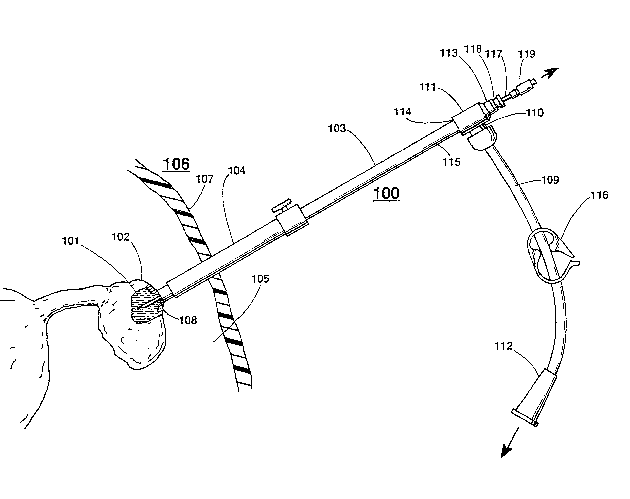
FIG. 1 depicts the endoscopic aspiration instrument of
the present invention inserted -through a trocar sheath into
a patient;
FIG. 2 depicts a partial cross-sectional view of the
endoscopic instrument of FIG. l; and
:,
`' ' ,
` Topel-Foster 1-3
-
FIG. 3 depicts a view of the distal end o-f the
endoscopic instrument along the line 3-3 of FIG. 2.
Detailed Description
Depicted in FIG. 1 is endoscopic instrument 100 such as
a pelviscopic cyst aspirator for aspirating fluid 101 from
an ovarian cyst 102. The instrument includes an elongated
member 103 such as a stainless steel suction tube which is
passed through the passageway of trocar sheath 104 and into
the peritoneal cavity 105 of patient 106. The trocar sheath
is inserted through the abdominal wall 107 of a patient for
performing a minimal.ly invasive laparoscopic or pelviscopic
,~ procedure usually associated with the reproductive organs of
a female patient. The distal end 108 of the elongated
suction tube member is positioned to engage the outer wall
of the ovarian cyst and maintain purchase of the cyst by a
vacuum introduced via suction connecting tube 109 attached
to side port 110 of connector 111 and a vacuum source (not
shown) attached to connector 112 of the connecting tube. T-
type connector 111 has three ports 110, 113, and 11~ of
which the proximal end 115 of suction tube 103 is connected
to access port 114. A well-known regulating clamp 116 is
positioned about suction connecting tube 109 and regulates
the amount of vacuum maintained on cyst 102. Aspiration
needle 117 is inserted through sealing cap 118, access port
113, and suction tube 103 to puncture and aspirate the
ovarian cyst. When inserted into the cyst, another source
of vacuum is applied to the proximal end of the aspiration
30 needle through one-way check valve 119 to aspirate fluid 101
without leaking into peritoneal cavity 105.
Depicted in FIG. 2 is a cross-sectional view of
endoscopic instrument 100 with aspiration needle 117
positioned for insertion through sealing cap 118 and into
;~ 35 access port 113. Endoscopic instrument 100 basically
` comprises elongated suction tube member 103, connector 111,
' and a second elongated needle guide member 120
```` 6
. ~
~` " .
}``
Topel-Fc~ster 1-3
' ' : ";
interconnected as shown. sy way of example, elongated
suction tube member 103 comprises a type 302 stainless steel
tube approximately 31cm in length. The suction tube
includes a hollow passageway 121 approximately 3/8" in
diameter and wall 122 approximately .035" in thickness.
Distal end 108 of the tube engayes and maintains purchase of
the ovarian cyst with the aiA of a vacuum applied through
passageway 121 and connector 111. Proximal end 115 of the
suction tube is connected to the first port 114 of the
connector with the aid of a well-known threaded
interconnection.
Second elongated member 120, referred to as an
aspiration needle guide, ls centrally positioned within
passageway 121 of suction tube 103. By way of example,
needle guide 120 is approximately 40cm long and comprises a
15 gauge thin wall tube having an outside diameter of
approximately .072" and an internal passageway having an
inside diameter of .059" between distal end 124 and proximal
- end 125. The proximal end 125 extends centrally through
first access port 114 of connector 111 and connects to
second access port 113 as shown with the use of well-known
``~ silver solder to secure the proximal end to the access port.
Positioning device 126 is positioned about the distal end of
the needle guide to center the needle guide within
passageway 121 of the suction tube.
Depicted in FIG. 3 is an end view of suction tube 103,
needle guide 120, and positioning device 126 taken along the
line 3-3 of FIG. 2. Positioning device 126 comprises a
hollow sleeve 127 connected to distal end 124 of needle
guide 120 using, for example, well-known silver solder. A
plurality of arms 128 extends radially from sleeve 127 for
centering needle guide 120 within passageway 121 of the
suction tube. The ends of the arms make colltact with the
inside surface of tubular wall 122 to center needle guide
120 within passageway 121 of the suction tube. This allows
the suction tube to be removed from the connector for
~j .
: . ~
.
Topel-Foster 1-3
cleaning. The positioning device is also located a
predetermined distance from the distal end 108 of suction
tube 103 for preventing the engaged ovarian cyst from being
drawn too far into the suction tube.
5Referring again to FIG. 2, connector 111 resembles a T-
type fitting having three access ports 110, 113, and 114.
The main body 129 of the connector is approximately 1 1/2"
in length between directly opposed access ports 113 and 11~.
Tha outside diameter of the main body near first access port
10114 is approximately .625". The main body also includes an
inside cylindrical passageway 130 approximately .312" in
diameter. Proximal end 115 and access port 114 are threaded
to provide ready interconnection of the two components.
Passageway 130 tapers to a diameter of .111" at the proximal
15end thereof for receiving the proximal end of needle guide
120. Similarly, the outside diameter of the main body of
the connector reduces to an outside diameter of
approximately .375". The proximal end of the main body
includes a circular flange 131 for well-known sealing cap
20118 to engage and provide an air-tight seal. Sealing cap
118 is commercially available from Cook Urological, Inc.,
Spencer, Indiana. Proximal end 125 of needle guide 120 is
connected to access port 113 using well-known silver solder.
Side access port 110 opens into main connector
25passageway 130 via connecting tube adaptor 132 having an
outside diameter of approximately .495" and an internal
passageway 133 having a diameter of approximately .250".
Connecting tube adaptor 132 is either silver-soldered or
press-fit into the main body o~ connector 111. Vacuum
30connecting tube 109 connects between adaptor 132 and vacuum
suction equipment (not shown) to provide vacuum and
aspiration through access port 110 and passageway 121 of
` suction tube 103. Well-known connecting cap 134 secures
connecting tube 109 to adaptor 132 and access port 110.
35When the suction tube 103 is placed next to an ovarian
cyst wall, suction applied thxough connecting tube 109 and
~ - :
Topel-Foster 1-3 ~ ~ ,
passageway 121 draws the ovarian cyst wall into the end of
suction tube 103 up to positioning device 126. In addition,
the ovarian cyst wall is engaged against the distal end 124
` of needle aspiration guide 120.
Aspiration needle 117 is a well-known aspiration needle
approximately 40cm in length and is comprised of either a 14
or 17 gauge metal tube with a hub connector 135 at the
proximal end thereof. A standard lancet bevel is provided
- at the distal end 136 of the tube. A well-known one-way
,
check valve 137 is connected to the proximal end of the hub
typically with a well-known Luer lock connector. A second
source of vacuum or suction is applied to proximal end 138
for providing suction to aspirate fluid from the ovarian
cyst through the tube of the aspiration needle.
15 To briefly describe the procedure utilized with the cyst
aspiration instrument, the aspirating needle 117 is inserted
through sealing cap 118, access port 113, and into
passageway 123 of the needle guide 120. Suction is applied
to the distal end 138 of the check valve. The instrument is
then inserted into the peritoneal cavity through the trocar
sheath 104 as shown in FIG. 1. Suction via connecting tube
~- 109 is applied to passageway 121 of suction tube 103, which
engages the outside wall of the ovarian cyst. Sufficient
purchase is maintained to manipulate the cyst and the ovary,
if necessary. Well-known regulating clamp 116 is in a
generally full open position for maximum purchase. The
aspiration needle is then extended through the needle guide
to puncture the wall of the cyst. Fluid from the cyst is
- drawn through the aspiration needle to aspirate and deflate
the cyst. A saline lavage is also utilized with the
aspirating needle to further aspirate or evacuate the cystic
contents. The one-way check valve 137 prevents fluid from
flowing back through the needle and into suction tube 103.
Should any fluid leak from about the outer wall of the
aspiration needle, suction tube 103 aspirates any emerging
fluid.
'`
.
. , : .
; ~
`' ' ' ` ,' ' ' , ~, ~
Topel-Foster 1-3
-. : ,
'
The aCpiration reedle is removed, and the cyst is fully
deflated by aspirati~n through suction tube 103. Purchase
of the cyst wall is maintained within suction tube 103 and
positioning device 128. The surgeon then employs a well-
known tying off techn7que of the ruptured cysk end, and thecyst is removed using well~~nown surgical techniques.
:Suction is maintained while t~Je aspirating instrument is
withdrawn from the cavity. This is c~one to prevent any
backward draining or leakage of fluid down the shaft. The
aspiration instrument is then removed without any leakage or
drainage of possibly malignant cells into the peritoneal
cavity. As a result, ~he leakage of possibly .nalignant cyst
fluid into the peritoneal cavity during a minimally invasive
pelviscopic procedure is minimized if not completely
eliminated.
It is to be understood that the above-described medical
instrument for aspirating a cyst without fluid leakage is
merely an illustrative embodiment of the principles of this
-invention and that other apparatus may be devised by those
skilled in t.he art without deparking from the spirit and
scope of this invention. In particular, the endoscopic
inStrUmeTIt is comprised of basically three components
consisting of a suction tube, a needle guide and a connector
with three acce~s ports. Alternatively, the suction tube
can be provided with a single, side access port and a second
access port at the proximal end for inserting the aspiration
needle therethrough. The centering device may be attached
to the distal end of the suction tu~e wikhout the need for
the needle guide. In this embodiment, the aspiration needle
;30 is inserted through the sealing cap and the second access
port at the proxi~al end of the suction tube and centered by
the ph~sician for insertion into the cyst through
positioning d~vice 128. Although described for aspirating
an ovarian cyst, the aspiration instrument may also be
utilized to aspirate hile from the gallbladder, fluid from
~:kidney cysts, or fluid from other cavitie~ of the hody.
~,
--. - ~ ~ .