Note : Les descriptions sont présentées dans la langue officielle dans laquelle elles ont été soumises.
W O 91/04706 2 0 6 7 3 S 7 P~r/US90/oS687
. ,
N~ Apparatus and Method for Detect m g Cancer
The present application is a continuation-in-part
application of prior pending application U.S. Serial No.
325,773, filed as a file wrapper continuation of USSN 262,073,
now abandoned, filed as a file wrapper continuation of USSN
188,752, now abandoned, filed as a file wrapper continuation
of USSN 036,943, now abandoned, filed as a divisional of U.S.
Serial No. 833,840 to Eric T. Fossel, filed February 26, 1986,
now abandoned.
STATEMENT REGARDING FEDERALLY SPONSORED RESEARCH
The funding for work described herein was provided by the
Federal Government, under a grant from the Department of
Health and Human Services. The Government may have certain
rights in the invention.
BACKGROUND OF THE INVENTION
Field of the Invention
The present lnventlon relates to an apparatus and
diagnostic method for detecting cancer in a living patient.
. .. .
SU~STl'rUTE S~IEET
.... . . .. . . .
~ -: . .. : .- - . . ....
~,: . . , . ., :
,
. . .
.. . : .
w091/0~706 2 0 6 7 3 ~ 7 PCT~US90/05687 ~ `
Prior Art
Approaches utilizing the technique of nuclear magnetic
resonance (NMR) to aid in arriving at a clinical diagnosis of
cancer are well known in the prior art.
Damadian was the first to propose a medical use for NMR.
He suggested it be used for detecting malignancy in tissue.
See R. Damadian, "Tumor Detection by Nuclear Magnetic
Resonance," Science 171:1151-1153 (1971). U.S. Patent
3,789,832 issued to Damadian covers an apparatus and method
for applying nuclear magnetic resonance to surgically removed
specimens to measure T1 and T2 for proton relaxation times,
which values, compared to values for healthy tissue, were
taken as a means of diagnosing cancer. U.S. Patent Nos.
4,411,270 and 4,354,499 issued to Damadian cover apparatus and
method for cancer detection with NMR imaging and scanning of
whole-body specimens.
A number of other investigators also reported that
nuclear magnetic resonance relaxation times (T1) for water
protons in organs of tumor-bearing animals have higher values
than the corresponding T1 for water structure in organs of
healthy animals. See Frey et al, J. Natl. Cancer Inst. 49, ~ ~
903 (1972); Inch et al, J. Natl. Cancer Inst. 52, 353 (1974); ; ~ ;
Ii~ima et al, Ph~siol. Chem. and PhYsics 5, 431 (1973); and
Hazelwood et al, J. Natl. Cancer Inst. 52, 1849 (1974). -
SUBSTITUTE SHEET . ~:
. . .~ -.
- , . ., . . , . . ;
~, . . . .; .. .. . . .
. . . . . ~ . ~ .
.-~ '', .- ' - , - : - ' .
WO91/04706 2 0 6 7 3 5 7 PCT/US90/0s687
Today, despite uncertainty regardlng the mechanistic
details, it is well known that biophysical changes which occur
in malignant cells often alter the proton NMR signal. See
D.G. Taylor et al, "A Review of the Magnetic Resonance
Response of Biological Tissue and Its Applicability to the
Diagnosis of Cancer by NMR Radiology," Com~uted Tomoara~hv,
5:122-133 (1981). Such changes form the physical basis for
detecting tumors by proton NMR imaging. See
R. Zimmerman et al, "Cerebral NMR: Diagnostic Evaluation of
Brain Tumors by Partial Saturation Technique with Resistive
MMR," Neuroradioloov 27:9-15 (1985) and K. Ohtomo, "Hepatic
Tumors: Differentiation by Transverse Relaxation Time (T2) of
Magnetic Resonance Imaging," Radioloov 155:421-423 (1985).
Proton NMR studies on excised tumors, as well as on
plasma and serum, from experimental animals and patients have
often shown differences in the relaxation parameters T1, T2,
and T2~ (T2~ being a combination of T2 from intrinsic
relaxation and relaxation induced by magnetic field
inhomogenieties) as a function of malignancy. Such findings
have been reported by the following:
L. McLachlan, "Cancer-induced Decreases in Human
Plasma Proton NMR Relaxation Rates," Phvs. Med. Biol. -
25:309-315 (1980);
F. Smith et al, "Nuclear Magnetlc Resonance Imaglng
of the Pancreas," Radioloov 142:677-680 (1982);
SUBSTITUTE SHEET
_ , . . . .
-
. . . .
. . . . .
. ;. `
; - .
.~ . . . ...
.. . .
~0~73~ 7
W09l/04706 PCT/US90/05687
P. Beall et al, "The Systemic Effect of Elevated
Tissue and Serum Relaxation Times for Water in Animals
and
Humans with Cancers," NMR Basic Princi~les and Pro~ress,
P. Diehl et al, Eds., 19:39-57 (1981);
R. Floyd, "Time Course of Tissue Water Proton Spin-
Lattice Relaxation in Mice Developing Ascites Tumor,"
Cancer Res. 34:89-91 (1974);
C. Hazlewood et al, "Relationship Between Hydration
and Proton Nuclear Magnetic Resonance Relaxation Times in
Tissues of Tumor Bearing and Nont~mor Bearing Mice: ~
Implications for Cancer Detection," J. Natl. Cancer Inst. -
52:1849-1853 (1974); and
.
R. Klimek et al, "A Discussion of Nuclear Magnetic
Resonance (NMR) Relaxation Time of Tumors in Terms of ~
Their Interpretation as Self-organizlng Dissipative
Structures, and of Their Study of NMR Zeugmatographic
Imaglng," Ginekol Pol. 52:493-502 (1981).
However, due to extensive overlap of groups and small i
differences between the means of groups, these methodologles
are not clinically useful.
SUBSTITUTE SHE~:T
.~............................ . . ~ . .
~ . ~ ~ . ., ` ..
~. . ~ - , ` '- -' . ` : `' '
~- . ... . .
, . . , ., ~ . , . ~
> . `:
~ ` . .
`--- : -. . .
~ W09l/04706 2 0 ~ 7 ~ ~ 7 PCT/US90/05687
While most of the prior art mentioned above suggests
using NM~ to analyze tissue, it is also known that body fluids
are subject to such analysis, as discussed by Beall et al.,
su~ra.
The cited prior art NMR methods for detecting malignancy
rely on the interpretation of the composite NMR signal arising
from all protons in the tissue or blood derived samples. This
composite signal is dominated by the water protons thus
obscuring the NMR signal from other proton-containing sample
constituents. A commonly held belief in the prior art is the
apparent correlation between malignancy and observed changes
in NMR parameters was due to "changes in water structure,"
quoting Prey et al., su~ra.
In other applications of proton NMR spectroscopy, it was
known to suppress the signal from the solvent (such as water),
in a sample. It was discovered that the components of the NMR
spectrum which have significant predictive value may be masked
by other materials in the sample, By eliminating the water
signal, the previously masked spectrum of these components was
revealed. In the co-pending application of Eric T. Fossel,
entitled "Process for the Detection of Cancer Using Nuclear :~
Magnetic Resonance," U.S. Serial No. 303,586, filed January
27, 1989, the teachings of which are incorporated herein by
reference, the aforementioned discoveries were incorporated
into a reliable method of diagnosing the presence of cancer in
a living patient.
~U~ITUl'E Stl~E~
. .. , . . . . - . .;
-
.':, . ~ ' , '. :
.' ' ' - '
~. - ,
:.~ . ` . -. , . : :. . ,
.
':
.
WO 91/04706 ~ ~ ~ 7 3 5 7 PCT/US90/05687 ,-.
In accordance with that invention, a sample of a
patient's bodily fluid is subjected to nuclear magnetic
resonance spectroscopy to generate a nuclear magnetic
resonance spectrum. A resonance line generated by a non-water
component of the sample is selected, and the full width of
this resonance line at a given height, e.g., at half its
height, is measured. The full width so measured has proved to
be a statistically reliable measure of the presence or absence
of cancer in the patient. s
:. ,
The above-described test of water-suppressed proton MMR
of plasma discriminates between persons with untreated cancers
and others with better than 90% accuracy. As such, the test
was widely acclaimed as one of the most important inventions
of the decade. No prior non-invasive test for cancer has ~ ~
approached that level of accuracy. False positive results, ~ -
however, have been obtained.
In the co-pending application of Eric T. Fossel, entitled ;
"Process for the Detection of Cancer Using NMR (Carbon 13),"
USSN 295,746, the teachings of which are incorporated herein
by reference, lt was shown that the major source of false ~ -
positive results is people with high levels of plasma
triglycerides. That invention teaches a non-invasive method
and apparatus with improved accuracy over prior non-invasive
methods to determine the presence of cancer in a living
patient.
SUBSTITUTE SHEET
i .
.
~ . . . .
,: . . . ..
~ - -
,`.-', ' ' :`' '
:.'-' . ' . :.
wosl/w706 2 0 6 7 3 5 7 PCT/US90/05687
In accordance with that invention, the triglyceride level
is measured of those patients with a positive result on the
proton NMR diagnostic test. A normal triglyceride level
confirms the cancer diagnosis; however, the fluid samples of
patients wlth high triglyceride levels are subjected to C-13
NMR. An abnormal result in that test confirms the cancer
diagnosis, whereas a normal result indicates that the prior
diagnosis a false positive.
. - .
Nothing in the prior art, however, teaches an apparatus
which automates the processes of obtaining NMR spectra,
interpreting such spectra, and making diagnoses.
SUMMARY OF THE INVENTION
In accordance with the present invention, an apparatus
was designed that is an improved nuclear magnetic resonance
spectrometer. The present invention automates the process for
diagnosing cancer using NMR. The apparatus has a spectrometer
component capable of taking water suppressed proton MMR and
C-13 MMR readings of a fluid sample. AdditionallY, the
apparatus has computer means for processing the proton and
C-13 readings and for obtaining a numerical value
corresponding to those readings. The apparatus further
comprises memory means for storing a set of standard or normal
values. The apparatus also has means for comparing values
o~tained from the NMR spectra with the set of stored values in -
the memory means and for classifying the fluid sample on the
'
~U~TITUTE SH~ a
' . ' : : ~ . - .
. . : .,
-: - . . - - , . . . .
. . -
,. . - ~ '-. . .
wogl/04706 2 0 6 7 3 ~ 7 PCT/US90/05687 ,,~,
basis of that comparison. The apparatus has computer programs
which direct its function. Additionally, the programs analyze
the data and yield a diagnosis with a great degree of
accuracy.
In preferred embodiments, the sample fluid is blood,
spinal fluid, or bone marrow plasma; blood is especially
advantageous. The component of interest is lipidic, and is
preferably from the methyl and methylene groups of the
lipoprotein lipids.
: ..
Accordingly, an object of the present invention is to - - -
provide an apparatus and method of using an apparatus for
automatic diagnosis of the presence of cancer in a living
patient using NMR spectroscopy.
. . .
Other objects and advantages of the invention will become ~ -
apparent from the descriptions of the drawings and the - -
invention which follow. -
BRIEF DESCRIPTION OF THE DRAWING
FIG. 1 is a typical 360 MHz NMR spectrum for the
non-water components (water-suppressed) of a plasma sample
from a healthy control o~tained in accordance with the present
invention;
' ,, .
SUBSTITVTE SHEET
. . . - - . -
~, . - . . -: . - - - ' - - --- - . . . -
~; , . . . - - ~ .. .
. . .- . . . . .. .. .... . . ..
:: - . . - . . ' :
. .- . . - . . .
:: . .. , . - . - . ,
... . : ~-- ... . ~ : . .
~: .- . :
.,. - -.
W09l/0~706 2 ~ 6 7 3 5 7 PCT/US90/OS687
FIG. 2 is an NMR spectrum for the same plasma sample from
which the spectrum of FIG. 1 was obtained, using the same
equipment and pulse frequency, except without water
suppression;
FIG. 3 is an expanded view of the methyl and methylene
region of the reading of the sample of FIG. 1;
FIG. 4 is an expanded view of the methyl and methylene
region of an NMR spectrum for a plasma sample for a patient
with an untreated malignancy; -
. '.
FIGS. 5A and 5B are C-13 NMR spectra of a plasma sample
in the olefinic region for a normal control and an untreated
cancer patient, respectively, obtained in accordance wlth the :`
present invention; . ~ :
FIGS. 6A and 6B are views of the C-13 NMR spectral region .
between 10 ppm and 90 ppm, with particular inclusion of the
region between 48 ppm and 80 ppm, of the plasma samples shown
in FIGS. 5A and SB for a normal control and an untreated
cancer patient, obtained in accordance with the present
invention; ~: .
, ' ': "' .'
FIG. 7 schematically illustrates the apparatus of the
present invention;
SUBST)TUTE SHE~T
. . - . -
, .-` . : ~ . - ~ .
-
Wo9l/~706 2 0 6 7 3 S 7 PcT/us9o/os687J~?~
FIG. 8 shows the results of a study performed using themethod of the present invention;
FIG. 9 shows a flowchart diagramming the operations cf
the apparatus of the invention;
FIG. lOA shows a flowchart for shimming, a task carried
out by the apparatus which ensures reproducible results from
water suppressed readings; - .
. ': ' '.
FIG. lOB shows a sample program for shimming,
corresponding to the proton spectrum step of the flowchart in ~-
FIG. lOA;
FIG. lOC shows a sample program for shimming, ~- .
corresponding to a carbon-13 spectrum; .
FIG. lOD shows a sub-program of step 8, RJ FQSET, of the
shimming program shown in FIG. lOC;
~ ,...
FIG. lOE shows a sub-program of step 2, RJ TUNE, of the
program shown in FIG. lOB; and
'~' '.
FIG. lOF shows a sub-program of step 6, RJ FQSET, of the
proqram shown ln FIG. lOB.
SUl~STITUTE SHEET .
.,': ~ ' ' ,
.' ' '
,': ' ~ ,
''. . ': . ' ~. ' . , :
.. ~ . ' , ' ,
. W091/04706 2 0 ~ 7 3 5 7 PCT/US90/OS687
11
DESCRIPTION OF THE PREFERRED EMBODIMENTS
At the outset, the invention is described in its broadest
overall aspects with a more detailed description following.
The present invention is a method to detect the presence of
cancer in a living patient. In accordance with the invention,
a sample of a patient's bodily fluid is subjected to proton
nuclear magnetic resonance spectroscopy to generate a nuclear
magnetic resonance spectrum. Since components of the NMR
spectrum which have significant predictive value may be masked
by other materials in the sample, the masking is eliminated to
produce the NMR spectrum. A resonance line generated by a
non-water component of the sample is selected, and the full -
width of this resonance line at a given height, e.g., at half
lts height, is measured to provide a reliable measure of the
presence or absence of cancer in the patient. The theory of
the above procedure is described in co-pending application,
United States Serial No. 303,586, the teachings of which are ;
incorporated herein by reference. ~
In practice, a computer component of the apparatus makes `
the measurement, or measurements in the event that more than
one re80nance llne ls selected, and compares the average value
obtalned to a stored value which is indicative of the normal
value; i.e., for a cancer-free person.
SUBSTITUTE SHEET
e_- . .. . . . .
,~ . . - . . -
~: ... . - . . .:
....
,
:: - - --.
,`, ,. . .~ . ~ :
. . . . . ~
W~91/04706 ~ O ~ 7 3 S 7 12 PCT/US90/05687 ~j,
In the event that a positive reading is obtained, this
reading may indicate the presence of cancer in the patient, or
it may be a false positive reading. It has been discovered - -
that a major source of false positive readings are persons
with high levels of plasma triglycerides. Accordingly, if the
measured value is greater than the normal value, the program
will diagnose the patient as cancer-free. However, if the
measured value is less than the stored value, the program will ;~
direct the apparatus to obtain a measure of the patient's ~-
triglyceride level.
' ~ "~ ,' . ;~;
In order to differentiate between true and false positive ~ -
readings, the sample tested previously is subjected to C-13
NMR spectroscopy for those who have elevated triglyceride
levels. The false positive results due to
hypertriglyceridemia and, conversely, the presence of cancer ~ -
in the patient, can be reliably determined from certain
features of the C-13 spectra.
In one embodiment of this invention, proton NMR
spectroscopy is performed initially on the sample to be
tested. The water suppressed proton NMR spectrum obtalned on
human blood plasma is dominated by the resonances of the
plasma lipoprotein lipids. Without water suppression, these
non-water resonances are virtually overwhelmed by the water.
Signal averaging allows observation of resonances of some
moieties associated with non-water bodily fluid components, at
high masnetic fields, even in the presence of the water
SUBSTITUTE SHEET
~s . : ,, -
,- ~
. .: ' , - ` . . :
.` . . , , - . '. . .`~ . . : .,
.;, . . - '
~. . . . . , - . . - ..
,
wo9l/0~706 2 0 6 7 3 S 7 PCT/US~/05687
resonance. However, the capability of modern NMR
spectrometers to suppress nearly completely the water proton
resonance will facilitate this reading. The water suppressed
proton NMR spectrum of plasma is essentially that of plasma
lipoproteins and a few low molecular weight molecules. The
protons of the proteins of plasma are obscured because they
comprise a broad smear of unresolved resonances. The sharper
resonances of the more mobile lipoprotein protons are
superimposed on this broad background.
The apparatus of the present invention operates on any
lipid-containing body fluid. Whole blood, serum or plasma may
be used. While the test may be performed on any such
lipid-containing body fluid, work to date has focused on blood
plasma. In blood, the lipids, inclusive of cholesterol, -
triglycerides, and phospholipids, are present in the form of
lipoproteins. The test for cancer will typically be performed
in vitro, preferably on serum or plasma.
The selected fluid of a SusPect patient or other person
to be screened for cancer is exposed to both a magnetic field
and radio-frequency energy to generate a nuclear magnetic
resonance signal whlch is then processed by the apparatus
which obtalns a value for a selected parameter, e.g., W1/2,
for lipid methyl and/or methylene protons. A relatively broad
range of proton frequencies may be employed, e.g., 60 MHz and
higher; 360 MXz or above is a preferred frequency. If cost is
not a factor, 500 MHz may be the preferred frequency.
ITUT~ ~ff~ET
.. . .
-
~ , ` ,
- : - ~, -: ' :
` - ; i ` - .~.
wo 9l/04706 PCT/US90/OS687 r~~
2~73~7
14
FIG. 1 shows a water suppressed proton spectrum of a
healthy control, and FIG. 2 shows a proton spectrum of the
same sample without water suppression. The truncated
resonance line of water is denoted A ~n FIG. 2. The resonance
lines between 0.5 and 2 ppm (parts per million of resonance
frequency) arise from the methyl and methylene groups of the
lipoprotein lipids. An expanded view of this region of the
proton spectrum is shown in FIG. 3 for a normal control and in
FIG. 4 for a patient with untreated malignancy. Accordingly,
in its preferred embodiments, the present invention uses one
of a number of conventional water suppression techniques,
i.e., techniques for suppression of the water proton NMR
signal. Numerous techniques have been devised to suppress the
water proton NMR signal in other contexts. These may be
broadly divided into two categories: (1) those that aim not
to excite the water proton signal, e.g., rapid scan
correlation spectroscopy and the selective eXcttation
technique, and (2~ those that arrange for the water proton
magnetization to be extremely small at the time the observed
radio frequency (rf) pulse is applied, e.g., the lnveræion
recovery technlque and saturatlon. These and other solvent
8uppres8ion technlques are described by P.J. Hore ln "Solvent
Suppression in Fourier Transform Nuclear Magnetic Resonance,"
Journal of Maanetic Resonance 55:283-300 (1983) and the
references footnoted therein. Although the water suPPression
technique is preferred when using A conventional NMR apparatus
due to its inability to distinguish between the signal of the
~U~l'ITUTE SH~ET
- . -- .
~` ' - - .. , - ~ ' . '. .
.- - -
. .. ' :
i' - . ' ':
~ WO 91/~i706 2 ~ 6 7 3 ~ 7 PCT/US90/05687
solvent protons and those of the moiety or species of
interest, a sufficiently sensitive apparatus would eliminate
the need for water suppression altogether.
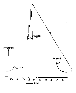
The linewidth at half-height of the resonances of
moieties, e.g., methyl and methylene groups. ~ssociated with
the lipids of plasma lipoproteins are treated as the variable
of interest. Full width at half-height W1/2 (linewidth) of an
NMR resonance line is inversely proportional to the apparent
spin-spin relaxation time (T2~), i.e. W1/2 =
2 -
The apparatus then compares the detected value for the
selected parameter with the corresponding parameter for the
healthy controls. In a preferred embodiment, values for
methyl and methylene are averaged and an average value of 33
Hz or less at a proton frequency of 360 MHz (8.45T) or 400 MHz
(9.40T) is taken as an indication of malignancy.
If a positive reading is obtained from the water
suppressed proton NMR spectrum of a plasma sample from a
patlont, a second level of testing to confirm the diagnosis is
performed. First, a conventional test, commonly called a
trlglyceride analysis, is performed to determine the
trlglyceride level of the patient. If the triglyceride level
ls normal. the posltlve readlng from the water-suppressed
proton NMR spectroscopy ls a true positive and indicates the
presence of cancer in the patient. If the tr~glyceride level
, ' ., ,
~U~1'11'UTE Stt~E7
?~ " ~ - ~
;~ ' : ' ;
f.' . , : .,,,, . ~ . :
-- . . .
... . . .
' -
wo gl/0~706 2 0 6 7 3 5 7 16 PCI'/US90/1)56117 ,~
is above normal, in order to differentiate between true and
false positive results, the program directs the apparatus to
obtain a C- 13 NMR spectrum of the patient's plasma sample
which is already available because of the earlier proton MMR
spectrum.
False positive results due to hypertriglyceridemia can be
reliably distinguished from true positive results by
substantial differences in certain features of C-13 spectra.
Accordingly, the plasma sample already obtained from the
suspect patient to be screened is exposed to a magnetic field
and radio frequency energy to generate a nuclear magnetic
signal which is then processed to obtain a value for C-13.
Initially, the olefinic region, 120-140 ppm, of the
spectrum is examined. Two peaks will appear, one at
approximately 128-129 ppm and another at approximately 130-131
ppm, about 2 ppm apart. The ratio of the resonance at the
general region of 128 ppm to that at 130 ppm is determinative
of cancer. In readings of plasma from normal controls and
from persons with non-cancer disease, the ratio of the hei~ht
of those two resonances ~128/130 ppm) is 0.9 or greater, l.e.
the refionance pea~ at 128 ppm is approximately equal to or
taller than that at 130 ppm. The heights of the peaks are
measured by computer from the center of the basellne noise to
the top of the pe~k. In readings of plasma from patients with
untreated cancer, the ratio of the peak helghts is less than
0.86, or the resonance peak at 130 p~m is taller, by at least ,
SUBSTITUTE SHEET
_ -. . .. ' ' . ` ': ' '. ~ . . ' ,'
~.; . . '' ' ' , . ' . '
.;: ' . , -.- ' - ' . , ~ ' ' :
& , ~ .`-~; , , ' , ' . . :'
''. " ' . ' " ' , . .. . '
.'':: ' ' . ' '
~- ~ ,. . , ' '
~ WOgl/04~06 2 0 ~ 7 3 ~ 7 PCT/US90/05687
5%, than that at 128 ppm. It should be noted that in patients
with hypertriglyceridemia, the ratio of the height of the
resonances (128/130 ppm) is the same as normal control values.
Accordingly, the computer will calculate the ratio of the peak
heights already measured, and if the ratio is greater than a
stored value will diagnose the patient as healthy, but
otherwise will render a diagnosis of cancer. In a preferred
embodiment, the stored value is 0.9.
FIGS. 5A and 5B show the olefinic regions of spectra
taken at 125.76 MHz with broadband proton decouPling from a
normal control patient and an untreated cancer patient. In
FIG. 5A, the ratio is 1.14 in the normal control patient and
in FIG . 5B the ratio is 0.70 in the untreated cancer patient.
In the patients with hypertriglyceridemia that were studied,
the ratio ranged from 1.05 to 1.68.
The changes in the olefinic region of the spectra of ~ ~-
untreated cancer patients can be explained by increases or
decreases in polyunsaturated fatty acid chains in the lipids.
The levels of oleic acid and linoleic acid are particularly
indicative.
Oleic acid is a monounsaturated fatty acid and is made by
the human body. Linoleic acid is a polyunsaturated fatty
acid, having two double bonds, and is not made by the human
body, but is obtained by consumption. Dietary fatty acids
include polyunsaturated acids, such as linoleic acid. A
SUE~STITUTE SHEET
., ~ - -,
,~ . ' ": '
; . ~~ - .. . .
ii s.
:- - . ?
W091/~706 2 0 ~ 7 3 S 7 PCT/US90/05687 ~?~
18
resonance peak in the general region of 128-129 ppm evidences
only linoleic acid in the patient. A resonance peak at the
general region of 130-131 ppm evidences both oleic and
linoleic acid in the patlent.
It has been discovered that the height of those resonance
peaks, relative to each other, are affected by certain
conditions of the patient. For example, persons with high
triglyceride levels usually have a high ratio of linoleic acid `
to oleic acid levels. Patients with untreated cancer are
found to have low levels of linoleic acid in their bodies,
presumably because cancer causes oxidation of polyunsaturated ~ -
fatty acids,-including linoleic acid. This is consistent with
the hypothesis that lipids are oxidized by hydroxyl free
radicals in cancer patients since polyunsaturated fatty aclds
are most susceptible to oxidation.
' '' '' '
Accordingly, if the sub~ect patient has both high
triglycerides and untreated cancer, the resonance peak at 130
ppm will be higher, reflecting the decreased linoleic acid in
both peaks. If, however, the peak at 128 ppm is not shorter
than that at 130 ppm by more than 7%, no depression, or an
lnsignl~icant de~resslon, o~ llnoleic acid levels has
occurred, and the posltive result obtained from the proton NMR
spectra is confirmed as a false positive. In that case, the
ap~aratus renders a diagnosis of no cancer present.
SUBSTITUTE SH~ET
~: . . . . .
.. .. ~ . . .
?~
_ ' '
20673~7
Wo91t04706 PCT/US90/0S687
In addition, the spectral region between 48 ppm and 80
ppm is far more complex in untreated cancer plasma than in
normal control or hypertriglyceridemia plasma. By "more ~ -
complex" is meant that there are more resonance peaks in the
region. A resonance peak is counted by the program if its
height is at least 50% greater than that of the bac~ground
noise during a normal testing period. As those skilled in
the art will know, the longer data is collected, the more
noise lessens and the more clearly peaks show. FIGS. 6A and
6B show this region for normal control and untreated cancer
plasma, respectively. These spectra were obtained at 125.76
MHz using a 5 mm sample tube and 14 hour accumulations. C-13
spectra with adequate information can also be obtained at 90.5
MHz in 10 mm or longer sample tubes. Of course, changes to
various parameters of the conditions under which the test can
be run will be evident to those skilled in the art.
These parameters include the size of the sample tube, the
pulse width, the pulse repetition rate, and the exponential
multiplication of the free induction decay by different
factors. For example, it is obvious to those skilled in the
art that the larger the sample tested, the faster spectra of
adequate quality will be obtained. Other changes to the
conditions given here will be evident to those skilled in the
art.
;Tl~UT~E SHE~T
.
.,. . ' .
-. : . ..
W091/04706 2 ~ ~ ~ 3 ~ 7 PCT/VS90/OS687 ~ -
C-13 MMR spectroscopy can be performed inltially on a
patient as a method to diagnose the presence of cancer.
without first performing a proton NMR spectroscopy as
described above. The C-13 NMR spectroscopy, however, is time
consuming, and therefore expensive to perform. While a proton
NMR spectroscopy generally takes 30 seconds to perform, C-13
spectroscopy may take anywhere from one to fifteen hours.
This increases costs accordingly. Accordingly, in a preferred
embodiment, C-13 spectroscopy is used to verify the positive
results obtained from the proton MMR spectra to illuminate
statistically and clinically significant differences in a
plasma C-13 spectra between true and false positive results
from the proton water suppressed NMR spectroscopy test.
In the preferred embodiments, an NMR spectrometer with a
magnet at constant field strength is used and the NMR signal
is Fourier transformed, with the full linewidth at half-height -~
for proton resonances of methyl and methylene groups, and then
C-13 resonances of linoleic and oleic acid, being the NMR
parameters of interest.
As noted in parent application, U. S. Serial No. 833,8gO,
correct sample preparatlon and executlon is essential to carry
out a successful measurement on plasma. Blood is collected in
tubes containing 70 microliters of a solution of 15% Na2EDTA.
8100d was maintained at 4C untll centrifugation. Plasma was
separated and stored at 4C until NMR analysis. Plasma
S~I~UT SH~E~
~ . . . . . ~ .. . . . .
, : . -
A
., ' .
WO9l/04706 2 0 ~ 7 3 ~ 7 PCT/~S90/OS687
21
samples were never frozen because freezing destroys
lipoprotein lipid structural integrity. Samples which showed
any visible sign of hemolysis were excluded.
All spectra were obtained at 20-22C at magnetic field
strengths of 360 MHz or greater. The samples were shimmed
individually by computer on the area of the proton free
induction decay until the full width at half height of the
water resonance was 4 Hz or less. Of course, careful shimming
is an assumed component of good NMR laboratory technique. ~ ~ -
It can be seen that of those experimental conditions,
temperature and shimming are not as critical with the C-13 NMR ,.
spectroscopy because a measurement of the linewidth is not
taken. Of course, the field strength used will determine the
length of time in which a sample is taken. In addition, to --
the experimental conditions, accurate results require careful
review of a patient's medical record to arrive at the patient
classification.
FIG. 9 shows a flowchart of the operation of the
Apparatus in whlch the apparatus first obtains a sample of
bodily fluid and then submits lt to NMR spectroscopy to obtaln
a water-suppressed H-1 spectrum. The apparatus then selects
and measures resonance lines and finds an average linewidth
whlch lt comPares with the value 33 Hz, a predetermined normal
value. If the average linewidth is greater than 33 Hz, the
apparatus will yield a negative diagnosis; otherwise, it will
SU~STITUTE SHEET
- - .
~ .
".- - . - . .............. ~ ,
- . ; .` :
W09l/04706 2 ~ ~ 7 3 ~ 7 PCT/US90/05687 ~ -
22
obtain a measure of the triglyceride level in the patlent. If
the triglyceride level is less than 190 mg/dl then it will
render a positive diagnosis; otherwise, it will obtain a C-13
spectrum of the sample. It will then analyze the C-13
spectrum by measuring the ratio of the peak at 128 ppm to the
peack at 130 ppm and by counting the number of peaks in the
range 48 ppm to 80 ppm. If the ratio is 0.9 or greater, the
machine will yield a negative diagnosis; however, if the ratio
is less than 0.9 or there is an abnormally high number of
peaks in the range 48 ppm to 80 ppm then the apparatus will
diagnose the patient as having cancer.
:~ :r.
Referring now to FIG. 7, there is illustrated a nuclear
magnetic resonance (MMR) spectrometer 2 which in the preferred
embodiment is capable of performing proton and C-13 NMR
spectroscopy and which is preferably, but not necessarily, of ~-
the type that suppresses the NMR signal of water. In order to
produce reproducible H-1 or C-13 spectra, it is necessary to
shim. The procedure for shimming is shown as a flowchart in
FIG. lOA in which a sample is placed into the machine, the
temperature is stabilized, the shimming parameters called, the
receiver gain is ad~usted, shimming is performed, the water
frequency is found for H-l water suppressed spectra, the
receiver gain is ad~usted on the suppressed proton frequency
lnduction decay area for water suppressed spectra, data is
acquired, a file is written, and the file is processed. FIG.
lOB shows a program ~written using the Bruker DISNMR software
packa~e) used to automatically perform the procedures depicted
$~ E ~T~ lE~T
~ .. . . . . .. . ,:
;.;~ .,..... -.: ~ ': :
,. ~, . , . -` .. . ; .
... . .. . . ..
. . .. .
~- . . ~ ; ..
~ . . . .. ,.. , . .. ~.. . .... . .. .
WOgl/04706 2 0 ~ 7 3 ~ 7 PCT/US90/05687
23
in FIG.lOA. FIG. 10C shows a sample program for shimming,
corresponding to a carbon-13 spectrum. FIG. lOD shows a
sub-program of step 8, RJ FQSET, of the shimming program shown
in FIG. lOC. FIG. lOE shows a sub-program of step 2, RJ TUNE,
of the program shown in FIG. lQB; and FIG. lOF shows a
sub-program of step 6, RJ FQSET, of the program shown in FIG.
lOB. The spectrometer 2 is adapted for examination of a
sample 4, which in this example is human blood plasma
contained within a test tube 6.
."~ ~
In accordance with the invention, the spectrometer 2
contains means 8 for selecting at least one and preferably a
plurality such as two NMR resonance lines in the NMR spectrum ~ ;`
of the sample 4 and measuring the linewidth of the line or
lines so selected. Preferably, the linewidth is measured at
half the height of the line, but this is not necessary and
linewidth can be measured at any predetermined fraction of the
height of the line in question. Measurement at half of line
height is preferred because this is a standard measurement
carried out in the field of NMR spectroscopy. Several
~ ':
commercially available computer programs can be used for
automatically measuring full linewidths at half height.
The means 8 of spectrometer 2 of the invention also `
measure selected pea~s for the examination of the C-13 NMR
spectra. The spectrometer 2 also is of conventional
construction and includes in addition to all lts other
structure a means 10 for storing a value or range of values.
~U8SS~TUT~ ~HEE~
. . .
. - . : ` ` ` `.,`~
.
, . .,. ` ~ .
W09l/04706 2 0 6 7 ~ ~ 7 PCT/US90/05687 ,~-
24
In accordance with the invention, the spectrometer 2 also
includes means 12 for comparing a linewidth which is either
measured directly or derived from a plurality of such direct
measurements with a value or range of values which represents
the value or range of values to be expected from normal
patients, i.e. patients who are free of cancer. Means 12 are
also used for classifying the measured or derived linewidths,
peak heights, and number of peaks as normal (i.e. cancer-free)
or abnormal (i.e. cancerous) based upon the stored
information. This may be done by comparison, subtraction, or
any other appropriate mathematical operation.
In a preferred embodiment, the selecting and measuring
means 8 is pre-ad~usted to measure the linewidths of the
methyl and methylene groups of the lipoproteln lipids, and the
pea~ heights and number of peaks in the C-13 NMR spectra.
~his may include suppressing the signal of water from the NMR
spectrum of the sample 4, or may alternatively be done
directly where the spectrometer 2 is sensitive enough to do
so .
In a preferred embodiment, the linewidths of the methyl
and methylene groups ~re averaged by the measuring means 8 to
produce a composlte linewidth which is the mathematical mean
of the two. This composite linewidth is compared with 33 Hz,
the value whi~ch is preferably stored $n the means 10, by the
SUE~STITUTE SHEET
';. . - - ~ - . . :
;. . .: ; - - , -~ :
': ' - . . .
.. ` . .. ~ `
-
. :, ... ... - -. . . :: - -
- . . ~. . : ..
- - . ~ ~ . . . . .. .
.
wo91/~1470~ 2067357 PCI/U590/05h87
classifying means 12. When the comparison shows that the
composite linewidth is less than 33 Hz, this indicates an
abnormal (i.e. cancerous) sample 4.
EXAMPLE
In this example, the method of the present invention was
applied to a group of 135 patients undergoing breast biopsy
for palpable and non-palpable breast lesions. For the
prospective breast study, blood was collected and maintained
at 4C until centrifugation. Blood was c~llected in
non-siliconized vacutainer tubes containing 70 microliters of
a solution of 15~ Na2EDTA. Plasma was separated and stored at
4C until NMR analysis. Plasma samples were never frozen
because freezing destroys lipoprotein lipid structural
integrity, Samples which showed any visible sign of hemolysis
were excluded.
Plasma triglyceride concentrations were measured (Damon
Clinical Laboratories, Westwood, Massachusetts) on all fresh
plasma samples. All spectra were obtained at 21C using an
improved spectrometer of the present inventlon operating at
360 MHz ~or proton (H-1) and 90.5 MHz for carbon (C-13). ! .
Additional C-13 spectra were obtained on an 11.8 T Bru~er AM
spectrometer operating at 125.7 M~z. All studies were carried
out in 5 mm OD sample tubes (Wilmad, Vineland, New Jersey;
#507PP or #528PP). ~ach sample, containing 0.6 ml plasma, was
shimmed individually on the area of the proton free induction
SUE~STITUTE SHEET
~;:- .. . . :
.~ . . . - - . . . ..
... .
- - , . : .
: - . : , ;::
;,
. . . . . . . .
;............ . - ` ~ ~
w091/0~706 2 ~ 6 7 3 ~ 7 PCT/US90/OS687 ,-~
26
decay (FID) until the full-width at half-height (FWHH) of the
water resonance was 4 Hz or less. An internal quality control
was found in the linewidth of the EDTA resonances. If all was
well with the sample preparation and shimming, the linewidth
(FWHH) of the EDTA resonances (without exponential broadening)
had to be 2 Hz or less and was often between 1.0-1.5 Hz. In
order to accomplish this, most H-1 probes require detuning to
avoid radiation damping. The probe was detuned until the 90
radio-frequency pulse became 20 msec. In the 8.45 T
spectrometer, this resulted in probe detuning of about 2 MHz.
The sample was spun during shimming of the Z shim coils and
during data acquisition. Our H-1 spectra were acquired using
presaturation to suppress water and an inversion-recovery
sequence to null any lactate methyl protons present. The
presaturation pulse was 4.0 sec, with a delay of about 0.8 sec
between the 180 and 90 pulse. Eight FIDs were signal
averaged and then Fourier transformed following multiplication
by an exponential resulting in 2 Hz line-broadening. The
portion of the spectrum form O.S to 1.6 ppm was phased so that
the baseline level at the edges of the plot was the same.
This resulted in defective phasing of other (non-plotted)
portions of the spectra.
C-13 spectra were obtained at 8.45 T and 11.5 T signal
wlth broadband proton decoupling bY averaging between 2,000
and 28,000 FIDs depending on slgnal-to-noise level and
resolution desired. The sample was identical to the samples
for H-1 spectra except 100 microliters of D20 was added for
SUE~3~TITUTE SHEET
;:: , ; : ........ : ~
, ` .~ . . . ~ . :- ;. . , ..
,-; ` - - . .
.- ` : ..
;-. : . ' . '. . '
w09l/04706 2 0 ~ 7 3 5 7 PCT/US90/05687
27
field lock. It was found that a minimum of 2,000 FIDs were
required to produce reliable resonance intensities.
Exponential multiplication equivalent to 25 Hz line-broadening
was used in the spectra obtained at 8.45 T .
In this study, we prospectively obtained plasma from a
series of women undergoing breast biopsy. Samples were drawn
prior to the biopsy, analyzed by NMR using the invention, and ~
results were then correlated with pathology reports. Two ~ -
groups of patients were included in this study; 63 patients
with palpable lesions and 72 patients with mammographically
discovered, non-papable lesions re~uiring wire localization.
Results of the H-1 NMR spectroscopic evaluation are shown in
Figure 8. In both groups, benign lesions were clearly
distinguished from malignant (p<0.0001) based upon the proton
values. For those values, triangles indicate patients who -
also had elevated triglyceride values. The open symbols
indicate samples in which the C-13 results conflicted with the
proton results. Thus, for the open symbols, the sample would
be changed from the benign column to the malignant column or
vice versa.
The patients in the study were a group of otherwise
healthy women, outpatients, referred for evaluation because of
an abnormality on a routine breast examination or a screening
mammosram. In this group, the sensitivity and specificity
were 93% and 95~, respectively. The predictive value of a
positive test was 84%, and the predictive value of a negative
SUB~;TITUTE SHEET
i. .. . . . . . . . . .. . . . .
~. - . .. .. . .. . . . , .- ~. .. .. ....
- . : , .. : ~ -. .. .
~,* . . . . . . . ..
.: - '-, . :. , ' .
~,, . .` ~ ' ` ' '' ,, ' - '-., - ,
Wosl/04706 2 ~ ~ ~ 3 ~ ~ PCT/US90/05687 ~
28
test was 98%. ~eclassifying patients on the basis of the C-13
data raises the sensitivity and specificity to 97~ and 98%,
respectively, and the predictive value of a positive test to
93%. These data would suggest that the H-1 NMR linewidth,
confirmed by a C-13 ratio, might be used as an aid in decision
making in patients with breast lesions.
There were five apparent false positive and two false
negative results. Elevated triglyceride levels (265 mg/dl and
206 mg/dl) were associated with two of the five false
positives and no false negatives. The C-13 ratio was negative
in three false positives, two of which also had elevated
triglycerides; and it was positive in two patients. The C-13
ratio was positive in all but one of the cases with malignant
breast biopsies. The one false negative by C-13 was also
negative by H-1. While patients wit~ elevated triglyceride
levels may need to be evaluated also by C-13, not all patients
with elevated triglycerides had narrowed linewidths.
The invention may be embodied in other specified forms
without departing from the spirit or essential characteristics
thereof. The present embodiments are therefore to be
considered in all respects as illustrative and not
restrictlve, the scope of the lnvention being indicated by the
appended claims rather than by the foregoing description, and
all changes whlch come within the meanlng and range or
equivalency of the claims are therefore lntended to be
embraced therein.
SUBSTITUTE SI~EET
.- . . . - . - : '. :- -
. . - . -
.
.; . . ` . .~
~: . . . ; ~.- ; ,
~. ~ -` "1' , .' ' ' " ' '