Note : Les descriptions sont présentées dans la langue officielle dans laquelle elles ont été soumises.
: ~ 2~g~
IMPLANTABLE ACCESS DEVICE
Thi~ i~vention is xela~ed ~o a pa~ient a~ce~s dsvice and
particularly ~o one which permits the ~ntroduc~ion o~ an external
~ilament such as a needle, ext~rnal ca~heter, guid~ wire, or
optical ~iber transGutan~ou~
This inven~ion xelat~s to a device ~o ~nable multiple
patient access proce~ures including in~using a therapeutic agent
to a desired site within a patient, ~eed~ng a ~ilament to a
desired internal site, or withdrawing a ~luid ~rom a patientJ and
more partiaularly, to such a deYice which is implanted ~u~h that
no portion i~ transcutaneous. It~ asc~ss portion is subcutaneous
but designed so as to racili~at~ repeated aacess by th~
percutaneous route.
In curren~ human and ani~al medical pra~ics, there are
numerous instanc~s where therapeutia agents must be delivered to
a speci~ic organ or tissu~ wlthin the-~ody. An example i~ the
infusion of chemotherapy into a central vein on a recurring ba~is
over a lengthy treatment period ~or widespread sites of malignant
........
'.
.
~ ~ $ ~
~umor. Withou~ an access d~vice ~or intravenous ~rug infusion,
multiple vein punctures over a lengthy period can result in
progressive thrombosis, venous sclerosis, and destruction of
small diameter pexipheral vessels. In other cases~ it may be
desirable to infuse chemotherapy to a localized malignan~ tumor
site. It may be difficult or impossible to ~eliver an agent
speci~ically to such a site on a regular repeti~ive basis without
surgically implanting an access system. Similarly, repeated
arterial access is occasionally needed for injection of an X-ray
dye or contrast agent into an artery ~or diagnos~ic purposes.
In other situations, there is a need to remove a body fluid from
a remote body ~ite repeti~ively for analysis. Finally, sensing
and physiological measuring devlces incorporated into small
diameter cathe~ers and small diameter optical fibers are
increasingly being utilized for monitoring body processes and
could be more easily implemented through a properly designed
access device with an adequate internal diameter.
In prior medical practice, peroutaneous catheters have been
used to provide vascular or organ access ~or drug therapy or
removing body fluids. Although such systems generally performed
in a satis~actory manner, numerous problems were presented by
such therapy approaches, including the substantial care
requirements by patients, è.g. dressing changes with sterile
techniques, a significant rate of infection o~ the catheter
because o~ its transcutaneous position, and a high rate of venous
thrombo~is, particularly if the catheter was located within an
extremity vein.
Implantable infusion devices or "ports" have recently become
available and are a significant advance over transcutaneous
catheters. Pres~ntly available in~usion ports havs a number of
common ~undamental design ~eatures. The ports themselves
comprise a housing which ~orms a reservoir which can be
constructed ~rom a varie~y o~ plastic or me~al materials. A
surface of the reservoir is enclosed by a high~density, salf
sealing septum, typically made o~ silicone xubber. Connected to
the port housi~g is an outflow catheter which communicates with
a vein or other site within the patient where it is desired to
infuse therapeutic agents. Implantation of such d vices
generally proceeds by making a small sub~utaneous pocket in the
patien~ under local anesthesia. The i~ternal out~low catheter
is tunnelled to the desired infusion site and is connecteclto the
infusion port. When the physician ~esires to infuse or remove
material through the port, a hypodermi~ needle is used which
pierces the skin over the infusion port and is placed into the
port.
Although presently avàilable implantable infusion ports
generally operate in a satisfactory mannex, they have a number
of shortcomings. S~nce these devices rely on a compressed rubber
septum for seallng, there are limitations in the diameter of
needles which can be used to penetrate the septum, since large
diameter needles can seriously damage the septum. These diameter
limitations severely restrict the flow rate o~ fluids passing
through the port. Moreover, the needles used must be of a
special design which minimizes septum damage.
--3--
For prolonged in~usion using a conventional port, the
`.infusion needle is taped to the patien~'s s~in to ~old it in
position. Conventional ports do not allow the needle to
penetrate deeply into the port; and consequently, a small
displacement o~ the needl~ can causc it to be pulled ~rom the
port, allowin~ ex~ravasation. In cases where locally toxic
material~ are being in~used, extravasation o~ such materials can
cause local tis~ue damage which can lead to a reguirement for
corrective surgery such as skin gra~ting or removal of tissue.
Presently available implantable in~usion devices mus~ also
have a signi~icant size to provide an acceptable target surface
area for the physician who mus~ locate the port and penetrate the
sep~um properly with a needle. ~ha port housing becomes bulky
as the septum size increases since s~ructure is re~lired to
maintain the septum in compr~ssion ~o provide el~-sealing a~ter
the needle is removed. Moreover, presently available infusion
ports are di~icult ~o ~lsar if thrombosis occurs within them or
in the implanted outflow catheter, since it is difficult if not
impossible to feed a cleaning wira through the penetrating
hypodermic needle in a manner which will clear the :Ln~usion
device and the i~ternal out~low catheter. Present inPusion ports
have a space which contains a retained fluid volu~e beneath the
self-sealing septum which increases the volume o~ drug which must
be administered to enable a desired quantity ts reach the
in~usion site. This retained volume also poses problems when a
physician desires to deliver di~ferent drugs to the same infusion
site which are incompatible or rendered less e~fective when
mixed. In addition, when it is desired to withdraw blood through
~ h~6l~8 '~
the port, the retained volume o~ the prior art in~usion ports is
an area where blood clotting can occur, ~hus in~er~ering with
future access to the site. And finally, ~or present infusion
ports, there is a risk that the physician attempting to pierce
~he port septum will not properly enter it, leading to the
po sibility o~ extravasation which can cause signi~icant
undesirable cons~guences as mentioned previously.
In applicants~ related patent application and issued
patents, various approaches toward permitting transcutaneous
access to implanted cathe~er are described. In accordance with
those devices, multiple sealing members are us d to provide an
adequate fluid seal across the access device, both when an
external filament i8 introduced into the dsvice and a~ter it is
removed. Th~ acce s ports in accordance with this invention
achieve simplicity in construction and reduce ~he number o~
components necessary to provide the ne~essary ~luid seal. In
those applications where it i8 desired to access a port using a
sharp needle, damage to elas~omeric sealing elements can occur
over repeated entries to the port in prior port designs. In
accordanc with this invention, the implanted port has an
articulating valve mechanism in which the accessing needle (or
other filament~ contacts a hard material such as a metal to open
the valve. ~ccordingly, a durable device is provided which is
not damaged through long term use.
~ he ~eatures of the present invention are primarily achieved
through use o~ a valve asse~bly in which a sealing elemen~ is
normally maintained in contac~ with a valve seat. When
introducing an external ~ilamen~, which may be a needle,
-5-
2 ~ 8 l~
catheter, wire, optical ~iber etc., ~he filament engages ~he
sealing element forcing it ~rom engagement with the valve s~at.
once fully inserted into the access device, fea~ures are providsd
to assure a ~luid ~eal around the introduced ~ilament.
Additional benefit~ and advantages o~ the present invention
will become apparent to those skillsd in the art to wh~ch this
invention relates from the subsequent description of the
preferred embodiments and the appended olaims t taken in
~onjunction with the accompanying drawings~
BRIEF DESCRIPTION OF THE DRA~WINGS
Figure 1 is a cross-sectional view through an access port
in accordance wi~h a first embodiment of this invention shown in
a normal condition in which an external ~ilament is not present
within the device.
: Figure 2 is a somewhat enlarged cros~-sectional view of the
access port of Figure 1 shown ~Yith an accessing needle
penetrating the device.
Figure 3 is an exploded pictorial view o~ the valve assembly
of the port shown in Figures 1 and 2.
Figure 4 is a cross-sectional view through an access port
according to a second embodiment o~ this invention showing a
valve assembly comprising metal seal elements affixed to a multi-
lea~ elastomeric valve disk.
Figure 5 is a ~rontal view of the valve assembly of the port
shown in Figure 4.
Figure 6 is an exploded pictorial view of a valve assembly
in accordance with a third embodiment o~ this invention
--6--
fil
incorporating a unl~ary seal member for sealing against the valve
seat formed by a sealing disk.
Figure 7 is a cross-sectional view of an access port
incorporating ~he valve assembly shown in Figure 6 and Purther
showing an acce~sing needle penetrating the devi~e.
Figure 8 is a cross-sectional view taken through an access
port in accordance with a ~ourth embodi~ent o~ this invention
shown with an accessing needle partially penetrating the deYice.
Figure 9 is a cross-sec~ional view o~ the access port shown
in Figure 8 but showing the accessing needle penetrating the
valve asse~bly to pe~mit access to an implanted catheter.
~1 '
~''' C~
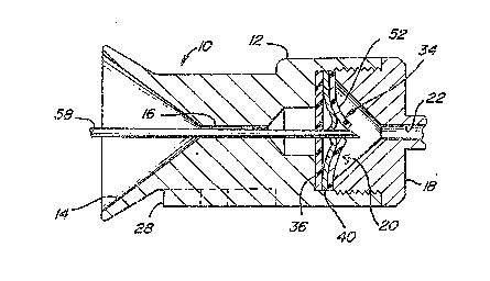
An access device in accordance with this invention is shown
in Figures 1 and 2, and is ~enerally designated by referen~e
number 10. As shown, access port 10 is similar to that described
in applicant~s issued patent nu~bers: 5,053,013 and 5,057,084,
to which the present applica~ion is rela~ed. Access port 10 is
designed to allow a sharp needle to access ~he device for
purposes including infusing drugs or other ~luids in the patient
or withdrawing ~luid~ from the patient. Access por~ 10 generally
has housing 12 which defines a generally funnel shaped entrance
orifice 14. Entrance orifice 14 has a decreasing cross-sectional
area which ends at housing passageway 16~ The shape of entrance
orifice 14 serves to guide a needle lnto passageway 16. To that
end, the sur~a-e o~ housing 12 ~orming orifice 14 is a hardened
material such as titanium which has been found to be acc~ptable
for this application.
,~
--7--
.~
~ g3 8 ~ ~ 8 ~
Housing 12 together with outlet plug 18 define valve chamber
~ located between passageways ~6 and 22. As shown, the
protruding catheter connector tube 24 of outlet plug 18 is bent
to provide a positive means ~or preven*ing an introduced needle
from passing e~tirely through the device and potentlally damaging
a soft elastomeric impla~ted cathe~er 26. Connec~or tube 24
does, however, permi~ ~ore ~lexible ~ilaments such as a catheter,
guide wire or optical ~iber to pass into implanted catheter 26.
~ounting pad 28 enables the device to he conveniently m~unted to
subcutaneous support tissue pre~erably using sutures, staples,
or other fasteners.
Valve asse~bly 34 is disposed within valve chamber 20 and
is best described with reference to Figure 3. Valve disk 36 is
made ~rom an elastomeric material such as silicone rubber and is
positioned in valve chamber 20 alosest ~o entrance orifice 14.
Disk 36 has a central aperture 38 de~ining a valve seat which is
intended to seal against the introduced needle ox ~ilament upon
insertion into access po~t lO, as wlll be described in more
detail as ~ollow~. Stacked directly against disk 36 is sealing
member 40 which is pre~erably made, at least partially, of a hard
material such as a metal. Sealing member 40 as shown in Figures
l, Z and 3 is a circular metal disk having three cuts
intersecting at the center o~ the disk and extending radially to
the outer perimeter but stopping short of the perimeter, thus
defining three 6eparate cantilever supported }eaves 42. Each of
leaves 42 is locally deflected ~rom the plane of ~he disk at the
disk center to define a segment 43 which combine to define
conical sealing plug 44. Plug 44 has an external generally
8-
:
.i,~,
conical surface 4~ with its oen~er de~ining~-~b~o~e sur~ace 48.
~ Sealing member 40 can be made ~rom a ~lat sheet metal stock which
.~ is locally deflected at the center area to define plug 44.
Alternatively, the disk can be machined or cast such that the
-, plug 44 is defined by a locally thickened region of the disk.
Valve asse~bly 34 also incorporates an additional lea~let
~ valve element 52 formed ~rom a ~lat sheet o~ elastomeric
j material. Valve el~ment 52 defines radial cuts which join at the
geometric center o~ the disk, defining separate valve leaves 54.
As shown 1n Figures 1 and 2, the three elements comprising
valve assembly 3~ namely, valve disk 36, sealing member 40 and
lea~let valve 52 are stacked directly against one another and are
trapped in position between access port housing 12 and outlet
.,
plug 18. As shown in the Figure~, housing 12 defines a
rela~ively smAll diameter passageway on the side of valve
assembly 34 closest to entrance passageway 16~ In this manner,
seal element 36 is constrained against de~lecting toward entrance
orifice 14 except at near its central area de~ining aperture 38.
On the opposite side of valve assembly 34, outlet plug 18 defines
a large diameter area ~or the deflection o~ the leaves of valve
elements 40 and 52.
The operation and cooperation of ~he elements defining
access port l0 will now be described with particular ref~rence
to Figures 1 and 2. Figure 1 shows ~he con~iguration of valve
assembly 34 when access port 10 is in its normal condition,
implanted within the patient and not being used for access. In
that condition, the segments o~ sealing member 40 making up
sealing plug 44 project into and seal against disX aperture 38
_g_
which acts as a valve seat. Plug 44, having a conical outside
sur~ace 46, presses against disk aperture 38, causing it to be
stretched and enlarged. ~u~ to the contact between disk 36 and
sealing me~ber 40, a seal against fluid leakage is provided.
` Leaflet valve element 52 ie provided to en~ance the level
-. o~ sealing by preventing fluid leakag~ between sealing member
leaves 42. In the normal condition o~ the device as shown in
Figure l, the valve leave~ 54 meet to provide a ~luid seal. As
; ' shown in Figure 3, as a means of providing enhanced fluid
' sealing, the orientation of the cu~s defining leaflet valve
i leaves ~4 and the cuts de~ining ~he ~ndividual sealing member
leaves 42 are off~set or indexed so that they are not in
: . registry.
Figure 2 shows the orientation of the elements o~ access
port 10 upon insertion of accessing ext~rnal needle 58. Housing
orifice 14 and passageway 16 serve to direct and orient needle
58 such that the sharp point o~ the needle strikes concave
surface 48 o~ plug 44. Due to the enlargement o~ valve disk
aperture 38 through i~s interaction with plug 44, the sharp point
of the needle does not strike valve disk 36. As needle 58 is
forced through the device, sealing member leave~ 42 are forced
to deflect in th~ direction of the outlet plug pa~sageway 22.
This movement o~ leaves 42 causes ~he segments defining plug 44
to move from engagement with disk aperture 38 which is allowed
to contract in diameter. The undeformed diameter of aperture 38
is selected so that it wi}l ~orm a fluid seal against needle 58
(or another introduced filament such as a catheter around the
Y, needle whi~h can be left in the dev1ce after the needle is
.i
~ -10-
'~ '
"~i
s removed~. Continued de~lec~ion of leaves 42 allows free passage
o~ the needle 58. Such de~lsctions also causes valve leaves S4
.' to separate, allowing passage of needle 58 but without beinq
damaged by contact with the needle point.
', ~s is eviden~ from ~he above descrip~ion of the operation
3 of access port 10, repe~ted access using needl~ 58 will not
t damage the device ~ince th~ needle r~peatedly strikes the hard
material forming plug 44~ ~ccess port 10 al~s permits the
introduction of other external ~ilaments~ such as an external
catheter, optical ~iber or gu~de wire, provided that it has
t sufficient xigidity to deflect the valve elements in the manner
previously described~ Acaess port 10 s~ould also enable external
filaments to be introduced via needle 58 ei~her as fed through
2 its center passageway, or introduced around the need~.e like a
¦ typical angiography catheter.
,', Figure 4 i}lustra~es an access port 60 inoorporating a valve
s a~sembly 6~ in accordance with the second embodiment o~ this
invention. This embodiment, al~ng With ~hose described elsewhere
in this specification have alements and features identical to
. those of the ~irst embodiment, and are identified with like
reference numbers. Figure 5 illustrates valve assembly 62 which
includes a valve disk 36 identical to that previously described.
The distinction of this embodiment over valve assembly 34 is that
the sealing member 64 which de~ines plug 70 is a composite
structure. Sealing element 64 is formed ~rom an elastomeric or
i flexible base disk 66 having a number of radially projecting cuts
:! defining individual leaves 68 as in the case o~ sealing member
~ 40 described previously. Attached to leaves 68 near the center
.,~ .
j -11-
.3
...... .
of base disk 66 ara plug segments 70 which together define a
sealing plug 72 as in the prior embodiment which are made of a
, hard material such as a metal. Plug elements 70 are bonded or
~ otherwise structurally a~fixed to disk 66.
'. In use, valve assembly 62 operates in a manner consistent
with the description of valve assembly 34. A principle advantage
~t of the con~iguration o~ val~e assemb}y 6~ is ~hat 6ealing element
' disk 66 performs the combined ~unctions o~ se~ling as with the
lea~let valve element 52 of the first embodiment, and *urther
supports plug segments 70.
Figures 6 and 7 illustrate an acoes~ port 78 in accordanc~
with a third embodiment of this invention, Access port 78 has
valve assembly 80 with a valve disk 36 identical to tha~ present
in the first and second embodiments. In this en~odiment,
: howeve.r, sealing member 8~ is a uni~ary s~ructure whia~l includes
plug element ~4 attached to a mounting ring 86 via a cantilever
arm 88. As with the prior embodiments, plug 84 de~ines an
external conical surfac~ 90 and a central concave surface 92.
In this design, however, the plug 84 is a unitary element.
In operation, valve assembly ~0 opera~es as like those of
;3 the prior embodiments in that in a normal condition without an
. external filame~t inserted within the access device, plug 84 is
in sealing engagement with disk aperture 38. Upon the
introduction of an extsrnal filament such as needle 58,
engagement between the needle and sealing plug 84 urges it out
of engagement with disk aperture 38, and deflects i~ suf~iciently
to allow passage of the needle, as shown in Figure 7. This
process also resu~ts in the contraction o~ the diameter of
-12-
- ~,
,
f ~
aperture 38, causing it to constrict around the introduced
filament. A signi~icant benefi~ of valve assemb~y 80 results
s from the fact that plug ~4 i8 a unitary structure and, therefore,
does not provide a fluid leakage path. In the normal condition
with plug 84 against disk aperture 38, a ~luid seal is provided,
and therefore, additional sealin~ elements such as a leaflet
valve 52 shown in the ~irst embodiment are unnecessary.
Figures 8 and g provide an illustration of acaess port 102
in accordance with a fourth embodiment o~ this invention. T~is
embodiment features a modi~ied housing 104 and outlet plug 106.
Housing 104 ~orms a small diameter counterbore 108 extending
toward entrance orifice 14. Piston element 110 is positioned
1, within housing cavity 112 and includes a central filament
passageway 114. Piston 110 butts against elasto~eric bushing 116
having passageway 117, which is trapped within counterbore 10~.
1 The head of piston 110 forms a dished concave surface 118 which
supports valve ball 120. Piston sur~ace 118 is ~ormed to
position ball 120 such that it is displaced ~rom alignment with
~, piston passageway 114. Outlet plug ~06 ~orms a generally flat
J surface 122 within housing cavity 112 which provides for movement
of ball 120, ac is described in more detail below.
Operation o~ access port 102 will be described with
reference to Figures 8 and 9. Figure 8 represents the
orientation of the elements comprising the device while inserting
access needle 58. As is shown in F1gure 8, access needle 58
engages ball 120 o~-centsr. Con~inued insertion o~ needle 58
causes ball 120 to be displaced upward to the position shown in
Figure 9. During such displacement, piston 110 is caused to move
-13-
toward entrance orifice 14 as ball 120 ~rides out" of concave
surface 118. This displacement o~ piston llO compresses bushing
116. Since bushing 116 is trapped within counterbore 108 its
axial compression causes bushing passageway 117 to cons~rict,
thus causing it to seal against the introduced needle or other
~ilament. As show~ in Figure 9, once ball 120 is fully
displaced, free passage to the exit passageway 1~4 is provided.
When needle 58 is completely removed from the device, ball lZ0
reseats in position within concave surface 118 which provides a
~luid seal. It would be possible to enhance the fluid seal
provided by ball 120 in i~8 normal position by providing an 0-
ring or other elastomeric valve seat (not shown) installed either
on outle~ plug 106 or a piston l~0 and engaging the ball.
While the abo~e descrip~ion constitutes ~he preferred
embodiments o~ the present inv~ntivn, it will be appreciated that
the invention is susceptible o~ modification, variation and
change without ~epart1ng from the proper scope and fair meaning
of the accompanying claims.
-14-