Note : Les descriptions sont présentées dans la langue officielle dans laquelle elles ont été soumises.
TECHNICAL FIELD
The present invention relates generally to medical
devices and more particularly to endosccpes. The present
invention is particul~rly although not exclusively related
to an improved endoscope construction such as for an
arthroscope in which a light source for the endoscope
assembly is internally mounted within a hand-held housing
of the arthroscop~.
,
BACKGROVND OF TH~ INVENTION
Modern medical techniques which are relatively non-
invasive have been recently developed for viewing and
performing different medical procedures on the interior
structuré of bo~y parts. Medical instrumcnts for
performing thcse procedures are known generally as
endoscopes and specifically include arthroscopes,
spinalscopes, laproscopes, esophagoscopes and others.
These instruments typically include a scope which is
inserted into the body part to be examined. ~ith an
arthroscope, for example, the scope is coupled to A camera
assembly and the camera assembly, in turn, is connected to
a vi~eo display for generating a picture of the interior
structure o~ t}~e joint. Consequently, the operator of the
arthroscope is able to view, in real-time, the interior
structure of the ~ody as the scope is inserted into the
body. By viewing the internal body structure, a diagnosis
can be made and appropriate treatment prescribed.
For illuminating the interior body structure, the
scope of an endoscope instrument or endoscope assembly of
an instrument includes optical illumination fibers that
terminate at the distal or viewing end of the scope and
direct light into the body. The opkical illumination
fibers in turn, are coupled to a high power light source
that includes a light bulb (i.e. xenon or metal halide).
Such a light source, because of its size, power and control
requirements is typically mounked externally to the probe
: . ~: . .
.: :.:: ,.:
2~9~633
and hand-held components of the endoscope. ~ith an
arthroscope, for instance, the scope of the endoscope and
a portion of the camera assembly are mounted in a hand held
housing, and the light source is external to this housing
in a separate console.
Such an arrangement necessitates a flexible light
transmissive conduit which connects the external light
source to the illumination fibers within the scope
assembly. These types of light transmissive conduits must
be precisely connected to the scope assembly and to the
external light source. This complicates the construction
of the arthroscope assembly. In addition, such a flexible
conduit can detract from the portability of the arthroscope
and can fimit the range of motion of the scope and hand
held housing of the arthroscope.
The present invention overcomes the typical problems
associated with an external light source by providing a
scope assembly that includes an internal light source.
Such a scope assembly allows a medical instrument such as
an arthroscope (or other types of endoscopes) to be
constructed with a light source that is mounted within the
hand held housing of the arthroscope. This eliminates the
bulky and complicated external light source and the light
transmissive conduits to the external light source.
In addition, with an endoscope assembly having an
i~ternal light source, medical instruments, such as
arthroscopes can be constructed with improved portability
and with far less power consumption. In particular the
endoscope can be more freely moved without an external
light source and can include a battery pack as an interna1
power source
l~oreover an ins~rument such as an arthroscope can
include an internal light source as well as a powcr supply
and a microscopic eyepiece. As an example, a microscopic
eyepiece can be used to focus on a lens assembly of the
endoscope for portable viewing of an image. Such an
'
,
~9~63~
endoscope can be totally self contained without any
external connections.
Accordingly it is an object of the present invention
to provide an endoseope assembly whieh ineludes an internal
light source. It is another object of the present
invention to provide an endoscope assembly in which an
external light souree and light transmissive eonduits for
connecting the endoseope assembly to such an external light
source are eliminated. It is a further object of the
present invention to provide an endoseope assembly with an
internal light source in whieh an image produeed by the
endoseope is substantially equivalent to that of an
endoscope having an external high power light souree. It
is yet another objeet of the present invention to provide
an endoscopc assembly constructed to optimize the
transmission of lignt from a miniature internal light
source. Finally, it is an object of the present inverltion
to provide an endoseope assembly with an internal light
source eonstrueted to dissipate heat generated by thc
internal light souree
SVMM~RY OF THE INVENTION
In aecordanee with the present invention an improved
endoscope assembly having an internal light souree is
provided. In an illustrative embodiment of the invention,
the endoseope is eonstructed as an arthroseope for
examining the jolnts of the body. Alternately the
inventive eo~eepts diselosed herein may be used in
eonneetion with other types of endoseopes and medical
instruments for viewing other body structures (i.e.
spinalscopes, laproseopes, esophagoscopes).
The improved endoscope assembly, generally stated,
includes, a hand held housing having an internal light
source, an arthroseope scope assembly coupled to the light
source, and a camera assembly. The internal light source
includes a micro light and light transmitting means for
:
,: : . . ; , :. .
:.,. :., .:: , . . :
:, . . .
: ~ : , - . ; . ~ ' ' '
2~633
efficiently transmitting light from the micro light to
light illumin~tion ~ibers mounted within the arthroscope
scope assembly. The light transmitting means may include
an integrating sphere, a parabolic reflecting mirror, a
tapered light conduit, and an illumination GRIN lens. This
construction utilizes essentially all of the light
gcnerated by the micro light and eliminates a sep;lrate
ei~ternal light source and a light conduit from the housing
to the light source. Heat elimination means is also
provided for eliminating heat generated by the micro li~ht
within the housing.
The scope assembly of the arthroscope provides a
conduit for light for illuminating the internal body
structuré and a return conduit for images from the internal
body structure. The scope assembly is disposable and
detachably mounted to the housing and may be malleable,
flexible or semi-flexible in construction depending on the
application. The scope assembly includes a cylindrically
shaped GRIN lens attached to an image guide. The image
guide collects the image and transfers thls im~ge from a
front plane (i.e. image at the GRIN lens of the internal
body structure) to another plane depending upon the len~th
of the image c3uide. The image guide is optically coupled
to focusing optics which in turn are optically coupled to
the camera assembly. An externally mounted camera control
unit controls operation of the camera assembly. In
addition, the camera control unit can control a CRT or
other visual display device to display an image of the
internal structure of the joint.
An arthroscope constructed in accordance with the
i~ention is portable and can be hand held by the physician
or other medical personnel. In several alternate
embodiments of the invention arthroscopes can be
constructed to be totally portable and may include an
3~ internal power source. Moreover arthroscopes may be
constructed with an internal source, an internal power
., . ~ , , .
' ' : '~ '' '
''' ' ~ -
source, and a mieroseopic eye piece and be totally self
contained.
BRIEF DESCRIPTION OF THE DRAWINGS
The novel features of this in~e~tion, as well as the
invention itself, both as to its structure and its
operation, will be best understood from the accompanying
drawings, taken in conjunction with the accompanying
description, in which sirnilar referenee characters refer to
similar parts.
Figure 1 is a perspeetive view of a portable endoscope
eonstructed in aeeordance with the present invention with
an internal light source and showing a prior art external
light source in phantom;
Figure 2 is a partial eross~sectional view taken along
section line 2-2 of Figure 1;
Flgure 2A is an enlarged portion of E`igure 2 of a
front tube component of the housing for mounting the light
source;
Figure 3A-3D are schematie views illustrating various
light sources for an endoseope eonstructed in accordance
with the present invention;
Figure 4A-4D are schematie views illustrating various
heat dissipation means for the arthroscope o~ Figure 1;
Figure 5A-SC are schematic views illustrating several
alternate embodiments of arthroseopes constructed in
accordanee with the present invention; and
Figure 5D is a prior art arthroscope having an
external light source showing a light transmittive
connection to the external light source
DESCRIPTION OF PREFERRED EMBODIMENT
With reference to Figure 1, an arthroscope 10 is
adapted to be inserted into an entry site 12 such as into
a knee 14 of a patient 16 to examine the internal structure
of the knee 14. A prior art arthroseope would be ln light
:, . ,: .. : "
. , , .,,, , .; ,:
; '' ' ,, . ;. ,,:" :.. ~.'. :
209~33
communication ~ia optical conduit 18 to an external light
source 20 that may include a high powered quartz, halogen,
or metal halide lamp. These optical conduit 18 and light
source 20 components (18,20) are shown in dashed lines
because they are replaced by an internal light source of
the invention.
Figure 1 also shows that the arthroscope 1o is
electrically connected via electrical line 22 to a camera
control unit 24. Camera control unit 24, is in turn,
electrically connected to a cathode ray tube (CRT) 26 and
to a video camera recorder (VCR) 28, for respectively
displaying and recording a video image of the internal
structure of the knee 14. ~`
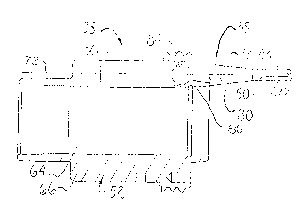
Referring now to Figure 2, the improved arthroscope 10
is shown in cross section and includes a housing 30, a
disposable arthroscope scope assembly 32 detachably mounted
to the housing 30, and a camera assembly 34 mounted within
the housing 30. An internal light source 35 including a
micro light 36 or miniature light bulb is also mounted
within the housing 30 in light communication with the
arthroscope scope assembly 32. The internal light source
3S also includes light transmitting means 39 for
transmitting light from the micro light 36 to the
arthroscope scope assembly 32. The light transmitting
means 39 is adapted to maximize transmission of the light
f~om the micro li~h~ 36 to the scope assembly 32 and may
include a number or all of the components to be hereinafter
described.
The construction of the scope assembly 32 and camera
assembly 34 are essentially the same as the arthroscope
described in more detail in application serial number
07/651,746 entitled "Portable Arthroscope With Disposable
Probe", assigned to the assignee of the present application
and included herein by reference.
Briefly stated, the arthroscope scope assembly 32
includes a scope 38 that is adapted for insertion at the
- : . , . ~ ................................. , , , :
: -
~ -
~Q~33
ent.-y site 12 of a joint such as th~ knee 12 (Figure 1).
The ~cope 38 may be constructed as a r..alleable, flexible or
semi-flexible member. As such it may include a metal
cannula, or a flexible polyimide tube as an outer member.
In addition, the arthroscope scope assembly 32 is
disposable and may be removably attached to the housing 30
using a threaded ring (lamp 40~ Such a threaded ring clamp
40 is more particularly described in the above cited patent
application. In general such a ring clamp 40 provides a
removable attachment 'or attaching the arthroscope scope
asscmbly with the optical components in a proper alignment.
The arthroscope scope assembly 32 also includes a
plulality of illumination fibers 42 (Figure 2A) mounted
within the scope 38. The illumination fibers 42 are
embedded within an epoxy material 44, and terminate at a
viewing or distal end 46 of the scope 38 of the arthroscope
scope assembly 32 for providing illumination for viewing
the interior of a joint such as the knee 1~ (Figure 1).
The illumination fibers ~12 are joined in a fiber bundle 48
(Figure 2). .
A front tube 52 is mounted within the housing 30. The
front tube 52 is generally cylindrical in shape and is
removably attached to the housing 30. The light source 35
is mounted within the front tube 52. The front tube 52
also houses a sapphire window 54 in light communication
with an image guide 58 of the scope 38. ~he sapphire
window 54 in turn, is in light communication with focusing
optics 56 mounted within the front tube 52.
The focusing optics 5G are mounted in an optics base
60 slidingiy mounted in the front tube 52. Thus, an image
from the image guide 58 which has passed through the
sapphire window 54 can enter the focusing optics 56. ~s
previously stated the ~mage guide 58 functions to transfer
an image from the pla~e of a GRIN lens attached to the
distal or vie;ling end oL` the image guide 58 to the focusing
optics 56. Preferabl~, the focusing optics 56 magnify
,, ^~ . ' ' ' : ' . '. ' ~ '
~9~3
light from the sapphire window 54 several times (i.e. 7X)
and focus this light onto a camera head 62 which is also
mounted within the housing 30. In a preferred embodiment
the camera head 62 is a charged coupled device (CCD).
Referring now to Figure 2A the ront tube 52, light
source 3S, and light transmitting means 39 are shown in
schematic form. The internal light source 35 includes the
micro light 36 mounted along a center line 72~ The
internal light source 35 also includes the light
transmitting means 39 for transmitting light from the micro
light 36 to the illumination fibers 48 of the illumination
fiber bundle ~8 of the scope 38.
The light transmitting means 39 may include a fiber
optic taper 30 that funnels light from the micro light 36
into the illumination fiber bundle 48. The light
transmitting means may also include a parabolic reflecting
mirror 84 for reflecting some of the light output from the
micro light 36 onto the illumination fiber bundle. In
addition, the light transmitt:ing means 39 may include an
illu~ination GRIN lens 50 juxtaposed to a proximal end of
the illumination fiber bundle 42 for collecting and
channeling light into the fiber bundle 42. ~ith a
disposable scope 38 such a GRIN lens 50 would be integrated
into illumination optics located within the housing 30.
Finally the light transmitting means may include an
intcgratinc3 sphere 86. Such an integrating sphere 86 may
be in the form of a coated spherical bulb that functions to
collect all of the light from an internal light source and
allows the light to escape through a single opening.
The micro liqht bulb 36 is preferably a miniature
light bulb of the type that produces a relatively high
output for its size. By way of example and not limitation,
a 5VDC, 1.4 A, Halogen technical lamp such as a focuscd
lens end lamp, that produces approximately 20,000 LUX
output is suitable for this application. An external power
linc 112 (Fic~ure 2) may be provided for the micro light 36.
. :
:
With such a micro light 36 as a light source it is
essential that most of the output of the micro light 36 be
transmitted to the illumination fiber bundle 48.
Figure 3A 3D illustrate various constructions for the
micro light 36 and light transmitting means 39. with
reference to Figure 3A, a parabolic reflecting mirror 84 is
positioned around a micro light 36' to reflect light from
the micro light 36'. The reflecting mirror 84 is shaped as
a paraboloid of revolution. The parabolic reflecting
mirror 84 forms the reflected light into a column of light
which is passed through a focusing lens 68 and focused on
the illumination fiber bundle 48. The focusing lens 68 may
be in the form of an aspherical lens. ` ;-
With reference to Figure 3B a micro light 3611 may be -
in light communication with a taper section 80 which is
adapted to funnel light from the micro light 36" into the
illumination fiber bundle 48. The taper section 80 may be
formed of a fiber optic material having and an
antireflection (AR) coating. Such an AR coating may be
selected to reduce reflection losses (i.e Fresnel)
occurring at the surface of the ta~er section 80. Moreover
the taper section 80 may be formed with a taper angle which
closely matches the numerical lperture (N.~.) of the
illumin~tion fiber bundle 48 (~,r a G~IN lens rod 50
attached to the illumination fibe~ bundle 48). This taper
angle enables an illumination oulput cone from the taper
section 80 to be directed at the lluminating fiber bundle
48 at a known angle of illumination. As an example such a
micro liqht 36" may be a commercially available bulb
having an integral focusing lens.
~ ith reference to Figure 3C, a focusing lens light
bulb 36"' may be adapted to focus light trough a focal
plane 49 and a GRIN rod 50 and onto the illumination fiber
bundle 48. Such a focusing lens light bulb 36''' may also
be a bulb with an integral focusing lens.
' ~
;~'
::
': ; .'.'.,~ ,! .
, ',' ' `' ~
With reference to Figure 3D a light bulb 36"'' may
include an integrating sphere 86 coated inside with a
highly re1ective white coating. A small uncoated area 88
on the spherical bulb allows light to escape only at
selected angles. These angles are chosen to be within the
N.A. of the illuminating fibers 42 of the illumination
fiber bundle 48.
With any of the arrangements shown in Figures 3A-3D,
essentially all of the light output from the micro light 36
is transmitted to the illumination fiber bundle 48. The
objective is to c_llect the maximum light energy and
transfer this light to the illumination fiber bundle 48.
This light then travels through the individual illumination
fibers 42 of the fiber bundle 48 and out the viewing end 46
of the flexible scope 40 to illuminate the internal
structure of the bod~. This light is then collected by the
image guide 58 of the scope 38 and transmitted through the
sapphire window 54 and focusing optics 56 to form an image
of the body.
In addition to providing the above structures for a
micro light 36 and light transmitting means 3~ shown in
Figure 3A-3D for transmitting light from the micro light 36
to the illumination fibe~ bundle 48, it is also
advantageous to overdrive the micro light 36 into the color
;~5 spectrum best suited for the arthroscope 10. In general
such a shift is into the blue visible light area and a~ay
from the infra-red light area. This shift also increases
the total photon flux.
Using a micro light 36 and light transmitting means 39
(Figure 3A-3~) constructed in accordance with the invcntion
the final light output at the viewing end 46 of the
flexible scope assembly 32 is equivalent to that of an
external ight source which uses a high powered Xenon or
l~etal Halide light bulb. With such external systems it is
not unusual to lose up to 98% of the original light energy
.. .
~9~33
by the time it has been transmitted to the point of
application.
'lo enable the use of a micro light 36 in an endoscope
system such as arthroscope 10 having a relatively small
S illuminating bundle 48 and to produce a high quality image,
it is also necessary that the light collecting system
(i.e., image guide 58) and imaging system relating to the
camera assembly 62 (CCD) be optimized. Some system
parameters which can be controlled to achieve these results
include the following.
1. Utilizing an image collecting lens such as a GRIN
lens that has a matching index with the image guide 58
(Figure 2) at their common interface.
2. Coating the sapphire lens 54 (Figure 2) with an
anti-reflection material (AR) to reduce surface reflection
losses.
3. The focusing optics 56 (Figure 2) or relay lens
system, either fixed or zoom design, must be optically
designed to suit all the input and output parameters of the
imaging system.
4. To balance the illumination system micro computer
electronic shuttering of the CCD 62 (Figure 2) video output
must be accomplished. This enables the optical system to
be more sensitive to changes in illumination on the CCD G2
surface, and yields an improved image onto a CRT 26 for
recording onto the VCR 28 (Figure 1).
5. White balance selection, either automatic or
manual, can be used for electronic comparison of this color
to set the spectral color content (or spectral balance) of
the picture. This feature is essential if a micro light 36
not having the ideal light spectrum in the visual range, is
to be made acceptable.
6. The image level signal/noise ratio must be
optimized for producing a high quality video image.
In addition to the above-described structure and
operational parameter for a light source 35 and light
.~.
. .: , :: , ,. . . ~: i - : : .
.~ . . . . . i , ~
":1'; .' ' . . '
, ~ ' , '' ' , '' ~: i,
3 3
1~ ~
transmitting means 39, an essential part of the present
invention is to be able to dissipate hcat generated by the
micro light 36 in its confined space within the housing 30.
This can be done passively by conducting the heat away or
by cooling with a coolant. Four methods are illustrated in
Figures 4A-4D for dissipating heat.
1. As shown in Figure 4A, heat can be dissipated by
using conventional methods of convecting the heat away.
These conventional methods include:
lo A. Conducting heat from the micro light 36 to the
housing 30 where it is eonvected away using a
conductive medium such as thermal grease.
B. Seleetively shaping the housing 30 (i.e. added
thickness) to spread heat more evenly anc to
conduct the heat away.
C. Ribbing the housing 30 for dissipating and
radlati~g the heat away.
D. Coating the inside of the housing 30 with a black
material for heat absorption.
E. Plating the outside of the ~ront tube 52 with a
black surface for radiation of heat.
F. Plating the outside of the camera assembly 34
bright aluminum for reflection of heat from the
eamera assembly 34.
2. As shown in Figure 4B heat can also be dissipated
by using a ehemieal transfer method such as a hollow tube
or a heat pipe 90 filled with a low vapor pressure, low
boiling point liquid that is adapted to carry heat away
from the housing. ~`
3. ~s shown in Figure 4C heat can also be dissipated
by installing a Peltier semiconduetor heat pump chip 92 and
heat pipe 114.
4. As shown in Figure 4D heat can be dissipated by ;
using a separate lamp holder 116 that clips to external
cables 96 connected to the housing 30.
AI,TERNATE EMBODIMENT
Re~erring now to Figures 5A-5C several alternate
embodiment endoseope assemblies or arthroscopcs constructed
in aceordance with the present invention are shown.
.. . ..
.
,,
, ,
,: `' ~ `,
-
2 ~ 3 ~
In general to make a cableless, totally self contained
endoscope, a battery pack may be utilized together with a
microscopic eyepiece.
With reference to Figure 5A, an arthroscope 100
includes battery pack 102 as an internal power source and
an eyepiece 104. Such an arthroscope 100 may also include
an internal light source 3G as previously explained. With
referance to Figure SB arthroscope 106 may include an
eyepiece 104 and a battery pack 108 formed as a handle.
With reference to Figure 5C an arthroscope 110 may
include an eyepiece 104 and a power line 112 to an external
power source. As comparison and with reference to Figure
5D the cable assembly of a prior art arthroscope 114
typically includes;
a. camera control unit cable 118 to the camera
assembly;
b. a heavy fiber optics illuminatiol~ cable 120.
With an arthroscope constructed in accordance with the
invention the heavy and expensive ~iber optic cable 118 is
eliminated as the internal light source 35 provides the
light. With an externally powered internal light source 35
this fiber optic cable 120 can be replaced by a small ~ower
cable 112 (Figure 5C). With the embodiments havin-~ an
internal powe~ source (100-Fig 5A, 106-Fig 5B) the power
cable is also eliminated
Thus an endoscope can be constructed in accordance
with the invention with an internal light source. Such and
endoscope may be constructed and produce an image that is
equivalent to images produced with endoscopes having large
external light sGurces.
While the particular endoscope as herein sho~;n and
disclosed in detail is fully capable of obtaining the
objects and providing the advantages herein before stated,
it is to be understood that they are merely illustrative of
the presently preferred embodiments of the invention and
that no limitations are intended to the details of
,, ~
. .. : :~
3 3
14 :
construction or design herein shown other than as described
in the appended claims.
.
. ,. i - , ,
: : . -
.
': '' ,