Note : Les descriptions sont présentées dans la langue officielle dans laquelle elles ont été soumises.
WO 92/12734 PCI`/US92/0~43~
., ~ I
7 ~
DescriptiQn
ANrI-GROs~iJI~ FACIOR ANTIBODIES lN THE TREAl~ENT OF VASCULAR Sl~OSIS
Development of this invention was supported in
part by National Institutes of Health Grants lP50HL 42270-
01, HL 03174-35, HL 41103 and HL 18645. The Government
may have certain rights in this invention.
Technical Field
The present invention relates to methods for
inhibiting stenosis in a mammal following vascular in~ury,
and to compositions useful within those methods.
Backqround of the Invention
Proliferation of smooth muscle cells (SMCs) in
the vessel wall is an important event in the formation of
vascular lesions in atherosclerosis or in response to
vascular injury. Treatment of atherosclerosis frequently
includes the clearing of blocked vessels by angioplasty,
endartarectomy or bypass surgery, surgical procedures in
which atherosclerotic plaques are compressed or removed
through catheterization (angioplasty) or stripped away
from the arterial wall through an incision
(endartarectomy) or bypassed by anastomising a vein or
artery proximal or distal to the site of occlusion
(bypass). These procedures remove the vascular
endothelium, disturb the underlying intimal layer, and
result in the death of medial SMCs. This injury is
'followed by medial SMC proliferation and migration into
the intima, which characteristically occurs within the
first few weeks after injury and stops when the overlying
endothelial layer is reestablished.
In 30%-40% or more of patients treated by
angioplasty, endartarectomy or bypass surgery, thrombosis
and/or SMC proliferation in the intima causes re-occlusion
of the vessel and consequent failure of the angioplasty,
, . : . . . . . . .
. - , . . .
: .
;: , ., ~ ~ - . . ,
. . . . . . :
WO92/12734 PCT/U~92/00438
.
0 ~ 7 ~ 2
endartarectomy or bypass procedure. This closure of the
vessel subsequent to surgery is known as restenosis.
A similar process of SMC proliferation has also
been observed in vascular grafts at the perianastomotic
site where the graft is surgically joined to the artery
wall, as well as with organ transplants, and may
contribute to transplant rejection.
It has been postulated that growth factors, such
as platelet derived growth factor (PDGF), play a role in
the development of atherosclerotic plaques (reviewed by
Ross et al., Cell 46: 155-169, 1986). PDGF has been
detected within macrophages in all stages of lesion
development in both human and nonhuman primate
atherosclerosis (Ross et al., Science 248:1009-1012,
1990). One proposed mechanism for plaque formation is the
release by platelets, at sites of endothelial denudation,
of growth factors that stimulate SMC growth (Ross and
Glomset, N. Enq. J. Med. 295: 369-377, 420-425, 1976;
Ross, Arteriosclerosis I: 293-311, 1981). Moore et al.
(Thrombos. Haemostas. tstuttq-) 35: 70, 1976) and Friedman
et al. (J. Clin. Invest. 60: 1191-1201, 1977), using an
indwelling catheter injury model, reported an inhibition
of experimentally induced intimal lesion formation in
rabbit arteries by prolonged thrombocytopenia induced by
administration of anti-platelet serum. It has also been
postulated that SMCs may the~selves produce PDGF which
stimulates lesion development through an autocrine
mechanism (Ross et al~, ibid; Walker et al., Proc. Natl.
Acaq. Sci. USA 83: 7311-7315, 1986). Fingerle et al.
(Proc. _Natl. Acad. Sci. USA 86: 8412-8416, 1989)
investigated intimal lesion formation in thrombocytopenic
rats and concluded that platelets do not play a role in
the initial SMC proliferation after balloon injury but may
regulate SMC migration into the intima. Platelets are now
known to release a number of growth -Eactors, including
PDGF, transforming growth factors alpha and beta (TGF~ and
TGF~), insulin-like growth Eactor I (IGF-I) and platelet
. . . .
.
: ' ' .
,
.' . : ~ ' : , ' .
WO92/12734 PCT/US92/00438
,. . .
~,, .
3 2 ~ 7 6
derived endothelial cell ~rowth factor. However, there
has been no direct evidence to demonstrate that a
particular mitogen or mitogens is responsible for the
development of arterial lesions.
Removal of atherosclerotic plaques by
angioplasty, endartarectomy or treatment by bypass surgery
has limited efficacy, and no effective treatment for
restenosis has been developed. There is therefore a need
in the art for methods of reducing or preventing stenosis
of blood vessels ~ollowing vascular injury, such as injury
due to balloon catheterization, endarterectomy or bypass
surgery, as well as in vascular grafts and organ
transplants. The present invention provides such methods
and fulfills other, related needs.
Disclosure of the Invention
The present invention provides methods for
inhibiting vascular stenosis, including restenosis
following angioplasty, endarterectomy, bypass surgery
(including arterial bypass surgery) or other procedures
whereby atherosclerotic plaques are removed from blood
vessels. These methods generally comprise admlnistering
to a mammal an anti-growth factor antibody in an amount
sufficient to inhibit mitogenesis and/or migration of
smooth muscle cells. Within preferred embodiments, the
antibody is an anti-fibroblast growth factor antibody or
an anti-platelet derived growth factor antibody.
~onoclonal antibodies are preferred.
In a related aspect, the present invention
provides methods for inhibiting restenosis in a mammal
following angioplasty, endarterectomy or bypass surgery
comprising administering to the mammal an anti-growth
factor antibody in an amount sufficient to inhibit
restenosis. Anti-fibroblast growth factor antibodies and
anti-platelet derived growth factor antibodies may be
used. In one embodiment, the antibody is administered
prior to angioplasty, endarterectomy or bypass surgery.
... . . .. . . .
::
: . : . . . i:
. ~
-:
.
:
WO92/12734 P~T/US92/00438 3
2100876
In another embodiment, the antibody is administered
subsequent to angioplasty, endarterectomy or bypass
surgery. In yet another embodiment, a panel of anti
growth factor antibodies is used, such as a panel of
antibodies capable of neutralizing the AA, AB and BB
isoforms of platelet derived growth factor.
Another aspect of the invention provides a
method of inhibiting restenosis following angioplasty,
endarterectomy or bypass surgery, wherein an anti-
fibroblast growth factor antibody is administered to a
mammal prior to angioplasty, endarterectomy or bypass
surgery in an amount sufficient to inhibit restenosis, and
an anti-platelet derived growth factor antibody is
administered to the mammal subsequent to angioplasty,
endarterectomy or bypass surgery in an amount su~ficient
to inhibit restenosis.
These and other aspects of the invention will
become evident upon reference to the following detailed
description and the attached drawings.
Brief Descri~tion of the Drawings
Figure 1 illustrates the effect of an anti-bFGF
antibody on proliferation of 3T3-D1 cells in response to 5
ng each of bFGF, PDGF-BB and EGF, ~nd to calf serum (5~)O
Figure 2 illustrates the replication of medial
smooth muscle cells 41 hours after balloon catheter
denudation in animals treated with anti-bFGF antibocly or
control (nonimmune) antibody. Data represent means +/-
SEM .
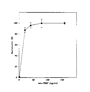
Figure 3 is a graph illustrating the effect of
; goat anti-PDGF on PDGF activity in rat whole blood serum.
The results are expressed as the mean +/- SEM for
triplicate determinations.
Figure 4 is a graph illustrating the anti-PDGF
IgG inhibition of the chemotactic response of rat platelet
releasate. Samples are identified as follows: (1), PDGF-
AA (5 ng/ml); (2), PDGF-BB (10 ng/ml); (3), TGF~ (300
.
.
.
W~g2/1~734 PCT/US92/00438
. ~
2 ~ 7 6
pg/ml); (4), bFGF (500 pg/ml); (5), rat platelet releasate
(RPR); (6), RPR plus 500 ug of anti-PDGF per milliliter;
(7), RPR plus 1 mg of antiwPDGF per milliliter;
(~), nonimmune IgG (1 mg/ml); and (9), control media. The
results are e~pressed as fold increase (mean ~/- SEM) in
cell migration above a buf~er control.
Figure 5 is 2 pair of photographs comparatively
illustrating the inhibition of the accumulation of intimal
smooth muscle cells 8 days after balloon catheter injury.
Panel A represents treatment with nonimmune goat IgG;
Panel B represents treatment with anti-PDGF IgG.
Figure 6 is a graph illustrating the effect of
nonimmune IgG and anti-PDGF IgG on the accumulation of
intimal smooth muscle cells after balloon catheter injury.
Measurements were made on intimal cross-sectional areas
and the results expressed as the mean ~/- SEM.
Figure 7 is a graph illustrating the clearance
of anti-PDGF IgG from the plasma of a nude rat after
administration of a single intra-peritoneal injection (60
mg/lOOg body weight).
Figures 8A, 8B and 8C illustrate the effects of
anti-PDGF on the intimal and medial response to a filament
loop injury of the rig~t carotid artery of a nude rat.
Figure 8A depicts the effect on the intima-media cross-
sectional area; Figure 8B, the effect on intimalcellularity; and Figure 8C, the effect on intimal and
medial proliferation at 8 days after injury measured by
H-thymidine incorporation and autoradiography.
Detailed Descri~tion of the Invention
As noted above, restenosis of blood vessels is a
common problem in patients who have undergone angioplasty,
endartar~ctomy or arterial bypass surgery. Resten~sis is
believed to proceed via a process that includes both
proliferation (mitosis) and migration of vascular smooth
muscle cells in the area damaged by the surgical
procedure.
. , , ; . ~ :
'
W092/12734 PCT/US92/~438
21~8`7~ ~
The present invention provides methods for
inhibiting vascular stenosis through the use of antibodies
against basic fibrobla~t growth factor (basic FGF or
bFGF), acidic fibroblast growth actor (acidic FGF or
aFGF) and/or platelet derived growth factor (PDGF). As
used herein, "vascular stenosis" denotes the partial or
complete blocking of a blood vessel through intimal
thickening due to cellular migration and/or mitosis.
Inhibition of stenosis will be understood to include
interfering with the stenotic process by reducing or
preventing cell migration, cell mitosis, or both. The
inventors have found that therapeutic use of anti-growth
factor antibodies can inhibit vascular stenosis by
reducing the migration and/or mitosis of vascular smooth
muscle cells (SMCs).
Antibodies useful within the present invention
may be produced by conventional procedures of imm~mization
and purification. Briefly, a purified growth factor is
administered to an animal such as a mouse, rat, rabbit or
goat in an amount sufficient to cause an immune response.
It is preferred to administer the growth factor in
combination with an adjuvant, such as ~reund's adjuvant,
in order to enhance the immune response. Although a
single injection of growth factor may be sufficient to
induce antibody production in the animal, it is generally
preferred to administer a large initial injection followed
by one or more booster injections over a period of several
weeks to several months. See, e.g., Hurrell, J.G.R., ed.,
Monoclonal HYbridoma_ Antibodies: _ Techniques and
Applications, CRC Press Inc., Boca Raton, FL, 1982, which
is incorporated herein by reference. Blood is then
collected from the animal and clotted, and antibodies are
isolated from the serum using conventional techniques such
as salt precipitation, ion exchange chroma~ography,
affinity chromatography or high performance liquid
chromatography. Growth factors for use in i~munization
are prepared from natural sources or genetically
~'~'''' " '
-.
WO92/12734 - PCT/US92/00438
, ......................... ..
7 ~ '7 ~
engineered cells according to conventional methods such as
those described by Raines and Ross (J. Biol. Chem. 257:
5154-5160, 1982)~ Antoniades (U.S. Patent No. 4,479,896),
Murray et al. (U.S. Patents Nos. 4,801,542, 4,845,075 and
4,889,919), Bohlen et al. (FEBS Lett. 185: 177-181, 1985),
~arr (W0 90105184), Fiddes et al. (W0 87/01728) and
Moscatelli et al. (EP 226,181), which are incorporated
herein by reference. In the alternative, purified growth
factors can be obtained from commercial suppliers (e.g.,
Genzyme Corp., Boston, MA; Collaborative Research,
Bedford, MA).
Within one embodiment of the invention,
monoclonal antibodies are used. Monoclonal antibodies
provide the advantages of ease of production and lower
therapeutic doses as compared to polyclonal antisera,
since only antibodies of the desired specificity are used.
Methods for producing monoclonal antibodies are well known
in the art and are disclosed, for example, by Kohler and
Milstein ~Nature 256: 495, 1975; Eur. J. Immunol. 6: 511-
519, 1976). See also Hurrell, J.G.R., ed., Monoclonal
Hybridoma Antibodies: Techni~ues and Ap~lications, CRC
Press Inc., Boca Raton, FL, 1982. As will be appreciated
by those skilled in the art, antibody fragments, such as
Fab fragments, may also be used.
It is generally preferred to use antibodies that
are syngeneic with the patient or that contain syngeneic
constant regions. For this reason, genetically engineered
antibodies will generally be used in the treatment of
humans. Methods for producing recombinant human
antibodies or humanized non-human (i.e., chimeric)
antibodies are disclosed by Cabilly et al. (U.S. Patent
No. 4,816,567), Robinson et al. (WO 87jO2671) and Neumaier
(W0 90/00616~, which are incorporated herein by reference.
Briefly, human constant region genes are joined to
appropriate human or non-human variable region genes. The
joined genes are then transfected into host cells, which
are cultured according to conventional procedures. In the
~ :,
.
:
W~92/12734 PCT/US92/00438
2~876 --
alternative, monoclonal antibody producing cells may be
transfected with cloned human constant region genes, and
chimeric antibody genes generated by homologous
recombination. Thus ik is possible to assemble monoclonal
antibodies with a significant portion of khe structure
being human, thereby providing antibodiles that are more
suitable for multiple administrations to human patients.
Within the present invention it is preferred to
use neutralizing antibodies. "Neutralizing antibody" is
used herein to designate an amount of an antibody
sufficient to block essentially all of the biological
activity of an antigen in an in vitro test sy~tem.
Suitable ln vitro test systems include, in:ter alia,
mitogenesis assays and receptor binding assays. For
example, 200 ~g/ml of a polyclonal anti PDGF IgG described
herein is able to block the mitogenic and chemotactic
activity of 2 ng/ml of each of the dimeric forms of PDGF.
As will be understood by those skilled in the art, the
amount of antibody needed to neutralize a given amount of
antigen will depend on such factors as antibody
specificity and affinity.
Because PDGF is a mixture of the three possible
dimer combinations (isoforms) of its component chains
(known as A-chain and B-chain), the anti-PDGF antibodies
used within the present invention will preferably be a
panel of antibodies capable of neutralizing all three
isoforms (AA, BB and AB). Monoclonal antibodies are
preferred. Methods of making isoform-specific anti-PDGF
monoclonal antibodies are disclosed by Hart et al. (U.S.
Patent Application Serial No. 07/139,960; Biochemistry 29:
166-172, 1990). Hybridomas producing isoform-specific
anti~PDGF monoclonal antibodies have been deposited with
the American Type Culture Collection, Rockville, MD, under
accession numbers HB 9610, HB 9611, HB 9612 and HB 9613.
Anti-FGF and anti-PDGF antibodies may be
administered in combination or in overlapping or
sequential schedules. When used in combination, the
WO9~/12734 PCT/US92/0~438
9 21~7~
antibodies will generally be administered prior to surgery
and continuing after surgery at intervals of from several
hours to several days over the course oE one to two weeks
or more. In many cases it will be preferable to
administer daily doses d~ring a hospital stay, followed by
less frequent bolus injections during a period of
outpatient treatment. In the alternative, an anti-FGF
antibody is administered prior to surgery, either alone or
in combination with anti-PDGF antibody, and the patient is
treated as described above with anti-PDGF antibody
following surgery.
Doses of antibody will be selected on the basis
of neutralization criteria as described above. Dosage
levels are calculated from neutralization data after
determining clearance of antibody from the blood. In
general, dosage is selected with the goal of maintaining
circulating levels of antibody sufficient to neutralize
- any released growth factors. In general, doses will be in
the range o~ about 20 ~g to 600 mg or more of antibody per
kg of patient body weight, preferably about Ool mg to 20
mg/kg, more preferably about 1 mg-10 mg/kg. Somewhat
higher doses may be required if two or more antibodies are
administered in combination.
For use within the present invention, anti-
growth factor antibodies are formulated into injectable
compositions according to conventional procedures and
packaged in sterile containers. The antibodies may be
combined with a suitable diluent such as sterile saline or
sterile water. The antibody compositions may further
contain carriers, stabilizers and excipients such as
sugars (e.g., mannitol) or albumin. In the alternativ~,
the antibodies may be provided in lyophilized form and
reconstituted in a suitable diluent prior to use. These
compositions may be packaged in single or multiple dosage
; 35 form, for example in sealed ampoules or vials. Packages
may contain one antibody, a mixture of antibodies (e.g.,
` :
. :, , ' . :': ' '
, , .
' , , . ,. ~ . ~, : , ,
WO92/1273~ PCT/US92/00438
2~0~8 ~ 10
anti-PDGF and anti-bFGF) or combinations of individual
antibodies in separate containers or compartments.
For inhibition of stenosis in vascular grafts,
anti-growth factor antibodies are covalently attached to
the graft through their constant regions or incorporated
into the graft in slow-release formulati.ons.
The ~ollowing examples are offered by way of
illustration, not by way of limitation.
EXAMPLES
EXAMPLE 1
Female New Zealand rabbits (3 kg body weight~
were immunized with recombinant human bFGF (pharmaceutical
grade; obtained from Synergen, Inc., Boulder, CO).
Immunization was by intradermal injection of 120 ~g of
bFGF in combination with Freund's adjuvant (Sigma Chemical
Co., St. Louis, MO). A booster immunization (60 ~g of
bFGF intradermally~ was given three weeks later, and
immune serum was obtained five weeks after the first
immunization. The IgG fraction of the immune serum was
obtained by chromatography using Protein ~ Sepharose
(Pharmacia LKB, Uppsala, Sweden). Pre-immune serum was
obtained from the rabbits prior to i~munization.
The specificity of the anti-bFGF IgG was tested
in a mitogenesis assay on 3T3-Dl cells (a subclone of
Swiss mouse 3T3 fibroblasts~. The cells were plated at a
density of 4 x 10⁴ cells/well in a 24-well tray in
Dulbecco's modified Eagle's ~edium supplemented with 10~
calf serum. After three days, the cells reached
confluence and were made quiescent by incubation in medium
containing 0O5~ calf serum for an additional two days.
Recombinant PDGF-BB (prepared in yeast essentially as
disclosed by Murray et al., U.S. Patent No. 4,845,075,
incorporated herein by reference), EGF (culture grade;
obtained from Collaborative Research, Bedford, MA) and
bFGF were preincubated for 10 minutes at 37C with either
-;
.
~ .
WO92/12734 PCT/US92/00438
,
7 ~
100 ~g/ml of anti-bFGF IgG or preimmune IyG. The
quiescent cells were then incubated in the presence of 5
ng of growth factor or 5% calf serum for 20 hours. [3H]-
thymidine (6.7 mCi/mmol, DuPont-New ~ngland Nuclear)
incoxporation into DNA of the cells (1 ~Ci/ml, 1 x 10⁵
cells/well) was measured in a liquid scintillation counter
following a two hour pulse. As shown in Figure 1, the
anti bFGF IgG neutralized the mitogenic effect of bFGF but
did not significantly reduce the mitogenic response to
lO PDGF, EGF or calf serum. The antibody also showed no
cross-reactivity with acidic fibroblast growth factor in
an immunoblot assay and had no effect on the initial
platelet adherence to denuded arteries.
Male Sprague-Dawley rats (3.5 months old, 350-
15 400 grams body weight) were obtained from Tyler
Laboratories (Bellevue, WA). The animals were
anesthetised with an initial intramuscular injection of
0.06 mg/kg fentanyl (Innovar-Vet, Pitman-Moore, Mundelien,
IL) and additional injections when necessary. The distal
2U left common and external carotid arteries were exposed
through a midline wound in the neck. The endothelium was
removed from the left common carotid artery using a
filament loop essentially as described by Fingerle et al.
(Arteriosclerosis 10: 1082, 1990) and Lindner et al. (Lab.
25 Invest. 61: 556, 1989). The monofilament suture loop was
introduced into the left external carotid artery via a
trocar made of polyethylene tubing. The device was pushed
through the trocar into the common carotid, then steadily
pulled back along the carotid with constant rotation.
30 Anti-bFGF antibody (10 mg/animal) was administered via the
tail vein. Five minutes after denudation with the
filament loop, balloon catheter denudation of the same
artery was carried out essentially as described by Clowes
et al. (Lab. Invest. 4~: 327, 19833. A 2 French balloon
35 catheter was introduced through the external carotid
artery and passed through the common carotid three times
with the balloon distended sufficiently with saline to
. . , ' : - ~ ~' -
: . . , :
: : '
WO92/12734 P~/V~92/00438
~2
generate slight resistance and produce distension of the
carotid itself. The external carotid was ligated after
removal of the catheter, and the wound was closed. After
surgery, five additional intravenous injections o~ anti-
bFGF antibody (2.5 mg/animal) were given at eight hourintervals. Control animals were treate~d in an identical
fashion with the exception that matching concentrations of
nonimmune IgG were injected at the same times.
At 24, 32 and 40 hours after balloon catheter
injury, all animals were injected with tritiated thymidine
(50 ~Ci/l00 g body weight). Forty-one hours after injury
the animals were perfused-fixed. Briefly, the animals
were anesthetized and killed by injection of sodium
pentobarbital. A catheter was placed in the carotid
artery and the animal perfused with Ringer's lactate, then
fixed with 2% glutaraldehyde, 1% paraformaldehyde in
cacodylate buffer at physiologic pressure for five
minutes. The denuded carotid arteries were excised and
further fixed by immersion in the same fixative as was
used for perfusion. Tissue samples were embedded in
paraffin for cross-sectioning. One-micrometer cross-
sections were dipped in Kodak NTB-2 emulsion, stored at
4C for two weeks, and developed using ~odak D l9
developer. Under these conditions the background was
negliyible. The thymidine index was determined by
counting cells under oil immersion. As shown in Figure 2,
the proliferation of medial SMCs was significantly reduced
in those animals that received the anti-bFGF antibody
(1.5% vs. 7.6% in controls).
EXAMPLE 2
Balloon catheter in~ury was induced in rats as
described in Example l. The animals were each given a
single injection of l0 mg anti-bFGF IgG or nonimmune IgG
prior to surgery. Post-surgical administration of
antibody was omitted. All other procedures were carried
out as described in Example l. As shown in Figur~ l, the
`
.
WO92/1~734 PCT/US92/00438
.,
13 2~ 7~
prolifexation of medial SMCs was reduced by a single
injection of anti-bFGF antibody prior to injury (1.4% v9.
16.8% in controls).
~X~MPLE 3
Goat antisera were raised to purified human PDGF
(Raines and Ross, ibid). Six injections of approximately
75 ~g were given a~ two-week intervals. The initial
injection was in complete Freund's and subsequent
lo injections in incomplete Freund's adjuvant. All
injections were given subcutaneously at lO to 15 sites.
The first positive bleed was 3 months following the
initial injection. Antibody was obtained by
plasmapheresis of the animal to allow frequent collection
of large amounts of antibody and to prevent release of
endogenous PDGF from platelets. All of the antibody used
in these studies was obtained following the additional
injection of 350 ~g of PDGF which resulted in a 5 20 fold
increase in serum titre. Typical titre of the animal
removed the mitogenic activity of 2 ng/ml purified PDGF on
3T3 cells at a plasma dilution of 1:320. The IgG ~raction
was prepared by 18% sodium sulfate precipitation of plasma
followed by DEAE-Sephacel chromatography. Normal goat IgG
was prepared by the same procedure. Protein concentration
of both preparations was determined by the method of Lowry
et al., (J. Biol._Chem. lg3: 265, 1951).
Three different methods were used to evaluate
the specificity of anti-PDGF antibody; immunoprecipitation
of 125I-test substances, including insulin, EGF, platelet
factor 4, ~-thromboglobulin, FGF, TGF-~, and TGF-~;
inhibition of the mitogenic activity of test samples on
3T3 cells as described in Example 1 using insulin, EGF,
FGF, and IGF-l; and inhibition of PDGF competitive
acti~ity to evaluate species specificity, in which a serum
concentration which resulted in approximately 75%
competition in a PD~F radioreceptor assay (performed
essentially as described by Bowen-Pope and Ross, Methods
: ' '. . :' . ,.
'
.
.
, , . , : : : . -.
WO92/12734 PCT/US92/00438
2 ~ 14
Enzymol. 109: 69-100, 1985) was pre;ncubated with 400
~g/ml anti-PDGF IgG for 1 hour at 37C' prior to addition
to the 3T3 cells. Anti-PDGF only immunoprecipitated
dimeric forms of PDGF and only neutra]ized the mitogenic
activity of dimeric forms of PDGF. PDGF competitive
activity in serum from humans, pigs, dog, horse, mouse,
rat, chicken, rabbit and non-human primate were completely
neutralized by anti-PDGF. Only PDGF competitive activity
in serum from sheep, cows and goat were not neutraliæed by
the anti-PDGF.
The anti-PDGF completely neutralized the PDGF
binding competitive activity present in rat whole blood
serum at 50 ~g/ml (Figure 3). PDGF binding activity in
rat whole blood serum (WBS) was assessed by radioreceptor
assay with human foreskin fibroblasts (SK5 cells) as the
target cell and PDGF-AB as a standard (Bowen Pope and
Ross, Methods Enzymol. 109:63, 1985). A constant amount
of rat WBS (25% ~y volume, which is equivalent to 2.5 ng
of PDGF-BB per milliliter) was incubated with increasing
concentrations of anti-PDGF before evaluation by
radioreceptor assay. The specific binding of l25I-labeled
PDGF-AB was determined ~or each sample, and the data were
expressed as the percent neutralization of PDGF
competitive activity in rat WBS in the absence of anti-
PDGF IgG.
Furthermore, the anti-PDGF substantially
prevented PDGF-induced [3H]thymidine incorporation by rat
~mooth muscle cells and inhibited 50% of the mitogenic
activity of rat platelet releasate (Ferns et al., Am. J.
Pathol. 138:1045, 1991). The anti-PDGF also inhibited
chemotaxis of rat carotid smooth muscle cells to purified
PDGF and inhibited most of the chemotactic activity in rat
platelet releasate (Figure 4). In contrast, the control
medium, the nonimmune IgG, and PDGF AA exhibited no
chemotactic activity (Figure 4).
To determine dosage levels of the antibody in
vivo, the clearance of the anti-PDGF was evaluated.
WO92/12734 PCT/US~2/00438
- 2 ~
Plasma antibody concentrations in rats treated with anti-
PDGF determined by enzyme-linked i~munosorbent assay
(ELISA) were reduced by 50% a~ter 30 hours, and daily
intraperitoneal administration of anti-PDGF IgG ~60 mg per
100 g of body weight) maintained plasma concentrations of
1000 ~g/ml during the 9-day duration of the experiment.
At these concentrations, there was no significant effect
on platelet counts or complement levels and ln vitro
chemotaxis to rat platelet releasate was completely
inhibited (Figure 4).
Homozygous nude rats aged 4-5 months of age
tapproximately 200 g weight) were obtained from the
National Institutes of Health, Bethesda, MD and housed in
a pathogen-free facility. Either goat anti~PDGF or non-
immune goat IgG were administered daily by IP injectionstarting the day before surgery. An antibody dose of 60
mg/100 g body weight was sufficient to achieve antibody
levels between 1.5 and 2 mg/ml 24 hours after
administration. On the day of surgery, animals were
anesthetized using ketamine and Rompùn, and both common
carotid arteries were balloon catheterized using a 2
French embolectomy catheter. Blood samples were taken
during the operative procedure and at sacrifice to
determine circulating blood levels of antibody in each
animal. Animals were sacrificed 8 days after surgery.
Se~enteen, 9 and 1 hour prior to sacrifice, animals were
injectsd with [3H~thymidine (50~Ci/100gm) to label
proliferating intimal and medial cells. At the time of
sacrifice, animals were anesthetized with ketamine and
Rompun. Blood was taken for antibody levels and the
animals injected with Evans Blue. After 10-15 minutes,
the jugular veins were isolated for perfusion run-off with
a cannula introduced into the abdominal aorta. The
animals were injected with a lethal dose of sodium
pentobarbital before perfusion with Ringer's lactate and
fixation ln situ with 4% paraformaldehyde. Both carotids
. . , . , :
~ , . - : . : :
. . ~ . . : . : . : .
: ~ . : .
.
.: ~.
.
WO92/12734 PCT/US92/00438
~?
~ 16
were divided into three segments so that the uniformity of
the lesion could be assessed.
Administration of anti-PDGF before and after
balloon catheter deendothelialization reduced the
thickness and cellular content of the neointima (Figure
5). Quantitative image analysis of the neointima of the
19 animals in each experimental group demonstrated that
administration of anti-PDGF resulted in a 40.9% reduction
in the area of the neointima (P < 0.01 by two tailed t
test) (Figure 6).
EXAMPLE 4
An anti-human PDGF polyclonal antibody was
obtained as generally described in Example 3. Briefly, a
goat was immunized with PDGF purified from outdated human
platelets tRaines and Ross, ibid.) emulsified in Freund's
incomplete adjuvant and administered subcutaneously every
two weeks for 3 months. Once a titre was established~
plasma was collected by plas~apherPsis every week with
periodic boosts of PDGF in incomplete Freund's. A
concentrated IgG fraction was prepared by sodium sulphate
precipitation and DEAE-Sephacel (Pharmacia, Piscataway,
NJ) column chromatography. Purified IgG was obtained by
elution with 0.01M phosphate buffer ~pH 6.8). The pooled
void volume and pH 6.8 wash was concentrated by
ultrafiltration (PM-10, Amicon Corp., Danvers, MA) and
dialyzed against phosphate buffered saline to a protein
concentration of approximately 60-90 mg/ml as determined
by the method of Lowry et al. (J. Biol. Chem. 193-265,
1951). Bovine serum albumin was u~ed as a standard and a
correction factor applied for immunoglobulin (Klosse
et al., Clin._Chim. Acta 32:321, 1971). Before use, the
Ig~ was filter-sterilized through a 0.22 ~m filter
(Millipore, Bedford, MA) and stored at 4C. A non-immune
goat IgG was prepared following the same method, using
commercially available goat plasma. The specificity of
WO9~/12734 PCT/US92/00438
17
the anti~ody was evaluated in the same manner a~ described
in Example 3, with similar r~sults.
Homozygous nude rats (nu/nu~ (Ferns et al., Am.
J. Path. 138:1045, 1991) were obtained from the breeding
colony at the National Institutes of Health (Bethesda,
MD), and were maintained in a pathogen-free facility.
Animals were administered a single dose of anti PDGF
antibody (60 mg/100 g body weight) i~p. when they were 20-
24 weeks of age (250-350 g). Blood samples were then
drawn at various times afterwards into tube~ containing
approximately 0.38% sodium citrate (final concentration).
The plasma was separated and storad at -20C until
analyzed by ELISA.
Micro-titre plates (96 well, Nunc) were coated
with 10 ng PDGF-AB per well for 18 h. Non-specific
protein binding. was reduced by blocking with 2~ bovine
serum albumin (Sigma Chemical Co., St. Louis, MO)/0.2~
Tween-20 in PBS for 1 h at 37C. Dilutions of standards
and samples were prepared in a 1:16 ~ixture of rat plasma-
derived serum and PTB (0.05% Tween 20/0.2% bovine serum
albumin in PBS). One hundred microliter aliquots of
sample or standards of known concentrations of anti-PDGF
IgG were incubated in wells for 90 min at 37C. The
plates were rinsed 5 times in wash buffer (0.05% Tween
20/0.9% NaCl), then incubated with lO0 ~l/well 1:1000
biotinylated anti-goat IgG (Tago Diagnostics, Burlingame,
CA) in PTB for 60 min at 37C. Unbound secondary antibody
was removed by rinsing in wash buffer followed by
incubation with avidin/bioti~ylated peroxidase. Substrate
(o-phenylenediamine, Sigma Ch~mical Co.) was dissolved in
0.05M citrate/0.1 M Na2HPO~ (pH 5.0) and incubated for 15
min at room temperature. The reaction was terminated by
adding 4N sulphuric acid, and the absorbance of the
: reaction prod~ct read at 490 nm.
Anti-PDGF IgG injected i.p. had a half life of
approximately 24 hours (Figure 7); peak levels were
attained within 10 hours after administration. All
.: ,: ' ' . ' : :
W~92/1~ PCT/US92/00438
~ 18
animals included in the study had plas~a levels of anti-
PDGF in excess of 1000 ~g/ml at the time of carotid injury
and sacrifice; most had levels greater than 2000 ~g/ml.
Platelet counts at the time of surgery and sacrifice were
unaffected by anti-PDGF antibo~y treatment. On the basis
of tha clearance data, a daily i.p. injection of antibody
was found to be adequate to maintain antibody levels at
approximately 10-20 times higher tham necessary to
substantially prevent the effects of PDGF contained in rat
serum.
Approximately 18h after a priming dose of
antibody, rats were anesthetized with intraperitoneal
xylazine (Rompun, Miles Laboratory Inc., Shawnee, XA;
40 mg/kg) and ketamine (Vetalar, Parke-Davis, NJ;
10 mg/kg) and the right carotid bifurcation exposed
through a paramedian incision. The endothelium was
removed using a nylon filament catheter (Fingerle et al.,
Arteriosclerosis 10:1082, 1989). A blood sample was
obtained perioperatively, anticoagulated with 0.38% sodium
citrate and kept on ice until the plasma was separated by
centrifugation at 1000 x g and stored at -20C. This
sample was used for the determination of plasma anti-PDGF
levels at the time of surgery using the ELISA method.
Following balloon catheter de-endothelialization, the skin
and deep fascia were closed with metal clips. Each animal
then received a further injection of either anti-PDGF
(n=19) or non immune IgG (n=16) post-operatively and then
daily until sacrifice. In order to label cells, 17, 9 and
1 h before sacrifice each animal was injected i.p. with 50
uCillOO g 3H-thymidine (New England Nuclear, Boston, MA).
Eight days post-operatively, animals were
sedated with xylazine and ketamine. Both jugular veins
were exposed and a paramedian abdominal incision was made
to access the aorta for in~ertion of a cannula. A blood
sample was taken from the cannula for determination of
plasma anti-PDGF immunoglobulin levels and a platelet
count. The cannula was then connected to a perfusion
~, .. . ..
WO92/12734 PCT/US92/00438
.. f~.`. i
2 ~
apparatus. The animals were given a lethal dose of sodium
pentobarbital, the jugulars were cut and the animals were
perfused with Ringer's lactate at a pres~sure of 120 mm Hg
- until the run-off was clear. This was then replaced with
4% paraformaldehyde in isotonic PBS (pH 7.4), which was
perfused at the same pressure for 15 minutes. Following
fixation in-situ, the carotids were isol~ted and dissected
free of adherent fat and connective tissue. Mid-carotid
segments were embedded in paraffin.
Five micron sections of carotid were dipped in
NTP-2 photographic emulsion (Kodak, Rochester~ NY) (Clowes
et al., Lab Invest. 49:327, 1983). The autoradiographs
were exposed for 14 days at 4C in a light-tight box and
were then developed using Kodak D-l9 developer and fixed
with Kodak Rapid Fix. Cell nuclei were stained with
hematoxylin. Cells with more than five silver grains
above the nucleus were considered positive. Duplicate
sections from two levels of each carotid were examined
under oil immersion using a Zeiss Axioskop microscope with
a X100 objective. Approximately 600 cells per wall
compartment were counted t~ determine the proportion of
labeled cells.
The intimal and medial cross-secticnal areas
were measured using an image analysis system consisting of
a Leitz microscope fitted with a X25 objective, a
digitizing pad and IBM PC with Vids-V software (Ai
Cambridge, Papworth, U.X.). Comparisons of the group
means were made using the Mann-Whitney U test.
At eight days after injury, carotid injury by
the nylon filament loop resulted in a thickened intima
which (in the control animals) constituted approximately
one fifth the total cross-sectional area of the carotid
artery. Neo-intimal cellularity and intima: media cross-
sectional area were reduced by 33.2~ (p<0.025) and 33.8~
(p<0.025) respectively in the anti-PDGF antibody treated
animals (Figures 8A and 8B) as compared to controls.
.
: , , .
W092/l2734 PCT/VS92~00438
~ 20
Loop injury induced a marked proliferative
response in the neo-intimal cells. Eight days after
injury approximately 30% of intimal cells were thymidine
labeled. By this time, medial cell proliferation had
fallen to approximately 2%. In contrast to its effect on
intimal thickening and cellularity, the anti-PDGF antibody
had no significant effect on intimal nor medial cell
proliferation (Figure 8C).
EXAMPLE 5
Normal male Sprague-Dawley rats were
anesthetized with ether, and were injected
intraperitoneally with phosphate-buffered saline (PBS),
with normal goat IgG, or with goat anti-PDGF IgGO The
dosage volume was 9 ml/kg, and the dose of IgG was 600
mg/kg. The anti-PDGF and the normal IgG were both
prepared as described in Examples 3 and 4. Twenty-four
hours after the first injection, animals were anesthetiæed
with ketamine/xylazine and both common carotid arteries
; 20 were injured by 3 passes of a saline-filled, size 2 French
balloon catheter. Another injection of PBS or IgG was
given immediately after catheterization. Further
injections were given under ether anaesthesia 1, 2 and 3
days after catheterization. On the fourth day after
catheterization, animals were sacrificed by injection of
sodium pentobarbital followed by exsanguination by
perfusion with Ringer's buffer. The left common carotid
artery was removed for biochemical analysis. The right
common carotid artery was perfused at 120 ~mHg with Chi's
fixative (2% glutaraldehyde, 1% paraformaldehyde in
phosphate buffer). This vessel was prepared for en face
scanning electron microscopy.
In a separate procedure, normal male Sprague-
Dawley rats were a~esthetized with ketamine/xylazine, and
both common carotid arteries were denuded of endothelium
using a filament loop catheter. PBS, rabbit anti-basic
FGF IgG, or normal rabbit IgG was injected intravenously
W092/l2734 PCT/U592/~0438
~ 2~ O~37~ l
in a dosage volume of 1.34 ml/rat: the dose of IgG was 10
mg/rat. The anti-basic FGF IgG and the normal rabbit IgG
were both prepared as described in Æxample 1. Five
minutes after injection, both common carotid arteries were
re-injured by 3 passes of a saline-filled, size 2 French
balloon catheter. Further injections of PBS, anti-basic
FGF or normal IgG (0.67 ml/rat; 5 mg IgG/rat) were given
under ether anaesthesia 1, 2 and 3 days after
catheterization. On the fourth day after catheterization,
animals were sacrificed by injection of sodium
pentobarbital followed by exsanguination by perfusion with
Ringer's buffer. The left common carotid artery was
removed for biochemical analysis. The right common
carotid artery was perfused at 120 mm Hg with Chi's
fixative (2% glutaraldehyde, 1% paraformaldehyde in
phosphate buffer). This vessel was prepared for en face
scanning electron microscopy.
Vessels from the above procedures were opened
longitudinally and were pinned out on Teflon cards. Thay
were dehydrated through an ethanol series, and then were
dried at the critical point of carbon dioxide in a
critical point drier. The dried specimens were mounted on
aluminum stubs with colloidal silver paste. After sputter
coating with gold/palladium, the specimens were examined
in a JEOL 35C scanning electron microscope at an
accelerating voltage of 15 kV and at 86-fold
magnification. An acetate sheet bearing a ruled grid was
placed over the microscope screen. Each square of the
grid had an area of 81 mm2, corresponding to 4133 ~m2 on
the specimen. The total area of the specimen, and the
area occupied by intimal smooth muscle cells, were
determined by counting squares. The extent of smooth
muscle cell migration was expressed as the percentage of
the total intimal area that was occupied by smooth muscle
cells. The results are depicted in Tables 1 and 20
WO92/12734 PCT/US92/00438
22
TABLE 1
PDGF
SMOOTH MUSCLE CELL
MIGR TION t%)
GROUPSTANDARD
TREATMENT SIZE MEAN ERROR
PBS 10 4.~63 1.293
Normal IgG 8 3.513 0.806
Anti-PDGF IgG 8 0.737 0.310
As shown in Table 1, there was no significant
difference between PBS and normal IgG groups, hut
treatment with anti-PDGF IgG reduced the extent of
migration by 79.0% ~p<0.01~.
TABI,E 2
BASIC FGF
SMOOTH MUSCLE CELL
MIGRATION (%)
GROUPSTANDARD
TREATMENT SIZE MEAN ERROR
PBS 6 11.794 3.806
Normal IgG 7 20.261 3.225
Antl-FGF IgG 8 4.001 1.061
~s shown in Table 2, there was no significant
difference between PBS and normal IgG-treated groups, but
treatment with anti-FGF IgG reduced the extent of smooth
muscle cell migration by 80.3% (p<0.001).
From the foregoing it will be appreciated that,
although specific embodiments of the invention have heen
described herein for purposes of illustration, various
modifications may be made without deviating from the
spirit and scope of the invention. Accordingly, the .
invention is not limited except as by the appended claims.
:
~.