Note : Les descriptions sont présentées dans la langue officielle dans laquelle elles ont été soumises.
CA 02117477 1997-11-03
WO 94/13806 PCT/US93/12039
~A. ~ i 7+77
MULTIVALENT SINGLE CHAIN ANTIBODIES
The present invention relates to single chain multivalent antibodies.
Antibodies are proteins belonging to a group of immunoglobulins elicited by
the
immune system in response to a specific antigen or substance which the body
deems foreign.
There are five classes of human antibodies, each class having the same basic
structure. The
basic structure of an antibody is a tetramer, or a multiple thereof, composed
of two identical
heterodimers each consisting of a light and a heavy chain. The light chain is
composed of one
variable (V) and one constant (C) domain, while a heavy chain is composed of
one variable and
three or more tonstant domains. The variable domains from both the light and
heavy chain,
designated V~ and VH respectively, determine the specificity of an
immunoglobulin, while the
constant (C) domains carry out various effector functions.
Amino acid sequence data indicate that each variable domain comprises three
complementarity determining regions (CDR) flanked by four relatively conserved
framework
regions (FR). The FR are thought to maintain the structural integrity of the
variable region
domain. The CDR have been assumed to be responsible for the binding
specificity of individual
antibodies and to account for the diversity of binding of antibodies.
As the basic structure of an antibody contains two heterodimers, antibodies
are
multivalent molecules. For example, the IgG classes have two identical antigen
binding sites,
white the pentameric IgM class has 10 identical binding sites. .
Monoclonal antibodies having identical genetic parentage and binding
specificity
have been useful both as diagnostic and therapeutic agents. Monoclonal
antibodies are
routinely produced by hybridomas generated by fusion of mouse lymphoid cells
with an
appropriate mouse myeloma cell line according to established procedures. The
administration
of murine antibodies for in vivo therapy and diagnostics in humans is limited
however, due to
the human anti-mouse antibody response illicited by the human immune system.
Chimeric antibodies, in which the binding or variable regions of antibodies
derived from one species are combined with the constant regions of antibodies
derived from a
different species, have been produced by recombinant DNA methodology. See, for
example,
Sahagen et al., J. Immunol., 137:1066-1074 (1986); Sun et al., Proc. Natl.
Acad. Sci. USA,
82:214-218 (1987); Nishimura et al., Cancer Res., 47:999-1005 (1987); and Lie
et al. Proc Natl.
Acad. Sci. USA, 84:3439-3443 (1987) which disclose chimeric antibodies to
tumor-associated
antigens. Typically, the variable region of a murine antibody is joined with
the constant region
of a human antibody. It is expected that as such chimeric antibodies are
largely human in
composition, they will be substantially less immunogenic than murine
antibodies.
Chimeric antibodies still carry the Fc regions which are not necessary for
antigen
binding, but constitute a major portion of the overall antibody structure
which affects its
pharmacokinetics. For the use of antibodies in immunotherapy or
immunodiagnostics, is it
CA 02117477 1997-11-03
desirable to have antibody-like molecules which localize and
bind to the target tissue rapidly and for the unbound material
to quickly clear from the body. Generally, smaller antibody
fragments have greater capillary permeability and are more
rapidly cleared from the body than whole antibodies.
Since it is the variable regions of light and heavy
chains that interact with an antigen, single chain antibody
fragments (scFvs) have been created with one VL and one VH,
containing all six CDR's, joined by a peptide linker (U. S.
Patent 4,946,778) to create a VL-L-VH polypeptide, wherein the
L stands for the peptide linker. A scFv wherein the VL and VH
domains are oriented VH-L-VL is disclosed in U.S. Patent
5,132,405.
As the scFvs have one binding site as compared to
the minimum of two for complete antibodies, the scFvs have
reduced avidity as compared to the antibody containing two or
more binding sites.
It would therefore be advantageous to obtain
constructions of scFvs having more than one binding site to
enhance the avidity of the polypeptide, and retain or increase
their antigen recognition properties. In addition, it would
be beneficial to obtain multivalent scFvs which are bispecific
to allow for recognition of different epitopes on the target
tissue, to allow for antibody-based recruitment of other
immune effector functions, or allow antibody capture of a
therapeutic or diagnostic moiety.
It has been found that single chain antibody
fragments, each having one VH and one VL domain covalently
_2_
64693-5016
CA 02117477 1997-11-03
linked by a first peptide linker, can be covalently linked by
a second peptide linker to form a multivalent single chain
antibody which maintains the binding affinity of a whole
antibody. In one embodiment, the present invention is a
multivalent single chain antibody having affinity for an
antigen wherein the multivalent single chain antibody
comprises two or more light chain variable domains and two or
more heavy chain variable domains; wherein, each variable
domain is linked to at least one other variable domain.
In another embodiment, the present invention is a
multivalent single chain antibody which comprises two or more
single chain antibody fragments, each fragment having affinity
for an antigen wherein the fragments are covalently linked by
a first peptide linker which contains an amino acid sequence
of Leu Ser Ala Asp Asp Ala Lys Lys Asp Ala Ala Lys Lys Asp Asp
Ala Lys Lys Asp Asp Ala Lys Lys Asp Leu and each fragment
comprising=
(a) a first polypeptide comprising a light chain
variable domain;
(b) a second polypeptide comprising a heavy chain
variable domain; and
(c) a second peptide linker linking the first and second
polypeptides into a functional binding moiety.
In another embodiment, the invention provides a DNA
sequence which codes for a multivalent single chain antibody,
the multivalent single chain antibody comprising two or more
-3-
64693-5016
CA 02117477 1997-11-03
single chain antibody fragments, each fragment having affinity
for an antigen wherein the fragments are covalently linked by
a first peptide linker and each fragment comprising:
(a) a first polypeptide comprising a light chain
variable domain;
(b) a second polypeptide comprising a heavy chain
variable domain; and
(c) a second peptide linker linking the first and second
polypeptides into a functional binding moiety.
The multivalent single chain antibodies allow for
the construction of an antibody fragment which has the
specificity and avidity of a whole antibody but are smaller in
size allowing for more rapid capillary permeability.
Multivalent single chain antibodies also allow for the
construction of a multivalent single chain antibody wherein
the binding sltes can be two different antigenic determinants.
BRIEF DESCRIPTION OF THE DRAWINGS
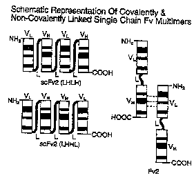
Figure 1 illustrates covalently linked single chain
antibodies having the configuration VL-L-VH-L-VL-L-VH(LHLH)
and VL-L-VH-L-VH-L-VL(LHHL) and a noncovalently linked Fv
single chain antibody (Fv2).
Figure 2 illustrates the nucleotide sequence of CC49
VL (SEQ ID NO: 1).
Figure 3 illustrates the amino acid sequence of CC49
VL (SEQ ID NO: 2).
Figure 4 illustrates the nucleotide sequence of CC49
VH (SEQ ID NO: 3).
-3a-
64693-5016
CA 02117477 1997-11-03
Figure 5 illustrates the amino acid sequence of CC49
VH (SEQ ID NO: 4).
Figure 6 illustrates the nucleotide sequence and
amino acid sequence of the CC49 single chain antibody LHLH in
p49LHLH (SEQ ID NO: 6).
Figure 7 illustrates the nucleotide sequence and
amino acid sequence of the CC49 single antibody LHHL in
p49LHHL (SEQ ID NO: 8).
Figure 8 illustrates construction of plasmids
pSL301T and pSL301HT.
Figure 9 illustrates construction of plasmid
p49LHHL.
Figure 10 illustrates construction of plasmid
p49LHLH.
Figure 11 illustrates the results of a competition
assay using CC49 IgG, CC49 scFv2, and CC49 scFv using
biotinylated CC49 IgG as competitor.
Nucleic acids, amino acids, peptides, protective
groups, active groups and such, when abbreviated, are
abbreviated according to the IUPAC IUB (Commission on
Biological Nomenclature) or the practice in the fields
concerned.
The term "single chain antibody fragment" (scFv) or
"antibody fragment" as used herein means a polypeptide
containing a VL domain linked to a VH domain by a peptide
linker (L), represented by VL-L-VH. The order of the VL and
VH domains can be reversed to obtain polypeptides represented
as VH-L-VL. "Domain" is a segment of protein that assumes a
-3b-
64693-5016
CA 02117477 1997-11-03
discrete function, such as antigen binding or antigen
recognition.
A "multivalent single chain antibody" means two or
more single chain antibody fragments covalently linked by a
peptide linker. The antibody fragments can be joined to form
bivalent single chain antibodies having the order of the VL
and VH domains as follows:
-3c-
64693-5016
CA 02117477 1997-11-03
WO 94/13806 PCT/US93/12039
~A~' ~ i 1477
VL-L-VH-L-VL-L-VH; VL-L-VH'L-VH-L-VL~ VH-L-VL-L-VH-L-VL~ or VH-L-VL-L-VL-L-VH.
Single chain multivalent antibodies which are trivalent and greater have one
or more antibody
fragments joined to a bivalent single chain antibody by an additional
interpeptide linker. In a
preferred embodiment, the number of VL and VH domains is equivalent.
The present invention also provides for multivalent single chain antibodies
which
can be designated VH-L-VH-L-VL-L-VL or VL-L-VL-L-VH-L-VH.
Covalently linked single chain antibodies having the configuration VL-L-VH-L-
VL-L-
-VH (LHLH) and VL-L-VH-L-VH-L-VL (LHHL) are illustrated in Figure 1. A
noncovalently linked Fv
single chain antibody (Fv2) is also illustrated in Figure 1.
The single chain antibody fragments for use in the present invention can be
derived from the light and/or heavy chain variable domains of any antibody.
Preferably, the
light and heavy chain variable domains are specific for the same antigen. The
individual
antibody fragments which are joined to form a multivalent single chain
antibody may be
directed against the same antigen or can be directed against different
antigens.
To prepare a vector containing the DNA sequence for a single chain multivalent
antibody, a source of the genes encoding for these regions is required. The
appropriate DNA
sequence can be obtained from published sources or can be obtained by standard
procedures
known in the art. For example, Kabat et al., Sequences of Proteins of
Immunological Interest
4th ed.~ (1991), published by The U.S. Department of Health and Human
Services, discloses
sequences of most of the antibody variable regions which have been described
to date.
When the genetic sequence is unknown, it is generally possible to utilize cDNA
sequences obtained from mRNA by reverse transcriptase mediated synthesis as a
source of DNA
to clone into a vector. For antibodies, the source of mRNA can be obtained
from a wide range
of hybridomas. See, for example, the catalogue ATCC Cell Lines and Hybridomas,
American
Type Culture Collection, 20309 Parklawn Drive, Rockville Md., USA (1990).
Hybridomas
secreting monoclonal antibodies reactive with a wide variety of antigens are
listed therein, are
available from the collection, and usable in the present invention. These cell
lines and others of
similar nature can be utilized as a source of mRNA coding for the variable
domains or to obtain
antibody protein to determine amino acid sequence of the monoclonal antibody
itself.
Variable regions of antibodies can also be derived by immunizing an
appropriate
vertebrate, normally a domestic animal, and most conveniently a mouse. The
immunogen will
be the antigen of interest, or where a hapten, an antigenic conjugate of the
hapten to an
antigen such as keyhole limpet hemocyanin (KLH). The immunization may be
carried out
conventionally with one or more repeated injections of the immunogen into the
host mammal,
normal ly at two to three week i ntervals. Usually, three days after the last
challenge, the spleen
is removed and dissociated into single cells to be used for cell fusion to
provide hybridomas
from which mRNA can readily be obtained by standard procedures known in the
art.
-4-
CA 02117477 1997-11-03
When an antibody of interest is obtained, and only
its amino acid sequence is known, it is possible to reverse
translate the sequence.
The VL and VH domains for use in the present
invention are preferably obtained from one of a series of CC
antibodies against tumor-associated glycoprotein 72 antigen
(TAG-72) disclosed in published PCT Application WO 90/04410 on
May 3, 1990, and published PCT Application WO 89/00692 on
January 26, 1989. More preferred are the VL and VH domains
from the monoclonal antibody designated CC49 in PCT
Publications WO 90/04410 and WO 89/00692. The nucleotide
sequence (SEQ ID NO: 1) which codes for the VL of CC49 is
substantially the same as that given in Figure 2. The amino
acid sequence (SEQ ID NO: 2) of the VL of CC49 is
substantially the same as that given in Figure 3. The
nucleotide sequence (SEQ ID NO: 3) which codes for the VH of
CC49 is substantially the same as that given in Figure 4. The
amino acid sequence (SEQ ID NO: 4) for the Vg of CC49 is
substantially the same as that given in Figure 5.
To form the antibody fragments and multivalent
single chain antibodies of the present invention, it is
necessary to have a suitable peptide linker. Suitable linkers
for joining the VH and VL domains are those which allow the VH
and VL domains to fold into a single polypeptide chain which
will have a three dimensional structure very similar to the
original structure of a whole antibody and thus maintain the
binding specificity of the whole antibody from which antibody
fragment is derived. Suitable linkers for linking the scFvs
-5-
64693-5016
CA 02117477 1997-11-03
are those which allow the linking of two or more scFvs such
that the VH and VL domains of each immunoglobulin fragment
have a three dimensional structure such that each fragment
maintains the binding specificity of the whole antibody from
which the immunoglobulin fragment is derived. Linkers having
the desired properties can be obtained by the method disclosed
in U.S. Patent 4,946,778. From the polypeptide sequences
generated by the methods described in the 4,946,778, genetic
sequences coding for the polypeptide can be obtained.
Preferably, the peptide linker joining the VH and VL
domains to form a scFv and the peptide linker joining two or
more scFvs to form a multivalent single chain antibody have
substantially the same amino acid sequence.
It is also necessary that the linker peptides be
attached to the antibody fragments such that the binding of
the linker to the individual antibody fragments does not
interfere with the binding capacity of the antigen recognition
site.
A preferred linker is based on the helical linker
designated 205C as disclosed in Pantoliano et al. B.iochem.,
10117-10125 (1991) but with the first and last amino acids
changed because of the codon dictated by the Xho I site at one
end and the Hind III site at the other. The amino acid
sequence (SEQ ID NO: 5) of the preferred linker is as follows:
Leu-Ser-Ala-Asp-Asp-Ala-Lys-Lys-Asp-Ala-Ala-Lys-Lys-Asp-
Asp-Ala-Lys-Lys-Asp-Asp-Ala-Lys-Lys-Asp-Leu.
-5a-
64693-5016
WO 94/13806 PCT/US93/12039
"A2ii7477
The linker is generally 10 to 50 amino acid residues. Preferably, the linker
is 10 to
30 amino acid residues. More preferably the linker is 12 to 30 amino acid
residues. Most
preferred is a linker of 15 to 25 amino acid residues.
Expression vehicles for production of the molecules of the invention include
plasmids or other vectors. In general, such vectors contain replicon and
control sequences
which are derived from species compatible with a host cell. The vector
ordinarily carries a
replicon site, as well as specific genes which are capable of providing
phenotypic selection in
transformed cells. For example, E. toll is readily transformed using pBR322
[Bolivar et al., Gene,
2, 95- (1977), or5ambrook et al., Molecular Cloning, Cold Spring Harbor Press,
New York, 2nd
Ed. (1989)].
Plasmids suitable for eukaryotic cells may also be used. 5. cerevisiae, or
common
bakes s yeast, is the most commonly used among eukaryotic microorganisms,
although a
number of other strains, such as Pichia pastoris, are available. Cultures of
cells derived from
multicellular organisms such as SP2/0 or Chinese Hamster Ovary (CHO), which
are available from
the ATCC, may also be used as hosts. Typical of vector plasmids suitable for
mammalian cells
are pSV2neo and p5V2gpt (ATCC); pSVL and pKSV-10 (Pharmacia), pBPV-1/pML2d
(international Biotechnology, Inc.).
The use of prokaryotic and eukaryotic viral expression vectors to express the
genes for polypeptides of the present invention is also contemplated.
It is preferred that the expression vectors and the inserts~which code for the
single
chain multivalent antibodies have compatible restriction sites at the
insertion junctions and
that those restriction sites are unique to the areas of insertion. Both vector
and insert are
treated with restriction endonucleases and then ligated by any of a variety of
methods such as
those described in Sambrook et al., supra.
Preferred genetic constructions of vectors for production of single chain
multivalent antibodies of the present invention are those which contain a
constitutively active
transcriptional promoter, a region encoding signal peptide which will direct
synthesis/secretion
of the nascent single chaff n polypeptide out of the cel 1. Preferably, the
expression rate is
commensurate with the transport, folding and assembly steps to avoid
accumulation of the
PolYPeptide as insoluble material. In addition to the repiicon and control
sequences,
additional elements may also be needed for optimal synthesis of single chain
polypeptide.
These elements may include splice signals, as well as transcription promoter,
enhancers, and
termination signals. Furthermore, additional genes and their products may be
required to
facilitate assembly and folding (chaperones).
Vectors which are commercially available can easily be altered to meet the
above
criteria for a vector. Such alterations are easily performed by those of
ordinary skill in the art in
light of the available literature and the teachings herein.
-6-
WO 94/13806 PCT/US93/12039
~'~Z I 17477
Additionally, it is preferred that the cloning vector contain a selectable
marker,
such as a drug resistance marker or other marker which causes expression of a
selectable trait
by the host cell. "Host cell" refers to cells which can be recombinantly
transformed with vectors
constructed using recombinant DNA techniques. A drug resistance or other
selectable marker
is intended in part to facilitate in the selection of transformants.
Additionally, the presence of
a selectable marker, such as a drug resistance marker, may be of use in
keeping contaminating
microorganisms from multiplying in the culture medium. In this embodiment,
such a pure
culture of the transformed host cell would be obtained by culturing the cells
under conditions
which require the induced phenotype for survival.
?0 Recovery and purification of the present invention can be accomplished
using
standard techniques known in the art. For example, if they are secreted into
the culture
medium, the single chain multivalent antibodies can be concentrated by
ultrafiltration. When
the polypeptides are transported to the periplasmic space of a host cell,
purification can be
accomplished by osmotically shocking the cells, and proceeding with
ultrafiltration, antigen
affinity column chromatography or column chromatography using ion exchange
chromatography and gel filtration. Polypeptides which are insoluble and
present as refractile
bodies, also called inclusion bodies, can be purified by lysis of the cells,
repeated centrifugation
and washing to isolate the inclusion bodies, solubilization, such as with
guanidine-HCI, and
refolding followed by purification of the biologically active molecules.
20 The activity of single chain multivalent anti bodies can be measured by
standard
assays known in the art, for example competition assays, enzyme-linked
immunosorbant assay
(ELISA), and radioimmunoassay (RIA).
The multivalent single chain antibodies of the present invention provide
unique
benefits for use in diagnostics and therapeutics. The use of multivalent
single chain antibodies
25 afford a number of advantages over the use of larger fragments or entire
antibody molecules.
They reach their target tissue more rapidly, and are cleared more quickly from
the body.
For diagnostic and/or therapeutic uses, the multivalent single chain
antibodies
can be constructed such that one or more antibody fragments are directed
against a target
tissue and one or more antibody fragments are directed againsta diagnostic or
therapeutic
30 agent.
The invention also concerns pharmaceutical compositions which are particularly
advantageous for use in the diagnosis and/or therapy of diseases, such as
cancer, where target
antigens are often expressed on the surface of cel Is. For diagnostic and/or
therapeutic uses, the
multivalent single chain antibodies can be conjugated with an appropriate
imaging or
35 therapeutic agent by methods known in the art. The pharmaceutical
compositions of the
invention are prepared by methods known in the art, e.g., by conventional
mixing, dissolving
or lyophilizing processes.
_7_
WO 94/13806 PCT/US93/12039
CA2ii7477
The invention will be further clarified by a consideration of the following
examples, which are intended to be purely exemplary of the present invention.
15
25
35
_g_
WO 94/13806 PCT/LTS93/12039
C" ? i i 74.77
ABBREVIATIONS
BCIP 5-bromo-4-chloro-3-indoyl phosphate
by base pair
Bis-Tris (1,3-bis[tris(hydroxymethyl)-methylamino]-
propane propane)
BSA bovine serum albumin
CDR Complementarity determining region
ELISA enzyme linked immunosorbent assay
Fv2 non-covalent single chain Fv dimer
IEF isoelectric focusing
Rbp kilo base pair
LB Luria-Bertani medium
Mab monoclonal antibody
MES 2-(N-Morpholino)ethane sulfonic acid
MW molecular weight
NHT vitro blue tetrazolium chloride
Oligo Oligonucleotides
PAG polyacrylamide gel
PAGE polyacrylamide gel electrophoresis
PHS phosphate buffered saline
PCR polymerase chain reaction
pSCFV plasmid containing DNA sequence coding
for SCFV
RIGS radioimmunoguided surgery
RIT radioimmunotherapy
scFv single chain Fv immunoglobulin fragment
monomer
scFv2 single chain Fv immunoglobulin fragment
dimer
covalently linked
SDS sodium dodecyl sulfate
THS Tris-buffered saline
Tris (Tris[hydroxymethyl]aminomethane)
TTHS Tween-20 wash solution
Vg immunoglobulin heavy chain variable domain
VL immunoglobulin light chain variable domain
_g_
CA 02117477 1997-11-03
Antibodies
CC49: A murine monoclonal antibody specific to the
human tumor-associated glycoprotein 72 (TAG-72) deposited as
ATCC NO. HB9459.
CC49 FAB: An antigen binding portion of CC49
consisting of an intact light chain linked to the N-terminal
portion of the heavy chain.
CC49 scFv: Single chain antibody fragment
consisting of two variable domains of CC49 antibody joined by
a peptide linker.
CC49 Fv2: Two CC49 scFv non-covalently linked to
form a dimer. The number after Fv refers to the number of
monomer subunits of a given molecule, e.g., CC49 Fv6 refers to
the hexamer multimers.
CC49 scFv2: Covalently-linked single chain antibody
fragment consisting of two CC49 VL domains and two VH domains
joined by three linkers. Six possible combinations for the
order of linking the VL(L) and the VH(H) domains together are:
LHLH, LHHL, LLHH, HLLH, HLHL, and HHLL.
Plasmids
pSCFV UHM: Plasmid containing coding sequence for
scFv consisting of a CC49 variable light chain and a CC49
variable heavy chain joined by a 25 amino acid linker.
p49LHLH or p49LHHL: Plasmids containing the coding
sequence for producing CC49 scFv2 LHLH or LHHL products,
respectively.
-10-
64693-5016
CA 02117477 1997-11-03
EXAMPLES
General Ex erimental
Procedures for molecular cloning are as those
described in Sambrook et al., Molecular Clonjng, Cold Spring
Harbor Press, New York, 2nd Ed. (1989) and Ausubel et al.,
Current Protocols in Molecular 91o1ogy, John Wiley and Sons,
New York (1992).
All water used throughout was deionized distilled
water.
Olictonucleotide Svnthesis and Purification
All oligonucleotides (oligos) were synthesized on
either a Model 380A or a Model 391 DNA Synthesizer from
Applied Biosystems (Foster City, CA) using standard J3-
cyanoethyl phosphoramidites and synthesis columns. Protecting
groups on the product were removed by heating in concentrated
ammonium hydroxide at 55°C for 6 to 15 hours. The ammonium
hydroxide was removed through evaporation and the crude
mixtures were resuspended in 30 to 40 uL of sterile water.
After electrophoresis on polyacrylamide-urea gels, the oligos
were visualized using short wavelength ultraviolet (UV) light.
DNA bands were excised from the gel and eluted into 1 mL of
100 mM Tris-HC1, pH 7.4, 500 mM NaCl, 5 mM EDTA over 2 hours
at 65°C. Final purification was achieved by applying the DNA
to Sep-PacTM C-18 columns (Millipore, Bedford, MA) and eluting
the bound oligos with 60 percent methanol. The
-l0a-
64693-5016
WO 94/13806 PCT/US93/12039
~A 2 i i 7477
solution volume was reduced to approximately 50 pL and the DNA concentration
was
determined by measuring the optical density at 260 nm (OD2~).
Restriction Enzvme Di4ests
All restriction enzyme digests were performed using Bethesda Research
Laboratories (Gaithersburg, MD), New England Biolabs, Inc. (Beverly, MA) or
Boehringer
Mannheim (BM, Indianapolis, IN) enzymes and buffers following the
manufacturer's
recommended procedures. Digested products were separated by polyacrylamide gel
electrophoresis (PAGE). The gets were stained with ethidium bromide, the DNA
bands were
visualized using long wavelength UV light and the DNA bands were then excised.
The gel slices
were placed In dialysistubing (Union Carbide Corp., Chicago) containing 5 mM
Tris, 2.5 mM
acetic acid, 1 mM EDTA, pH 8.0 and eluted using a Max Submarine
electrophoresis apparatus
(Hoefer Scientific Instruments, CA). Sample volumes were reduced on a Speed
Vac
Concentrator (Savant Instruments, Inc., NY). The DNA was ethanol precipitated
and redissolved
i n steri le water.
Enzyme Linked Immunosorbent Assay (ELISA)
TAG-72 antigen, prepared substantially as described by Johnson et al, Can.
Res.,
46, 850-857 (1986), was adsorbed onto the wells of a polyvinyl chloride 96
well microtiter plate
(Dynatech Laboratories, Inc., Chantilly, VA) by drying overnight. The plate
was blocked with
1 percent BSA in PBS for 1 hour at 31°C and then washed 3 times with
200 pL of PBS,
0.05 percent Tween-20. 25 pL of test antibodies and 25 pL of biotinylated CC49
(1/20,000
dilution of a 1 mg/ml solution) were added to the wells and the plate
incubated for 30 minutes
at 31°C. The relative amounts of TAG-72 bound to the plate,
biotinylated CC49, streptavidin-
alkaline phosphatase, and color development times were determined empirically
in order not
to have excess of either antigen or biotinylated CC49, yet have enough signal
to detect
competition by scFv. Positive controls were CC49 at 5 ug/mL and CC49 Fab at 10
pg/mL.
Negative controls were 1 percent BSA in PBS and/or concentrated LB. Unbound
proteins were
washed away. 50 pL of a 1:1000 dilution of streptavidin conjugated with
alkaline phosphatase
(Southern Biotechnology Associates, Inc., Birmingham, AL) were added and the
plate was
incubated for 30 minutes at 31 °C. The plate was washed 3 more times.
50 pl of a
para-nitrophenyl-phosphate solution (Kirkegaard & Perry Laboratories, Inc.,
Gaithersburg, MD)
were added and the color reaction was allowed to develop for a minimum of 20
minutes. The
relative amount of scFv2 binding was measured by optical density scanning at
404-450 nm
using a microplate reader (Molecular Devices Corporation, Manlo Park, CA).
Binding of the
scFv2 species resulted in decreased binding of the biotinylated CC49 with a
concomitant
decrease in color development.
SDS-PAGE and Western Blotting
Samples for SDS-PAGE analysis (20 pL) were prepared by boiling in a non-
reducing
sample preparation buffer-Seprasol I (Integrated Separation Systems (ISS),
Natick, MA) for
CA 02117477 2000-07-13
64693-5016
S minutes and loaded on 10-20 percent gradient polyacryiamide Daiichi Minigels
as per the
manufacturer's directions (ISS).
Electrophoresis was conducted using a Mini 2-gel apparatus (ISS) at 55 mA per
gel
at constant current for approximately 75 minutes. Gels were stained in
Coomassie Brilliant Blue
R-250 (Bio-Rad, Richmond, CA) for at least 1 hour and destained. Molecular
weight standards
were prestained (Mid Range Kit, Diversified Biotech, Newton Center, MA) and
induded the
following proteins: Phosphoryiase b, glutamate dehydrogenase, ovalbumin,
lactate
dehydrogenase, carbonic amhydrase, B-lactoglobulin and cytochrome C The
corresponding
MWs are: 95,500, 55,000, 43,000, 36,000, 29,000, 18,400, and 12,400,
respectively.
When Western analyses were conducted, a duplicate gel was also run. After
electrophoresis, one of the gels was equilibrated for 15-20 minutes in anode
buffer # 1 (0.3 M
Tris-HU pH 10.4). An Immobilon-P PVOF (polyvinylidene dichlorine) membrane
(Millipore,
Bedford, MA) was treated with methanol for 2 seconds, and immersed in water
for 2 minutes.
The membrane was then equilibrated in anode buffer #1 for 3 minutes. A
Milliblot SDE
apparatus (Millipore) was utilized to transfer proteins in the gel to the
membrane. A drop of
anode buffer #1 was placed in the middle of the anode electrode surface. A
sheet of Whatman
3MM filter paper was soaked in anode buffer #1 and smoothly placed on the
electrode surface.
Another fitter paper soaked in anode buffer #2 (25 mM tris pH 10.4) was placed
on top of the
first one. A sandwich was made by next adding the wetted PVDF membrane,
placing the
equilibrated gel on top of this and finally adding a sheet of filter paper
soaked in cathode
buffer (25mM Tris-HO, pH 9.4 in 40 mM glycine). Transfer was accomplished in
30 minutes
using 250 mA constant current (initial voltage ranged from 8-20 volts).
After blotting, the membrane was rinsed briefly in water and placed in a dish
with 20 mL blocking solution ( 1 percent bovine serum albumin (BSA) (Sigma,
St. Louis, MO) in
Tris-buffered saline (TBS)). TBS was purchased from Pierce Chemical (Rockford,
IL) as a
preweighed powder such that when 500 mLwater is added, the mixture gives a 25
mM Tris,
0.1 S M sodium chloride solution at pH 7.6. The membranes were blocked for a
minimum of
1 hour at ambient temperature and then washed 3 times for 5 minutes each using
20 mL
0.5 percent Tween-20 wash solution (TTBS). To prepare the TTBS, 0.5mL of Tween
20 (Sigma)
was mixed per liter of TBS. The probe antibody used was 20 mL biotinyiated
FAID14 solution
(10 gg per 20 ml antibody buffer). Antibody buffer was made by adding 1 g BSA
per 100 mL of
TTBS. After probing for 30-60 minutes at ambient temperature, the membrane was
washed
3 times with TTBS, as above.
Next, the membrane was incubated for 30-60 minutes at ambient temperature
with 20 mL of a 1:500 dilution in antibody buffer of streptavidin conjugated
with alkaline
phosphatase (Southern Biotechnology Associates, Birmingham, AL). The wash step
was again
repeated after this, as above. Prior to the color reaction, membranes were
washed for
2 minutes in an alkaline carbonate buffer (20 mL). This buffer is 0.1 M sodium
bicarbonate,
Trade-mark
-12-
64693-5016
CA 02117477 2000-07-13
1 mM MgCI~-HsO, pH 9.8. To make up the substrate for alkaline pho~sphatase,
nitroblue
tetrazolium (NBT) chloride (50 mg, Sigma) was dissolved in 70 percent
dimethylformamide.
5-Bromo-4-chloro-3-indoyl phosphate (BCIP) (25 mg, Sigma) was separately
dissolved in
100 percent dimethyiformamide. 5-Bromo-4-chloro-3-indoyl phosphate (BC1P) 25
mg, Sigma)
was separately dissolved in 100 percent dimethyiformamide. These solutions are
also
commercially available as a Western developing agent sold by Promega. For
color
development, 120 pL of each were added to the alkaline solution above and
allowed to react
for 15 minutes before they were washed from the developed membranes with
water.
Biotinyiated FAID14
FAID14 is a murine anti-idiotypic antibody (IgG2a, K isotype) deposited as
ATCC
No. CRL 10256 directed against CC49. FAID14 was purified using a Nygene
Protein A affinity
column (Yonkers, NY). The manufacturer's protocol was followed, except that
0.1 M sodium
citrate, pH 3.0 was used as the elution buffer. Fractions were neutralized to
pH -7 using 1.0 M
Tris-HC1 pH 9Ø The biotinyiation reaction was set up as follows. FAID14 (1
mg, 100 pL in
water) was mixed with t 00 pL of 0.1 M Na~C03 pH 9.6. Biotinyl-e-amino-caoroic
acid N-hydroxy
succinimide ester (Biotin-X-NHS) (Calbiochem, LaJolla, CA) (2.5 mg) was
dissolved in 0.5 mL
dimethylsulfoxide. Biotin-X-NHS solution (20 pL) was added to the FAID14
solution and
allowed to react at 22°C for 4 hours. Excess biotin and impurities were
removed by gel
filtration, using a Pharmacia Superose 12 HR10/30 column (Piscataway, NJ)_ At
a flow rate of
0~8 mUmin, the biotinyiated FAID14 emerged with a peak at 16.8 min; The
fractions making up
this peak were pooled and stored at 4°C and used to detect the CC49
idiotype as determined by
the CC49 V' and VN CDRs.
Isoelectric Focusing (IEF)
Isoelectric points (pl's) were predicted using a computer program called
PROTEIN-
-TITRATE, available through DNASTAR (Madison, WI). Based on amino acid
composition with
an input sequence, a MW value is given, in addition to the pl. Since Cys
residues contribute to
the charge, the count was adjusted to 0 for Cys, since they are all involved
in disulfide bonds.
Experimentally, pl's were determined using Isogel agarose IEF plates, pH range
3-10 (FMC Bioproducts, Rockland, ME). A Biorad Bio-phoresis horizontal
electrophoresis cell
was used to run the IEF, following the directions of both manufacturers. Tne
electrophoresis
conditions were: 500 volts (limiting), at 20 mA current and 10 W of constant
power. Focusing
was complete in 90 min. IEF standards were purchased from Biorad; the lut
included
phycocyanin, j3-lactoglobulin B, bovine carbonic anhydrase, human carbonic
anhydrase, equine
myoglobin, human hemogiobins A and C. 3 lentil lectins and cytochrome C, with
pl values of
4.65, 5.10, 6.00, 6.50, 7.00, 7.10 and 7.50, 7.80, 8.00, and 8.20 and 9.60,
respectively. Gels were
stained and destained according to the directions provided by FMC.
Trade-mark
-13-
64693-5016
CA 02117477 2000-07-13
Quantitation of CC49 Antibody Species
All purified CC49 antibodies including the IgG, scFv2 species and the
monomeric
scFv were quantitated by measuring the absorbence of protein diiutions at 280
mm using
matching 1.0 cm pathlength quartz cuvettes (Heilma) and a Perkin-Elmer UVNIS
Spectrophotometer, Model 552A. Molar absorptivities (Em) were determined for
each
antibody by using the following formula:
Em - (number Trp) X 5,500 + (number Tyr) X 1,340 +
(number (Cys)2) X 150 + (number Phe) X t 0
The values are based on information given by D. B. Wetlaufer, Advances in
Protein Chemistry,
.1 ~. 375-378).
High Performance Li4uid Chromatocrraohy
All high performance liquid chromatography (HPLC) was performed for CC49
scFv2 purification using an LKB HPLCsystem with titanium ortefion tubing
throughout. The
system consists of the Model 2150 HPLC pump, model 2152 controller, UV CORD
SII model 2238
detection system set at an absorbence of 276 nm and the model 2211 SuperRac
fraction
collector.
PCR Generation of Subunits
All polymerase chain reactions (PCR) were performed with a reaction mixture
consisting of: 150 picograms (pg) plasmid target (pSCFVUHM); 100 pmoles
primers; t pL
Perkin-Elmer-Cetus (PEC, Norwalk, CT) Ampii-Taq polymerase; t6 pL of 10 mM
dNTPs and 10 pL
of 10X buffer both supplied in the PEC kit; and sufficient water to bring the
volume to total
volume to 100 pl. The PCR reactions were carried out essentially as described
by the
manufacturer. Reactions were done in a PEC 9600 thermocyder with 30 cycles of:
~denaturation
of the DNA at 94°C for 20 to 45 sec, annealing from between 52 to
60°C for 0.5 to t .5 min., and
elongation at 72°C for 0.5 to 2.0 min. Oligonudeotide primen were
synthesized on an Applied
8iosystems (Foster City, CA) 380A or 391 DNA synthesizer and purified as
above.
Lioations
Ligation reactions using t 00 ng of vector DNA and a corresponding t :1
stoichiometric equivalent of insert DNA were performed using a Stratagene (La
Jolla, CA) T4
DNA ligase kit following the manufacturer's directions. Ligation reactions (20
pL total volume)
were initially incubated at 18°C and allowed to cool gradually
overnight to 4°C.
Transformations
Transformations were performed utilizing t00 pL of Stratagene E. toll AG 1
competent cells (Stratagene, La Jolla, CA) according to the directions
provided by the
manufacturer. ONA from the ligation reactions ( 1-5 pL) were used. After the
transformation
step, cells were allowed to recover for t hr in Luria broth (LB) at
37°C with continuous mixing
and subsequently plated onto either 20 pg/mL chloramphenicol containing (CAM
20) Luria agar
for pSCFVUHM, p49LHLH or p49LHHL or 100 ~g/mL ampicillin (AMP 100) Luria agar
plates
Trade-mark
-14-
CA 02117477 2000-07-13
64693-5016
(LB-AMP 100) for clones containing the plasmid pSL301 or
subsequent constructions derived from pSL301.
Screening of E. coli Clones
Bacterial plasmids were isolated from LB broth
culture containing the appropriate drug to maintain selection
pressure using Promega (Madison, WI) Magic mini-prep plasmid
preparation kits. The kit was used per the manufacturer's
specifications.
Plasmid Constructions
Two plasmids, designated p49LHLH and p49LHHL, were
constructed to produce multivalent single chain antibodies.
The host cell containing p49LHLH produced a polypeptide which
can be designated by VL-L-VH-L-VL-L-VH where VL and VH are the
light and heavy chain variable regions of CC49 antibody and
linker (L) is a 25 amino acid linker having the sequence (SEQ
ID NO: 5).
Leu-Ser-Ala-Asp-Asp-Ala-Lys-Lys-Asp-Ala-Ala-Lys-Lys-
Asp-Asp-Ala-Lys-Lys-Asp-Asp-Ala-Lys-Lys-Asp-Leu.
The host cell containing p49LHHL produced a
polypeptide which can be designated by VL-L-VH-L-VH-L-VL where VL
and VH are the light and heavy chain variable domains of the
CC49 antibody and L is a peptide linker having the amino acid
sequence indicated above.
The nucleotide sequence (SEQ ID N0: 6) and amino acid
sequence (SEQ ID NO: 7) of the CC49 VL-L-VH-L-VL-L-VH (p49LHLH)
are given in Figure 6. The nucleotide sequence (SEQ ID NO: 8)
and amino acid sequence (SEQ ID NO: 9) of the CC49 VL-L-VH-L-VH
L-VL (p49LHHL) are given in Figure 7.
- 15 -
CA 02117477 2000-07-13
64693-5016
Construction of pSL301 HT
The construction of pSL301 HT is illustrated in
Figure 8. The Bacillus lichiformis pencillinase P (penP)
terminator sequence was removed from the plasmid designated
pSCFV UHM by a 45 minute digest with Nhe I and BamH I, excised
from a 4.5 percent polyacrylamide gel after electrophoresis,
electroeluted, ethanol precipitated and ligated into the same
sites in the similarity prepared vector: pSL301 (Invitrogen,
San Diego, CA). A procedure for preparing pSCFV UHM is given
in U.S. Patent No. 6, 071, 515. In general, pSCFV UHM contains
a nucleotide sequence for a penP promoter; a unique Nco I
restriction site; CC49 VL region; Hind III restriction site; a
25 amino acid linker; a unique a Xho I restriction site; CC49
VH region; Nhe I restriction site; penP terminator; and BamH I
restriction site (see, Figure 8). The penP promoter and
terminator are described in Mezes, et al. (1983), J. Biol.
Chem., 258, 11211-11218(1983).
An aliquot of the ligation reaction (3 ~L) was used
to transform competent E. coli AG1 cells which were plated on
LB-AMP100 agar plates and grown overnight. Potential clones
containing the penP terminator insert were screened using a
Pharmacia (Gaithersburg, MD) T7
- 15a -
646~~-5D16
CA 02117477 2000-07-13
Quickprime'~P DNA labeling kit in conjunction with the microwave colony iysis
procedure
outlined in Buluwela et al., NudeicAcid Research, ,1~7 452 (1989). The probe,
which was the
penP-Nhe !-BamH I terminator fragment itself was prepared and used according
to the
directions supplied with the Quickprime kit. A done which was probe positive
and which
contained the 207 base pair inserts from a BamH I and Nhe I digest (base pairs
(bp) 1958 to
2165, Figure 6) was designated pSL30 t T and chosen to construct pSL301 HT
which would
contain the nudeatide sequence for CC49 VH. The reason the Nhe I-BamH 1 penP
terminator
was placed into pSL301 was to eliminate the Eco47 III restriction endonudease
site present in
the polyiinker region between its Nhe I and BamH I sites. This was designed to
accommodate
the subsequent build-up of the V~ and VH domains where the Eco47 III site
needed to be unique
for the placement of each successive V domain into the construction. As each V
domain was
added at the Eco47 III-Nhe I sites, the Eco47 III was destroyed in each case
to make the next
Eco47 III site coming in on the unique insert.
The VH sequence was made by PCR with oiigos 5' SCP1 and 3'oligo SCPS using
PSCFV UHM as the target for PCR amplification. The DNA sequence for SCP1 (SEQ
ID NO: 10)
and SCPS (SEQ ID NO: 71 ) are as follows:
SCP1: 5'-TAAA CTC GAG GTT CAG TTG CAG CAG -3'
SCPS: 5'-TAAA GCT AGC ACCA AGC GCT TAG TGA GGA GAC GGT GAC TGA GGT-3'
The underlined portion indicates the endonudease restriction sites.
The amplified VH DNA was purified from a 4 percent PAS, elettroeluted ethanol
precipitated and dissolved in 20 pL water. The VH sequence was digested with
Xho I and Nhe I
restriction enzymes and used as the insert with the pSL301 T vector which had
been digested
with the same restriction enzymes and subsequently purified. A standard
ligatiori reaction was
done and an aliquot (4 ~L) used to transform competent E. toll AG 1 cells. The
transformed cells
were plated onto LB AMP100 agar plates. Candidate doves were picked from a Nhe
I and Xho I
digest screen that revealed that the CC49VH insert had been obtained.
DNA sequencing was performed to verify the sequence of the CC49VH with
United States Biochemical (USB) (Cleveland, Ohio) Sequence kit and sequencing
primers
pSL301SEQB (a 21 by sequencing primer which annealed in the pSL301 vector 57
by upstream
from the Xho I site) and CC49VHP, revealed clones with the correct CC49VH
sequence in
p5L301 HT. This piasmid was used as the starting point in the construction of
both pSL30t-HHLT
and p5L30t-HLHT. The sequencing oligos used are shown here.
The nucleotide sequence of pSL30tSEQ B (SEQ ID NO: t2) and CC49VH (SEQ ID
No: 13) are as follows:
pSL301SEQB: S'-TCGTCCGATTAGGCAAGCTTA-3'
CC49VHP: 5'-GAT GAT TTT AAA TAC AAT GAG-3'
Trade-mark
-16-
CA 02117477 1997-11-03
Example 1 p49LHHL Construction
Using pSL301 HT (5 ug) as the starting material, it
was digested with Eco47 III and Nhe I and the larger vector
fragment was purified. A CC49VH insert fragment was generated
by PCR using SCP6B as the 5' oligo and SCP5 as the 3' oligo.
The nucleotide sequence (SEQ ID NO: 14) of SCP6H is as
follows:
SCP6B: 5'-TAAA TGC GCA GAT GAC GCA AAG AAA GAC GCA GCT AAA
AAA GAC GAT GCC AAA AAG GAT GAC GCC AAG AAA GAT CTT
GAG GTT CAG TTG CAG CAG TCT-G'
The oligo SCP6B also contains part of the coding region for
the linker (bp 8-76 of SEQ ID NO: 14). The portion of the
oligo designed to anneal with the CC40VH target in pSCFV UHM
is from bp77-90 in SEQ ID NO: 14.
The underlined sequence corresponds to the Fsp I
site. The resulting PCR insert was purified, digested with
Fsp I and Nhe I and used in a ligation reaction with the
pSL301 HT Eco47 III-Nhe I vector (Figure 7). Competent E.
cola AG1 cells were used for the transformation of this
ligation reaction (3 uL) and were plated on LB-AMP100 agar
plates. Two clones having the correct size Xho I-Nhe I insert
representative of the pSL301 HHT product were sequenced with
the oligo SQP1 and a single clone with the correct sequence
(nucleotides 1124-1543 of Figure 7) was chosen for further
construction. The nucleotide sequence of SQP1 (SEQ ID NO: 15)
is as follows:
SQP1:5'-TG ACT TTA TGT AAG ATG ATG T-3'
-17-
64693-5016
CA 02117477 1997-11-03
The final linker-VL subunit (bp 1544-1963, Figure 7)
was generated using the 5'oligo, SCP7b and the 3' oligo,
SCPBa, using pSCFV UHM as the target for the PCR. The
nucleotiode sequence of SCP7b (SEQ ID NO: 16) is as follows:
SCP7b: 5'-TAAA TGC GCA GAT GAC GCA AAG AAA GAC GCA GCT AAA
AAA GAC GAT GCC AAA AAG GAT GAC GCC AAG AAA GAT CTT
GAC ATT GTG ATG TCA CAG TCT CC
The underlined nucleotides correspond to an Fsp I site. The
nucleotide sequence of SCPBa (SEQ ID NO: 17) is as follows:
SCP8a: 5'-TAAA GCT AGC TTT TTA CTT AAG CAC CAG CTT GGT
CCC-3'
The first set of underlined nucleotides correspond
to an Nhe I site, while the other corresponds to an Afl II
site. Nucleotides 8-76 of SCP70 code for the linker
(nucleotides 1544-1612 of Figure 7) while nucleotides 77-99
which anneal to the VL correspond to 1613-1635 of Figure 7.
The primer SCPBa has a short tail at its 5' end, a Nhe I
restriction site, a stop codon, an Afl II restriction site and
the last 21 bases of the VL. After Fsp I and Nhe I digestion,
this resulting 420 by insert was purified and ligated into the
Nhe I and Eco47 III sites of the purified pSL301HHT vector,
candidate clones were screened with Nhe I and Xho I, the
correct size insert verified and sequenced with 49LFR2(-) and
SQP1 to confirm the newly inserted sequence in pSL301HHLT.
The nucleotide sequence (SEQ ID NO: 18) is as follows:
49LFR2(-): 5'-CTG CTG GTA CCA GGC CAA G-3'
-18-
64693-5016
CA 02117477 1997-11-03
The plasmid pSL301HHLT was digested with Xho I and
Nhe I, purified, and the resulting 1179 by VH-linker-VH-
linker-VL segment ligated into pSCFV UHM, which had been cut
with the same restriction enzymes and the larger vector
fragment purified, to form p49LHHL. The ligation reaction (4
ul aliquot) was used to transform competent E. cola AG1 cells
(Stratagene) and plated onto LBCAM20 agar plates. A single
clone which had a plasmid with the correct restriction enzyme
map was selected to contain p49LHHL. The p49LHHL contains a
penP promoter and a nucleotide sequence for the CC49
multivalent single chain antibody scFv2:
VL-L-VH-L-VH-L-VL or CC49 scFv2 (LHHL).
Example 2: p49LHLH Construction
The construction of p49LHLH is schematically
represented in Figure 10. A linker-VL subunit was generated
with the 5' oligo SCP7b and the 3' oligo SCP9 (SEQ ID NO: 19).
SCP9: 5'-TAA AGC TAG CAC CAA GCG CTT AGT TTC AGC ACC AGC
TTG GTC CCA G-3'
The SCP7b oligo (nucleotides 8-76) codes for the
linker in Figure 6 (corresponding to nucleotides 1124-1192)
and annealed to the pSCFV UHM target for the PCR (nucleotides
77-99) corresponding to nucleotides 1193-1215 of the VL in
Figure 6.
SCP9 has a Nhe I site (first underlined nucleotides)
and an Eco47 III site (second underlined nucleotides) which
are restriction sites needed for making the pSL301HLT ready to
accept the next V domain. Nucleotides 18-23 of SCP9
correspond to nucleotides 1532-1537 of nucleotides 1508-1531
-19-
64693-5016
CA 02117477 1997-11-03
of Figure 6 which was also the annealing region for SCP9 in
the PCR. The plasmid pSL301 HT was digested with Eco47 III
and Nhe I and the larger vector fragment was purified for
ligation with the linker-CC49VL DNA insert fragment from the
PCR which had been treated with Fsp I and Nhe I and purified.
The ligation mixture (3 uL) was used to transform E. cold AG1
competent cells and one colony having the correct Xho I-Nhe I
size fragment was sequenced using the oligo PENPTSEQ2. The
nucleotide sequence (SEQ ID NO. 20) is as follows:
5'-TTG ATC ACC AAG TGA CTT TAT G-3'
The sequencing results indicated that there had been
a PCR error and deletion in the resulting pSL301HT clone. A
five base deletion, corresponding to nucleotides 1533-1537 as
seen in Figure 6 had been obtained and nucleotide 1531 which
should have been a T was actually a G, as determined from the
DNA sequence data. The resulting sequence was
5'...G AAGC GCT T...etc.
wherein the underlined sequence fortuitously formed an
Eco47 III site. The AGCGCT sequence in Figure 6, would
correspond to nucleotides 1530, 1531, 1532, 1538, 1539 and
1540. This error was corrected in the next step, generating
pSL301 HLHT, by incorporating the 5 base deletion at the end
of oligo SCP6C (SEQ ID NO: 21).
SCP6C: 5'-TAAGCGCTGATGATGCTAAGAAGGACGCCGCAAAAAA
GGACGACGCAAAAAAAGATGATGCAAAAAAGGATCTGG
AGGTTCAGTTGCAGCAGTCTGAC-3'
The underlined sequence in SCP6C corresponds to an
Eco47 III site. SCP6C was used as the 5' oligo, with SCP10 as
-19a-
64693-5016
CA 02117477 1997-11-03
the 3' oligo in a PCR to generate a linker CC49 VL segment.
The nucleotide sequence (SEQ ID NO: 22) is as follows:
SCP10: 5'TTG TGC TAG CTT TTT ATG AGG AGA CGG TGA CTG AGG
TT-3'
The underlined sequence in SCP10 corresponds to the
Nhe I site found at nucleotides 1958-1963 in Figure 6. The
PCR insert was digested this time only with Nhe I and
purified. The vector (pSL301 HLT) was digested at the Eco47
III site (that had been formed) and Nhe I and purified. The
insert and vector were ligated and an aliquot (3 ~L) used to
transform competent E. coli AG1 cells. This was plated on LB-
AMP100 plates and candidate clones screened with Xho I and Nhe
I. Three clones having the correct size DNA were obtained.
Two of these clones were sequenced using the oligo 49VLCDR3(+)
and SQP1. The nucleotide sequence (SEQ ID N0: 23) of
49VLCDR3(+) is as follows:
49VLCDR3(+):
5'-CAG CAG TAT TAT AGC TAT-3'
One clone, with the correct sequence was obtained
and the sequence from nucleotides 1533 to 1963 in Figure 6
were verified, giving a correct pSL301 HLHL clone.
To generate the final plasmid, p49LHLH for
expression in E. cola, pSL301 HLHT (5 ug) was digested with
Nhe I and Xho I, and the smaller insert fragment containing
the VH-L-VL-L-VH sequence purified. It was ligated with the
larger purified vector fragment from a digest of pSCFV UHM
-19b-
64693-5016
CA 02117477 1997-11-03
(5 ug) with Xho I and Nhe I. An aliquot of the ligation mix
(4 uL) was used to transform competent E. cola AG1 cells. The
transformation mix was plated on LB-CAM20 plates, and a
representative clone for p49LHLH was selected on the basis of
a correct restriction enzyme map (see Figure 10) and
biological activity toward TAG-72.
Example 3: Purification of CC49 scFv2 LHLH and LHHL
Covalently Linked Dimers
For the purif icat ion of the CC49 covalent ly linked
single chain dimers, (scFv2), E. coli periplasmic fractions
were prepared from 1.0 L overnight cultures of both p49LHLH
and p49LHHL. Briefly, the culture was divided into 4 x 250 mL
portions and centrifuged at 5,000 rpm for 10 minutes in a
Sorvall GS-3 rotor. The pelleted cells were washed and
resuspended in 100 mL each of 10 mM tris-HC1 pH 7.3 containing
30 mM NaCl. The cells were again pelleted and washed with a
total of 100 mL 30 mM tris-HC1 pH 7.3 and pooled into one
tube. To this, 100 mL of 30 mM tris-HC1 pH 7.3 containing 40
percent w/v sucrose and 2.0 mL of 10 mM EDTA pH 7.5 was added.
The mixture was kept at room temperature, with occasional
shaking, for 10 minutes. The hypertonic cells were then
pelleted as before. In the next step, the shock, the pellet
was quickly suspended in 20 mL ice cold 0.5 mM MgCl2 and kept
on ice for 10 minutes, with occasional shaking. The cells
were pelleted as before and the supernatant
-19c-
64693-5016
CA 02117477 2000-07-13
64693-5016
containing the E. colt periplasmic fraction was clarified further by
filtration througtx a 0.2 pm
Nalge (Rochester, NY) filter apparatus and concentrated in Amicon (Danvers,
MA) Centriprep
30 and Centricon 30 devices to a volume of less than 1.0 ml.
The concentrated periplasmic shockates from either the p49LHLH or p49LHHL
Bones were injected onto a Pharmacia (Piscataway, NJ) Superdex 75 HR 10/30
HPLC column that
had been equilibrated with PBS. At a flow rate of 0.5 mUminute, the product of
interest, as
determined by competition ELISA, had emerged between 21 through 24 minutes.
The active
fractions were pooled, concentrated as before and dialyzed overnight using a
system 500
Microdialyzer Unit (Pierce Chemical) against 20 mM Tris-HCI pH 7.6 with 3-4
changes of buffer
and using an 8,000 MW cut-off membrane. The sample was injected on a Pharmacia
Mono Q*
HR 5l5 anion exchange HPLC column. A gradient program using 20 mM Tris-HQ pH
7.6 as
bufferA and the same solution plus 0.5 M NaCI as buffer B was employed at a
flow rate of
1.5 mUmin. The products of interest in each case, as determined by competition
EtISA,
emerged from the column between 3 and 4 minutes. Analysis of the fractions at
this point on
duplicate SDS-PAGE gels, one stained with Coomassie Brilliant Blue R-250 and
the other
transferred for Western analysis (using biotinyiated FAID 14 as the probe
antibody) revealed a
single band at the calculated molecular weight for the scFv2 (LHLH or LHHL)
species at 58,239
daltons. The active fractions were in each case concentrated, dialysed against
50 mM ME5 pH
5.8 overnight and injected on a Pharmacia Mono S HR 5I5 canon exchange column.
The two
fractions of interest from this purification step, as determined by SDS-PAGE
and ELISA, fractions
5 and 6, eluted just before the start of the gradient, so they had not
actually bound to the
column. Fractions 5 and 6 were consequently pooled for future purification.
A Mono Q column was again run on the active Mono S fractions but the buffer
used was 20 mM Tris-HCJ, pH 8.0 and the flow rate was decreased to 0.8
mUminute. The
pr~ucts emerged without binding, but the impurity left over from the Mono 5
was slightly
more held up, so that separation did occur between 5 and 6 minutes. After this
run, the
products were homogeneous and were saved for further characterization.
Isoelectric Focusing
The isoelectric points (pl) of the constructs was predicted using the DNASTAR
(Madison, WI) computer program Protein-titrate. Based on amino acid
composition, a MW and
pl value was calculated.
Experimentally, pls were determined using FMC Bioproducts (Rockland, ME)
Isogel*IEF plates, pH range 3-10. A Biorad (Richmond, CA) electrophoresis unit
was used to run
the IEF, following the directions of both manufacturers. The electrophoresis
conditions were as
follows: 500 V (limiting) at 20 mA and at 10 W of constant power. Focusing was
complete in
90 minutes. Biorad IEF standards included phycocyanin, beta ladogiobulin B,
bovine carbonic
anhydrase, human carbonic anhydrase, equine myoglobulin, human hemoglobins A
and C, 3
lentil lectin, and cytochrome C with pl value of 4.65, 5.10, 6.00, 6,50, 7.00,
7.50, 7.8, 8.00, 8.20
Trademark
-2 0-
WO 94/13806 PCT/US93/12039
('; 2 i i 7477
and 9.6, respectively. Gels were stained and destained according to directions
provided by
FMC. The DNASTAR program predicted values of 8.1 for the pl for both scFv2
species. A single,
homogeneous band for the pure products wasobserved on the gel at pl values for
both at 6.9.
Purified CC49 antibodies such as the IgG, scFv2 (LHLH and LHHL) were
quantitated
by measuring the absorbence spectrophotometrically at 280 nm. Molar
absorbtivity values, eM,
were determined for each using the formula cited above by Wettaufer.
Based on the amino acid composition, the E°~'% (280 nanometers) values
for CC49
IgG, CC49 scFv2 LH LH, CC49 scFv2 LH H L and CC49 scFv were 1.49, 1.65, 1.65
and 1.71,
respectively.
Example 4
Relative activities of the CC49 scFv2 species LHLH and LHHL, were compared
with
the IgG and a monomer scFv form with a FLAG peptide at the COOH terminus.
Percent competition was determined from the ELISA data by the following
equation:
Zero competition - sample reading (OD405-450 nm) ~ ~°°
zero competition - 100 percent competition
The "zero competition" value wasdetermined by mixing (1:1) one percent BSA
with the biotinylated CC49 (3 X 10-14 moles) while the 100 percent competition
value was
based on a S pg/mL sample of CC49 IgG mixed with the biotinylated CC49 IgG.
The data are
presented in Figure 11. Absorbence values for the samples were measured at 405
nm - 450 nm.
The average of triplicate readings was used. Initially samples (25 pL) were
applied to the
TAG-72 coated microliter plates at 1.0 X 10⁻¹⁰ moles of binding siteslmL.
Biotinylated CC49
(4 pglgl diluted 1:20,000-used 25 pL) diluted the samples by a factor of 2.
Serial dilutions (1:2)
were performed. Both forms of the scFv2 are approximately equivalent to the
IgG (see
Figure 11 ). In a separate experiment, a CC49 scFv monomer was compared to a
Fab fragment,
both of which are monovalent and these were also shown to be equivalent in
their binding
affinity for TAG-72. These results indicate that both forms of the covalently
linked dimers have
2 fully functional antigen binding sites. This is the same increase in avidity
as observed with the
whole IgG, relative to a monomeric species.
These data also indicate that the scFv2 molecules, tike their CC49 IgG parent
are
candidates for immunotherapeutic applications, but with the benefit of
increased capillary
permeability and more rapid biodistribution pharmacokinetics. The advantage
should allow
multiple injections of compounds of the present invention and give higher
tumoraissue ratios
in immunotherapeutic treatment regimens for cancer treatment, relative to the
existing IgG
molecules.
Other embodiments of the invention will be apparent to those skilled in the
art
from a consideration of this specification or practice of the invention
disclosed herei n. It is
_21_
413 PCT/US93/12039
C~~~ ~ ~ 4 7 ~
intended that the specification and examples be considered as exemplary only,
with the true
scope and spirit of the invention being indicated by the following claims.
15
25
35
_22_