Note : Les descriptions sont présentées dans la langue officielle dans laquelle elles ont été soumises.
CA 02141023 2001-09-21
f"
t
TITLE OF INVENTION:
PERCUTANEOUS GASTROSTOMY FEEDING TUBE APPLICATOR AND
METHOD
10
D E S C R I P T I O N
BACKGROUND OF THE INVENTION
Technical Field. This invention relates generally to
medical methods and apparatus and more specifically to
procedures for forming a channel through a stomach wall in
percutaneous gastrostomy.
Background Art. In recent years, the field of per-
cutaneous gastrostomy has emerged in veterinary medicine
as an effective technique for providing nutritional sup-
port for critically ill small animals. Animals that are
malnourished or unwilling or unable to eat may benefit
from this treatment, especially if nutritional support is
needed for longer than about one week.
Percutaneous gastrostomy is a procedure involving the
placement of a feeding tube through the skin, abdomen
wall, and stomach wall of a patient as a means of sup-
plying nutrients to the stomach without involving the head
or esophagus. Percutaneous placement of the gastrostomy
tube can be faster and involve less tissue trauma than the
alternative of surgical placement, which involves making a
WO 94/27524 ~ ) PC'Tf~S93/07737
~~~Y~~~~.~ 1
grid incision through the skin and abdominal wall to
locate and reach the stomach wall.
Percutaneous gastrostomy has been done in the past
with the aid of an endoscope, which is a fiber-optic
instrument that can be directed through the esophagus and
ir_to the stomach for viewing the inside of the stomach.
The endoscope typically has a forceps extending through it
and reaching to the distal end and a channel for delivery
of gas or liquid to the vicinity of the distal end.
Percutaneous Endoscopic Gastrostomy (PEG) fo:r veteri-
nary patients is discussed in the article "Enteral Feeding
of Critically I11 Pets: The Choices and Techniques," by
P. Jane Armstrong, Veterinary Medicine, September 1992.
Typically, the endoscope is introduced into the stomach
1~ and air is pumped through the endoscope to insufflate and
distend the stomach. As the endoscopist views the inside
i of the stomach wall, an assistant chooses a point on the
abdominal wall where the endoscope light can be clearly
seen through the abdominal wall. The location of that
point is confirmed by the assistant applying pressure to
the abdominal wall and the endoscopist observing the
resulting depression in the stomach wall. After good
visualization of this point is confirmed, the assistant
inserts a needle holding a suture strand through the skin,
the abdominal wall, and stomach wall, creating a channel
through these tissues. The endoscopist uses the endoscope
forceps to grasp the strand and pulls the endoscope out of
the stomach and esophagus and thus pulls the suture strand
out through the patient's mouth.
The end of the suture strand exiting the mouth is
attached to a pipette tip and then to a feeding tube such
as a mushroom-shaped catheter. The pipette tip is usually
threaded tip end first on to the suture strand to act as a
. :; . ; .. - ...... : : . . :,:.. ~: . .::; ,
..: -. ." . :.... :~ ~ . . ;.. " ...,. :.,.. . :... .... ". ..,~ ,.. . ,
..,..: : .
Wl~ 94127524 '~.'~ ~ ~ ~ ~ ~ PCT/US93I07737
- 3 -
smooth guide .for the end of the feeding tube as it travels
through the esophagus.
The end of t::e suture strand exiting the abdominal
wall is pulled so that the pipette and feeding tube move
through the esophagus, into the stomach, and into the
channel through the stomach wall and abdominal wall. The
suture strand and pipette may then be removed from the end
of the feeding tube which exits from the abdominal skin.
The feeding tube may be held in place by flanges, tape, or
other anchoring devices. The feeding tube then serves as
a conduit for nutritional supplements to flow into the
stomach.
Thus, PEG involves locating the site for the channel
by viewing the inside of the stomach and involves piercing
into the abdominal wall and stomach wall from the outside
of the body. PEG requires two people to perform the
technique and requires an expensive endoscopic instrument.
Similar PEG techniques are used in human gastrostomy
operations. Grobe (U.P. Patent 5,112,310) discusses the
~pull" PEG technique, which is similar to the veterinary
technique described above. Grobe also discusses the
similar ~ push" and ~ introducer" techniques and discloses
apparatus for use in PEG. All these techniques involve
the viewing of the irisid~ of the stomach with an endoscope
and an incision made from the outside toward the inside of
the body and stomach.
Several U.S. patents disclose apparatus for use in
PEG. Krol (U. S. Patent 4,573,576) discloses a PEG kit.
Picha et al. (U.S. Patent 5,007,900) discloses a T-bar
~. 30 device for anchoring a catheter in the abdomen wall.
Poirier et al. (U. S. Patent 4,897,081) discloses a button-
like device for anchoring a catheter.
CA 02141023 1998-12-10
- 4 -
Improved methods and devices, which are simple,
reliable, and safe, are needed for placement of a per-
cutaneous gastrostomy tube. Methods that can be done by
one person are needed. Apparatus that is simpler and less
expensive than an endoscope is needed.
DISCLOSURE OF INVENTION
The invented methods and apparatus allow percutaneous
gastrostomy to be performed by a single person and without
an endoscope. These methods and apparatus are especially
useful in the field of veterinary medicine, because inex-
pensive and simple apparatus is preferred in veterinary
clinics, and few of these clinics choose to invest in an
endoscope. These invented methods and apparatus may also
be useful in the field of human medicine, especially in
areas or situations where the lack of personnel or lack of
money for expensive equipment makes endoscope procedures
difficult to perform.
The invented method includes the insertion of a
device through the esophagus into the stomach so that the
distal region is inside the stomach and the proximal
region extends outside the mouth for access by the sur-
geon. The device has a removable needle near the distal
region and this needle is shielded during the insertion
into the stomach. The needle is placed in a desired
location in the stomach near the stomach wall by
manipulating the proximal region. The shield means is
remotely actuated to uncover the needle and the needle is
remotely actuated to move the needle forward to pierce
through the stomach wall and abdomen wall to reach the
outside of the body. The needle may be remotely actuated
to move backward to retract back into the stomach. Thus,
<IMG>
CA 02141023 1998-12-10
- 5 -
cuts or pierces a channel from the inside of the stomach
to the outside of the body. The channel may be used for
receiving a suture strand, a feeding tube, or other ap-
paratus and may be used for other access of the stomach.
The step of placing the needle in the desired loca-
tion may include the procedure of tapping a blunt end of
the device against the stomach wall so that the tapping
may be palpated or felt on the outside of the body.
Because the blunt end is a predetermined distance and
location relative to the needle, this tapping is used to
indicate where the blunt end is located inside the stomach
and therefore where the needle is located inside the
stomach.
Optionally, the method may include attaching a suture
strand to the needle after the needle pierces through to
the outside of the body and remotely actuating the needle
to move backward into the stomach to pull the suture
strand through the channel and into the stomach. Option-
ally, the method may include pulling the device out of the
stomach and esophagus to pull the suture strand out
through the esophagus and mouth for attachment to a feed-
ing tube or other apparatus. The end of the suture strand
exiting the channel may then be pulled to move the feeding
tube or other apparatus through the esophagus and the
stomach and into the channel.
The applicator device invented for this procedure has
an elongated body with a distal region and a proximal
region. The device includes a removable needle, a shield
means, and an actuating means. The shield means is for
covering the needle to prevent damage to the mouth,
esophagus, and stomach when the needle in being inserted
into the stomach. The actuating means is for remotely
moving the
CA 02141023 1998-12-10
- 6 -
needle forward to pierce through the body tissue and
backward to retract into the stomach.
The elongated body of the device may comprise a
probe, with a distal region having a blunt end, and a rod
that is generally parallel to and slidably attached to the
probe. The removable needle may be attached to the distal
region of the rod so that it slides forward and backward,
relative to the blunt end, to pierce the stomach wall and
abdomen wall and to retract away from the stomach wall,
respectively. Optionally, the probe may be a hollow tube
with open ends, and the rod may be slidably received
inside the tube.
BRIEF DESCRIPTION OF THE DRAWINGS
Fig. 1A is a side view of one embodiment of the in-
vented device, with the removable needle in the retracted
position.
Fig. 1B is a side view of the embodiment of Fig. 1A,
with the needle actuated into the extended position.
Fig. 2A is a detailed view of the removable needle
and the distal regions of the probe and rod from the
embodiment of Figs. 1A and 1B.
Fig. 2B is a cross-sectional view of the probe and
rod from Fig. 2A, as viewed along the lines 2B-2B.
Fig. 2C is a view similar to 2A showing how the
removable needle is attached to the rod in the preferred
embodiment.
Figs. 3A-3D illustrate the steps of one mode of the
invented method, using the device of Figs. 1A and 1B.
Fig. 3A shows the device inserted through the esopha-
gus and into the stomach, with the blunt end tapping
against the stomach wall to properly locate the needle.
CA 02141023 1998-12-10
- 6A -
Fig. 3B shows the needle actuated forward, piercing a
channel through the stomach wall and abdomen wall, exten-
ding outside the body, and receiving a suture strand.
'~ ~ ~ r '~ PCTIUS93/07737
WO X4/27524 ; E,, of
Fig. 3C shows the suture strand pulled through the
esophagus, attached to a feeding tube and pipette, and
ready to be pulled through the stomach and into the chan-
nel.
Fig. 3D shows the feeding tube anchored in place
after being pulled into the channel and ready for use as a
conduit for nutritional support.
Fig. 4 is a cross-sectional view of the stomach and
abdomen walls, with the needle of Fig. 3B piercing the
channel and receiving the suture strand.
BEST MODE FOR CARRYING OUT INVENTION
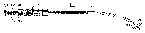
Referring to Figures 1 - 4, there are shown the
preferred but not the only: embodiments of the invented
device and method. The gastrostomy device 10 has an
elongated body, which has a distal region 14 for extending
into the patient's.stomach 16, and a proximal region 18
for extending out from the patient's mouth 20.
In the preferred embodiment, the elongated body
comprises an elongated rod 30 and an elongated probe,
which is a tube 32. The rod 30 has a distal region 34 and
a proximal region 36, and the tube 32 has a distal region
38 and a proximal region 40. The tube 32 has a hollow
interior' 42 and open ends that are referred to as the
opening 44 at the distal region 38 and the aperture 46 at
the proximal region 40. The rod 32 may be slidably
' received inside the tube 32, so that the rod distal region
34 may slide forward and backward through the opening 44
' 30 and the rod proximal region 36 may slide forward and
backward through the aperture 46. The terms "forward° and
"front" mean toward or past the distal region 38 of the
CA 02141023 1998-12-10
_ g _
tube 32 and the terms "backward" and "in back of" mean
toward or past the proximal region 40 of the tube 32.
A removable needle 50 is attached to the distal
region 34 of the rod 32 and 50 is removable and re-
attachable for easy cleaning, autoclaving, sharpening, or
replacement. Removable needle 50 can be attached to rod
32 by any conventional means, such as a bayonet mount or
friction fit. Here needle 50 has threaded extension 51
for threadable engagement in distal region 34 of rod 32.
The preferred needle 50 is a narrow arrow-head shape, with
a V-shaped cutting edge 52 oriented with the cutting edge
52 facing generally distally and generally parallel to the
longitudinal axis of the device 10. Other shapes and
orientations may be used to optimize the cutting edge 52
for a particular application. A thin, sewing-needle shape
or a scalpel-shaped blade could be used. The cutting edge
52 could face distally but at a 45° angle, for example, to
the longitudinal axis of the device 10. The limitation is
that the needle 50 should be a shape and orientation that
allows it to be shielded to fit through the mouth 20,
esophagus 54, and into the stomach 16.
The proximal region 36 of the rod 30 extends back
past the proximal region 40 of the tube 32 so that the
surgeon may access and push the rod 30 forward to slide
the removable needle 50 to an extended position and pull
the rod backward to slide the needle 50 to a retracted
position. When pushed forward, the distal region 34 of
the rod 30 and the needle 50 extend out from the opening
44 and in front of the blunt end 56 of the tube 32, thus
becoming unshielded and exposed. When the blunt end 56 of
the tube 32 has been placed in a desired location against
the stomach wall 58, this pushing of the rod 30 and needle
50 forward acts to force the needle 50 through the stomach
CA 02141023 1998-12-10
- 8A -
wall 58 and abdomen wall 60, piercing a channel 62
through these tissues. Therefore, grasping the rod proxi-
mal region 36 or optional handle 64 and the tube proximal
CA 02141023 1998-12-10
- 9 -
region 40 or optional grip 65 and pushing the rod 30
forward relative to the tube 32 is both the preferred way
of actuating the shield means to unshield the needle 50
and the preferred way of actuating the needle 50 to move
forward to pierce the channel. In the preferred embodi-
ment and preferred method, the tube distal region 38 acts
as the retractable shield means, because it covers the
removable needle 50 during the insertion through the
esophagus 54 and, in effect, retracts from the needle 50
when the needle 50 is pushed forward. Thus, the rod
proximal region 36, tube proximal region 40, and slidably
connection between the rod 30 and tube 32 cooperate to act
as the actuating means for moving the needle 50 forward
and backward.
Alternatively, other designs for the gastrostomy
device 10 may be used. For example, the rod could be
slidably connected parallel and beside, but not inside,
the probe. In such an embodiment, a shield plate could be
attached to the probe in such a way that it extends to
cover the removable needle during insertion through the
esophagus but allows the needle to slide forward and out
from under the plate when the rod is pushed. Another
shield means for this embodiment could be a hinged shield
plate that is biased to cover the removable needle until
the needle pushes the plate out of the way when the rod is
pushed forward. In another embodiment, the probe could
have the needle and a hinged shield attached to its distal
end and have linkage extending through or beside the probe
for actuating the hinged shield to unshield the needle.
In such an embodiment, after the unshielding of the
needle, the proximal region of the probe would be pushed
forward to actuate the needle to pierce a channel in the
stomach and abdomen
W~ 941~75Z4 PCTlUS93/07737
- 10 -
walls. Thus, the actuating means may be as simple as the
surgeon pushing the device forward into the tissue.
Tn another embodiment, the gastrostomy device may
include or be a part of an endoscope. The elongated body
may slide through a channel in the endoscope. Such em-
bodiment allows viewing of the inside of the stomach,
which is beneficial in human gastrostomy.
Preferably, a biasing means is included in the device
for biasing the rod 30 backwards relative to the tube
10 32, so that the needle 50 is shielded except when the
surgeon purposely pushes the rod 30 forward. In 'the
preferred embodiment, the biasing means is a coiled spring
66, which extends to force agart the rod handle 64 and the
tube proximal region 40.
The elongated body of the device 10 may be of various
degrees of flexibility, ranging from rigid to somewhat
flexible for allowing some bending when significant force
is placed on the device 10. Embodiments that are somewhat
flexible may aid in making easier the insertion of the
device 10 through the esophagus 54, however, flexibility
should be limited to a degree that assures efficient and
confident placement of the needle 50 without buckling,
bending, or crimping of the device 10.
In the preferred embodiment, the tube 32 is rigid and
curved,~resulting 'in the~tube distal region 38 and tube
proximal region 40 lying at an obtuse angle, of prefer-
ably, but not limited to, about 130° - 150° to each other.
This curve is slight enough and gradual enough to allow
easy insertion of the device 10 through the esophagus 54
but also is great enough to allow easy gointing of the
blunt end 56 toward the front or side of the stomach 16,
which are the preferred locations for the channel 62 for a
gastrostomy feeding tube 68. In embodiments having a
.. . '... . . ' : °.:
1° '7 'a pC°I°lIJS93/07737
dW0 94/27524 ~ ~ ~ ~ a~ !,, c)
e. 11
curve, the rod 30 should be flexible enough to follow the
curve of the tube 32 when pushed and pulled but rigid
enough to prevent buckling or bending that would bind the
rod 30 inside the tube 32 or interfere with the needle 50
piercing through the stomach wall 58 and abdomen wall 60.
The invented gastrostomy method involves making the
t channel through the stomach and abdomen walls 58,60 from
the inside out, as discussed in the above description of
the invented device. The method may be used to snake the
channel 62 for various medical uses, including the inser-
tion of a feeding tube 68 or other catheter.
The: preferred method involves tapping the blunt end
56 against the stomach wall 58 to determine when the blunt
end 56 and therefore the needle 50 are in a desirable
location for piercing the channel 62. The surgeon or
veterinarian may palpate the tapping from the outside of
the patient's body to accurately confirm the location of
blunt end 56 and needle 50 before actuating the needle 50
to pierce the channel 62. Thus, the invented method
provides a simple, accurate, and quick way of piercing the
channel 62 without expensive equipment and without the
assistance of a second person.
The method may optionally include other steps. A
elongated string may be attached
suture strand 70 or other
,
to the needle 50, for lexample, by threading the strand 70
through the eye 72 of the needle 50. The use of the term
' "suture strand" is not intended to limit the strand 70 to
a particular design or material. The needle 50 may be
' actuated backwards to draw the first end 74 of the suture
' strand 70 back into the stomach 16 and the device 10 may
be pulled out of the esophagus 54 to draw the first end 74
out of the mouth 20. The first end 74 may be attached to
a pipette 76 and feeding tube 68, as is done in the other
. :.:.:.~::: ... ::. .. ... ..: .. v. ,;:.. .. ,. .:~: . . ,..',: .. : : .:.
CVO 94/27524 . : PC7C/US93Itf7737
a
~ ~. ~ .~ s ? :~
_ 12
gastrostomy techniques discussed in the above section
"Background Art." The pipette 76 and feeding tube 68 may
then be drawn into the channel 62 by pulling the second
end 78 of the strand 70. The feeding tube 68 may then be
anchored in place and used for nutritional support as
described in the ~Background Art" section and the P. ,lane
Armstrong article.
The preferred materials for the invented device are
stainless or surgical steel. Other materials that fulfill
the sterility, strength and piercing requirements may also
be used.
While there is shown and described the present pre-
ferred embodiment of the invention, it is to be distinctly
understood that this invention is not limited thereto but
gray be variously embodied to practice within the scope of
the following claims.
I claim: