Note : Les descriptions sont présentées dans la langue officielle dans laquelle elles ont été soumises.
2143062
V~RlABLE-ANGLE SPINAL 5C~EW
The present invention relates to spinal fixation systems for use
in the treatment of spinal deformities and more particularly to an
5 apparatus for fixing a stabilizing appliance to spinal ve~tebrae
Spinal fusion, especially in the lumbar and s~ual region is
regL~larly used to correct and stabiliz~ spinal alrves and to stabilize
the lumbar and sacral spine tempor~rily while solid spinal fusions
10 develop in ~he treatrnent of othef spinal abnorrnalities. A spinal rod
systern is one of the stabilization systems currently in ~se. In spinal
rod systems, elongated ro~s are used to bridge across vario~s
portions of the spine Bone screlvs and co~pling devices ~re used
to attach the rods to various p~rtions of the spinal ~e~le~r~e. In
15 some situations one en~ of the elongat~d rod is anc~ored in the
~aGrsl region of the spine ~Ivith the other end being anchor~d in a
select~ lumbar vertebrae.
\Nhen spinal rod systems ~re anchored in the s~cral region,
20 the a~ility to achieve strong sacral fixation between the sacral and
lurnbdr ~er~eb~ b~c~ es ~irlicult. The ~n~umlQi posltlon of the
sacrum c~n c~use a 30 or mo~e difference between the angle of
implantation of bone screws in the sacral and lumbar ve~ le~rae.
When the spinal rod is not pfoperly contoured to a p~tient's lor~oti~
25 cur,ve, misalignrnent occurs be~ween the implanted bone scfews and
the coupling devices ~vhich causes inadequate fixation of the spinal
system.
A spinal rod system currently in use is shown in U.S. p~tent
30 5,102,412 to Rogozinski. In the Rogozinski Spin~l Syste~n, two
elongated rods afe used with cross-bars extendin~ laterally b~tween
the rods to fofrn ~ quadrilateral con~tr lct. Hooks, bone screYvs or a
combination of hooks and screws are attached to the vertebrae, ~nd
U-sh~ped couplefs are ~sed to sec~lre the hooks or screws to the
35 spinal rods. The bone screws each have a T-shaped he~d which is
piv~tally received in a coupler. The coupler h~s ~ U-shaped open
back with a recess for receiving either the T~ead of the screw or
the spinal rod. The bottom of ~he couplef h~s an opening for
214306~
receivin~ the threaded shank of the r-screw with the screw head
fitting bet~veen the side walls of the ~oupler. The side walls also
incl~-d~ aligned openings for re~eiving an elongate bar that extends
through the ali~nHd openings in a direction transverse to the r~cess.
5 The bar has set suews for holding the T-s~rew and spinal rod in
place in the coupler. Couplers are connected to each other ~hrough
the e~on~ate bar.
When a spinal rod system such as the Rogozinski spin~l
10 system is implanted in the saoral region of the spine, a need exists
for a bone screw which allows for the variability in angulation found
between the sacral and ll~mbar vertebrae.
A need also exists for bone screws having the ability to piYot in
15 the rnedial/lateral plane as ~ell as the ability to pivot and lock in the
cephalad/caudal plane ~hile maintaining the proper alignment
b~tw~cn an irnplanted bone screw, a coupler an~ a rod of a spinal
fixation system.
It is thus an object o~ the present invention to provide a
fastening apparatus for use in spinal rod systems that ~llows for
angulation in both the medialllateral an~ the ceph~ladlcaudal plane
and can then be locke~ into the selected angL~lar position ~he
fastening apparatus of the present invention incl~des ~ bone screw,
a coll~r and a locking pin. The bone screw has a threaded shaft
portion config~J~ed to be surgically implanted into a patient's bone
tissue and a head portion with ~n eyelet having a eentral bore.
The generally U-shape~ collar has a trans\~erse ~xis an~ ~ slot
th~t is placed over the eyelet portion of the scre~v. The elon~ated
lockin~ pin has longitudinal groo~les on at le~st a portion of the
outer surfa e and ;s sized and ~haped to be slidably inserted
through the eyelet bore Surface portions of the collar ~nd the
eyelst include reg~ rly spaced teeth, sized and shaped to engage
with the groo~es on the pin ~Ivhen the pin is inserted throu~h the
eyelet bore and the eyelet occ~pies the slot of the collar.
21~3062
When the fastening app~ratus is assembled th~ collar is
capable of pivoting in rel~tion to the ~yelet to adjust for an~ulation
l.etvl~cn ~ longitudinal axis of the threaded shaft and th~
transverse axis of the collar. The pin grooves and teeth on the
5 surfaces of the eyelet ~ore and collar en~age with each other for
locking the coll~ into a selected angled position The collar allows
for ~ngul~tion of the fxation apparatus in a rangQ of 140 in the
meclial/lateral plane and a range of 64 in the cephalad/ca~dal
plane.
In a pref~ d embo~iment, the fastening apparatus of the
present invention is use~ in the saorum of a p~tient in order to
maintain the coronal plane an~ulation in spinal rod systems th~t
have an attachment from the sacrum to the lumbar vertebrae
A better ul~de~"ding of the inven~ion can b~ obtained when
the ~etailed des~ i~lion of the exempl~ry embo~iments set fo,rth
below is reviewed in conjunction with the a~n".anying drawin~s,
in which:
Figure 1 is a p~rspective exploded view of the preferre~
embodiment of the apparatus of the present invention;
Figure 1A is 3 top plan vieu/ of the assembled apparatus of
25 Fig. 1;
Figu~e 1 B is a fr~gmentary side plan view of th~ assembled
a~,~.aral~Js of Fig. 1;
Figure 2 is a side plan view of the bon~ screw of the apparatus
of the present invention;
Figure 3 is an enlarge~ view of a portion of the bone screw of
Figure 2;
Figure 4 is a side pl~n view of tlle collar of the app~ratus of the
p~esent invention;
~ ~143062
Figure 5 is an enlarged fragmentary view of the wilar of Figure
4;
Figure 6 is a side plan view of the locking pin of the apparat~s
of the present invention;
Fig~e 7 is an enlarged crass-sectional vie~v looking along Ule
site line 7-7 of Figure 6;
Figure ~ is a schernatic side view of a partial spinal column
with an impl~n~ed fix~tion apparatus of the present invention
attache~ to a portion of spinal rod system;
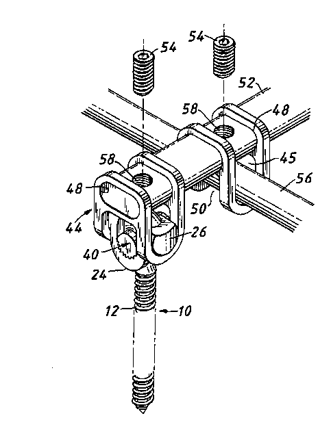
Figure g is a perspective exploded view of the pl~sent
invention is use with a portion of a spinal rod system; and
Figure 10 is a side plan view of the fixation apparatus of the
present in~Jention secured in a ~ouple~ of a spinal rod systern
2û
Figure 1 illustrates the preferred embo~lment of the fix~tion
al~p~r;~t.~s of the present invention desi~nated gene~ally by the
nurneral 10. The fixation apparatus 10 includes a bone scre-~v 11, 3
collar 20 and an elongated locking pin 40. Bone screw 11 has a
lon~;tlJdin~ cis 17 and a shank portion 12 ~v~th a bone engaging
course thread 13 thereon which ~llows it to be surgically impl~nted
into a patient's bone tiss~e, an~ in the preferred ernbodiment either
lumbar or sacral vertebrae (Figs. 1 and 2). A head por~ion 14 has
an eyelet 15 with a central bore 1~ (figs. 1 and 2). Bore 16 has a
surface 1 6a with a portion of bore surf~ce 16~ ~eing textured,
preferably at least 30%. In the preferred embodiment the textured
surface is forn~ed from ~egularly spaced teeth 18 as shown in Figs 1
~nd 3.
~ollar 20 is ~cnerally U-shaped ~vith a tr~nsve~se axis 21 an~
a oen~ally lo~ted slot 2~ sized an~ shape~ for plaoing over th~
eyelet 15 of t~e screw 11 (Figs. 1, 1 al 1 b ~nd 4). Collar 20 includes
2lq3o6~
a middle U-shaped portion 24 and extending end portions 26 with
the slot 22 positioned in the middle U-shaped portion ~4. As shown
in Fig. 1, the middle po~ion 24 includes inner ~Yalls 28 with flat
surfaces on ~ither side of the slot 2Z. A portion of the nat surface of
5 each of the inner w~lls 28 is textured, ~Jrefer~bly at least 30~ of tl~e
su~ce 28. In the preferred embodiment the textured s~ oe is
comprised of regularly spaced teeth 3~ ~vhidl m~t~ and int~,."esl.
the regula~ly sp~ teeth 18 on the bore surface 16a of eyelet 15.
The el~, Iydle~l locking pin 40 is generally cylindri~ in shape
and is sized and shaped to be inserted through the eyelet bore 16
(Fig. 1 b). Locl<in~ pin 40 has an oL~ter sutfa~e 41 with a portion of
the outer surface 41 being text~red to provide a gripping surfsce. In
the ~refe,.ed embodiment the tex~ured surface includes regularly
15 5p:~C~l longitt~dinal grooves 42"~, e~er~L Iy over at least 30% of the
outer $urface 41. The longitudinal grooves ~ of the locking pin 40
are sized and sl~laped to engage or intennesh with the teeth 18 on
the bore surface 1 6a and the teeth 30 on the inner wall surfaces 28
of the collar ~0 (Fig. 1 b). The teeth 30 are aligned on the wall
20 surfaces 28 and the grooves 42 are aligned on the pin surface 41 so
as to allow for locking the collar 20 in spaced i"~ erllel llS of between
about 10 to 20.
The flxation apparatus 10 of the ~Jresel 1l invention is used in
25 combination ~ith a spinal rod system and is an impro~ement on t~
bons sc~ews p~esently in use. In one prefe"ed em~odiment the
apparatus is used with a Rogozinski Spinal System in which
couplers are utilized to make the connection between the ve~teb~ae
and the spinal rods. The Rogozinski Spinal System is described in
30 U.S. patent 5,102,412 and sold by Smith & Nephew Spine The
fix~tion apparatus 10 of the present in~ention acc~r"",o~ s the
dirfiere"oe in angulation fo~lnd between the saua, ve, l~br~e S and
lumbar vei lebrae L, as illust~tsd in Fig. 8, where a difference of
angulation defined as X-X, ~n be approxima~ely 30.
A spinal syst~m s~ch as the Rogozinski Spinal System is
illust~ated in Figs. 9 ~nd 10, where a coupler ~4 ha~ a U-shaped
2143062
open ~ack ~itll aligned ~ide walls 4~ and a recess 45 b~tween them
for receiving a T-screw. ~n opening 50 is in the botton~ of the
FIcr 44 for receiving the threaded shank of ~ T~cre~,v with the
sc,rew head fitting between the side ~alls of the ~oupler 44.
The ~xation apparat~s 1 O ;s pre-assembled such that the collar
20 is placed over the eyelet 15 of the bone screw 11 with the eyelet
15 extending through the slot æ of the ~ollar 20. The pin 40 is
inserted through the eyelet bore 16 with the pin grooves 42
10 engaging with the teeth 18 on the bore surface 1 6a and the pin 40
extending o~t from botl~ sides of the eyelet bore 16 (Fig. 1 b). At this
pre-~sseril~led stage, the collar 20 is capable of pivoting up ~nd
down or in a ~ephala~/caudal plane in relation to the longit-Jdinal
axis 17 of t~le bone screw 11 (Fig. 1 b). The pre~ssembled rixati~l,
15 ~ ral-ls 10 is inserted into the coupler 44 with the collar 20
resting in the recess 45 between the side walls 46 and the thre~ded
shank 12 exten~ing through the opening 50. The thre~ded sh~nk
portion t2 of the bon~ screw 11 is then impl~nted into a paticnt's
ve"e l.rae.
The extending end portions 26 of the collar 20 allow the bone
scre~v 11 to pivot in the co~pler 44, in the medial/lateral plane,
within a range of generally 140. The collar 20 ;s ~3150 capable of
pivoting in l~ldtiOI~ to the eyelet 15 to adJust for angulation behveen
25 the longitudinal axis 17 of the implantecl bone ~crew 11 and the
transverse axis 21 of the collar 20 The adjustability of this
angulation is a range of generally ~ in th~ cephaladlcauclal plans.
An elonqated bar 52 is ins~rted through the aligned openings
30 48 in the side walls 4~ of the ~ouptef 44 holding the appar~tus 10 to
connect it witl~ ~ second ooupler 44 holding a spinal rod 56 (Fig. 9).
After the spinal rod system has been completely or partially installed
and ~ st~d for an~Jl~tion and alignment set screws 54 are
inserted into openings S8 in the elong~ted b~r 52 in ~rder to ti~hten
35 the assernbly. The tightening of the set screw 54 pushes down on
the screw he~d 14 an~ ~uses the coll~r 20 to pull 4p via the
couple~s 44 upwa~ force ~nabling the teeth 30 on the collar ~0 to
214306~
enga~e with the pin grooves 4~ on the pin 40 ~xtending thro~h the
~yelet borH 16. The collar teeth 30 and pin grooves 42 engage with
~ach othe~ and lock the collar 20 into a selected angled p~silio,~
within a range of ~4 (Fig 10). In a preferred embodirnent, the
S appa~ s 10 is formed from stainless steel or other biocom~ti~
material.
The apparatus 10 of the present invention allows for
angul~tion in three planes - the medial/lateral an~l cephalad/c~udal
10 plane with the third plane being the rola~io" of the screw itself for
the angle in which the screw is inserted into t~e sacrun~ or another
ve~e~,r~e. The intermeshing teeth 18 of the eyelet 14, the teeth ~0
of the ~ r 20 and the grooves 42 of the pin 40 provide a locking
mechanism that reslsts the rotational rnovement illlpossd durin~ in
15 vivo loading conditions in tlle cephal~d/caudal plane with an
ir,lpla,)ted spinal system.
It should be und~r~lood t~at there can be improvements an~
n)odifications made to the en~odiments of the invention described
20 in detail above without ~eparting from the spirit or scope of the
invention, ~s set forth in the ~ccompanyirlg clain~s.
