Note : Les descriptions sont présentées dans la langue officielle dans laquelle elles ont été soumises.
X149340
CVO 94/11734 PGT/DK93/00373
1
TWO-SITE INllNUNOASSAY FOR AN ANTIBODY WITH CHEMILUMINESCENT LABEL
AND BIOTIN BOUND LIGAND
The present i nvent i on rel ates to a method of detect i ng an ant i body
in a sample using a chemiluminescent labelling compound.
More specifically, the invention relates to the use of a chemilu-
minescent acridinium ester compound coupled to avidin or strepta-
vidin, and a ligand coupled to biotin in a two-site immunoassay
wherein the affinity complex is captured on paramagnetic particles,
which makes possible a rapid detection and/or quantification of
immunologically active substances, such as antibodies in samples
such as biological fluids and tissue samples, milk, food samples,
beverages, water or industrial effluents.
A method of detecting and quantifying immunoglobulin E-antibodies in
serum is disclosed in the brochure "Specific IgE, Magic~ Lite SQTM,
published by Ciba Corning Diagnostics Corp. and ALK Laboratories in
September 1990. In said method a specific allergen covalently bound
to paramagnetic particles reacts with the allergen-specific
IgE-anti body i n a serum or pl asma sampl e. After a fi rst i ncubati on
period and washing away unbound non-specific IgE, a chemiluminescent
acridinium ester-labelled monoclonal antibody against IgE is added.
Following a second incubation period the solid phase bound and
labelled antibody is measured in a Magic Lite~ Analyzer (Cat. No.
472733 or Cat. No. 472270) which automatically injects reagents,
which initiate the chemiluminescent reaction. When using said method
only the final step of initiating and measuring the chemilu-
minescence can be automated. The chemiluminescent acridinium ester
labelling compound is described in US patent No. 4,745,181.
US patent No. 4,946,958 discloses a chemiluminescent acridinium
ester linked to an N-succinimidyl moiety which can be further linked
to a protein or polypeptide to provide an immunologically reactive
luminescent reagent. Said reagent can be used in an immuno-assay,
which may involve the simultaneous binding of a solid phase
antibody, an antigen molecule and a labelled anti-antibody, separa-
tion and washing of the solid phase and quantifying the luminescence
of the solid phase.
WO 94/11734 ~ ~ ~ ~ ~ 2 PCT/DK93/003~''
Strasburger et al. discloses in Methods in Enzymology, 184(1990), pp
481-496 the use of a two-site chemiluminescent immuno-assay, wherein
hGH and hCG hormones are captured by antibodies immobilized on
microtiter plates and labelled with a chemiluminescent agent coupled
to avidin through a biotin labelled second antibody. Antigenic
analytes, such as protein hormones, may be assayed directly from
serum samples and compared to standard curves. The concentration of
immunoglobulins, such as IgE, is, however, extremely patient de-
pendent and an assay of a specific IgE must be compared with the
individual patient level of total IgE and therefore reuqires an
assay having a greater dynamic range than is obtainable in the assay
disclosed by Strasburger et al.
EP-A-0 425 217 discloses a hybridization assay wherein a chemilu-
minescent complex is formed comprising a nucleic acid hybridised
with a first labelled nucleotide probe coupled to paramagnetic
particles and a second nucleotide probe labelled with biotin and
coupled to an avidin-acridinium ester. However, the person skilled
in the art confronted with the problem of providing a fully auto-
mated method of detecting antibodies will search for a method which
can be carried out in one reaction container and preferably under
ambient reaction conditions. The assay represented here is
fundamentally different as it employs detection of specific
nucleotide sequences, which are not antigens towards which specific
antibodies can be raised. More detailed it is necessary in said
assay to use elevated temperatures for the hybridization and a
hapten-oligonucleotide probe which is not necessary nor desirable in
the immuno-assays.
Until now immuno-assays for the quantification of immunologically
active molecules, such as immunoglobulins (e. g. specific immunoglo-
bulin-E), in biological fluids, such as serum, have been manual,
e.g. the Enzyme Linked Immuno Sorbent Assay (ELISA), or semi-auto-
matical. And the typical duration of a semi-automatical immuno-
assay, such as the Magic~ Lite SQTM specific IgE assay referred to
above, is approximately two hours.
Moreover, commercial specific IgE assays (CAP,RAST, supplied by
Pharmacia, Uppsala) and the Magic~ Lite SQTM specific IgE assay use
-°~'O 94/11734 3 2 ~ ø~ ~ ~~ PCT/DK93/00373
a total-IgE reference assay having a non-identical protocol result-
ing in unprecise data. Only assays using the same catching and
detection procedures are directly comparable. For example all
speci fi c IgE cl ose response curves must be paral l el wi th total IgE
response curves. The reason is that the required dynamic range for a
specific (or total?) IgE assay is 2 decades and the required dynamic
range for a total IgE assay is from 2 to 7 decades, and the existing
immuno-assays do not allow concentration measurements over the
entire range. Thus, until now it has been necessary to use different
protocols for the specific and total immunoglobulin assays having
different reagents.
Because of the increasing interest in safe laboratory procedures
there is a need for a fully automated method of detecting substan-
ces, such as antibodies, in biological fluids, such as human serum,
plasma, blood, milk, urine or saliva, which method should provide a
minimised risk of contact with hazardous fluids. Also, the increas-
ing use of laboratory tests in diagnostics calls for methods of
short duration, preferably only a few minutes.
It is therefore an object of this invention to provide a method of
detecting an antibody in a sample, which method is safe, rapid, and
fully automatable.
This object is achieved by one method according to the invention
which method is characterised in:
a) mixing a ligand antigen, antibody, or hapten bound to biotin or
a functional derivative thereof; an antibody directed against the
antibody to be detected bound to paramagnetic particles; and a
chemiluminescent acridinium compound bound to avidin, streptavidin
or a functional derivative thereof with the sample to form a solid
phase bound complex,
b) magnetically separating the solid phase from the liquid phase,
c) initiating the chemiluminescent reaction, and analysing the
separated solid phase for the presence of chemiluminescent complex,
wherein the presence of chemiluminescence is an indication of the
2149340
(Amended page dated 24.11.94) 4 PCT/DK93/00373
presence of said antibody in said sample.
Although the method according to the invention can be carried out by
the above defined steps (a), (b) and (c) it is preferred to add the
labelling compound in a separate step, and the method is preferably
carried out according to the following steps:
i) mixing the ligand antigen, antibody, or hapten bound to biotin
or a functional derivative thereof with the sample and the antibody
directed against the antibody to be detected bound to paramagnetic
particles to form a first solid phase complex,
ii) adding a chemiluminescent acridinium compound covalently bound
to avidin, streptavidin or a functional derivative thereof '~o form a
second solid phase complex.
iii) magnetically separating the solid phase from the liquid phase;
iv) initiating the chemiluminescent reaction, and analysing the
separated solid phase for the presence of chemiluminescent complex.
A particular object of the invention is to provide a fully auto-
matable immuno-assay for the quantification of specific antibodies,
such as immunoglobulins, wherein a truly parallel reference immuno-
2~ assay using an identical protocol is used as the reference.
The object of quantifying specific antibodies using a truly parallel
reference immuno-assay is achieved by a method of measuring the
concentration and/or the relative contents of a specific antibody in
a liquid sample, wherein the measured light emission of a separated
solid phase comprising a captured specific antibody coupled to a
chemiiuminescent label is compared with the measured light emission
obtained in a parallel reference immuno-assay wherein the total
contents of the class of antibodies in the sample to which said
~ specific antibody belongs is measured, said method comprising the
steps of
a) mixing a ligand antigen or hapten towards which the specific
antibody to be measured is directed bound to biotin or a functional
AMENDED SHEET
S S
~~49340
(Amended page dated 24.11.94) S PCT/DK93/00373
derivative thereof; an antibody directed against the constant
portion of the antibody to be measured bound to paramagnetic par-
ticles; and a chemiluminescent acridinium compound bound to avidin,
streptavidin or a functional derivative thereof with the sample to
form a first solid phase complex,'
b) magnetically separating said first solid phase from the liquid
phase,
c) initiating a chemiluminescent reaction and measuring the light
emission of the separated first solid phase,
d) mixing a ligand antibody directed against the class of anti-
bodies to be measured bound to biotin or a functional derivative
13 thereof; an antibody directed against the constant portion of the
class of antibodies to be measured bound to paramagnetic particles;
and a chemiluminescent acridinium compound bound to avidin, strep-
tavidin or a functional derivative thereof with the sample to form a
second solid phase complex,
e) magnetically separating said second solid phase from the liquid
phase,
f) initiating the chemiluminescence reaction and measuring the
light emission of the separated second solid phase, and
g) comparing the light emission of the separated first solid phase
with that of the separated second solid phase.
In a preferred embodiment of the method described above step a) is
performed by
(i) mixing the ligand antigen or hepten bound to bioten or a
functional derivative thereof with the sample and the antibody bound
to paramagnetic particles to form a solid phase complex, and
(ii) adding the chemiluminescent acridinium compound bound to
avidin, streptavidin or a functional derivative thereof to form said
first solid phase complex,
AIvIENDE~ SHEET
(Amended page dated 24.:1.94) 6 ~ ~ 4 9 3 ~ o PCT/DK93/00373
and step d) is performed by
(i) mixing the ligand antibody bound to biotin or a functional
deri va t i ve t hereof w i th the sampl a and the an t i body bound to para-
magnetic particles to form a solid phase complex and
(ii) adding the chemiluminescent acridinium compound covalently
bound to avidin, streptavidin or a functional derivative thereof io
form said second solid phase complex.
T he speci fi c anti body to be measured i n the sampl a i s preferabl y a
specific immunoglobulin selected from the group consisting of IgA,
ig0, IgE, IgG, IgM, and isotypes thereof, and the ligand antigen,
antibody, or hapten directed against the variable portion of said
antibody is an allergen, and the class or~ antibodies is preferably a
class of immunoglobulins selected from the group consisting o.f total
IgA, total IgD, total IgE, total IgG, total IgM, and isotypes
thereof, and the ligand antigen, antibody or hapten is an antibody
directed against said class of immunoglobulins.
More preferably the specific immunoglobulin is a specific IgE, and
the class of antibodies is total IgE.
30
AMENDED SHEEN
~~-~VO 94/11734 ~ ~ ~~ ~ ~ ~ PCT/DK93/00373
7
The antibody directed against the antibody to be measured bound to
paramagnetic particles is preferably selected from the group con
sisting of polyclonal antibodies, monoclonal antibodies including
recombinant antibodies, fragmented antibodies, preferably a mono
clonal mouse anti-immunoglobulin.
The advantages of the invention are:
- All reagents can be mixed simultaneously in one reaction
container, which minimises the risk of contamination, errors
and operation steps, reduces significantly the duration of the
immuno-assay, and greatly facilitates automation of the pro-
cess and precision is improved, cf. example 6;
- Quantification of specific antibodies in, e.g. a serum sample,
can be performed with reference to a truly parallel total
antibody assay using an identical protocol, cf. example 3;
- The obtained greater capacity and sensitivity facilitates that
even very low concentrations of immunoglobulins and low concen-
trations of specific immunoglobulins can be detected, cf.
examples 1, 2 and 7.
A preferred embodiment of the invention is an immuno-assay for the
detection of antibodies, such as specific immunoglobulins (IgA, IgE,
IgG, IgM and isotypes thereof) in a sample, such as serum or saliva.
Particularly, the invention can be used in an assay for the detect-
ion and quantification of specific IgE in a sample. When the sample
is liquid, e.g. serum or plasma, it can be added directly to a
reaction container comprising preferably a monoclonal mouse anti-IgE
antibody bound to suspended paramagnetic particles and a specific
allergen (ligand) bound to biotin in an aqueous medium. The biotin
is preferably biotin amidocaproate N-hydroxysuccinimide ester (Sigma
Catalog No. B2643) When the sample is non-liquid, e.g. a tissue
sample, it is preferably homogenised and suspended in an aqueous
liquid. A simultaneous reaction between specific IgE in the sample,
allergen and monoclonal anti-IgE antibody in the aqueous medium
results in the formation of a conjugate. A chemiluminescent la-
CA 02149340 2003-11-05
belling compound, preferably an acridinium ester coupled to strep-
tavidin (Sigma Catalog No. S4762) (avidin-DMAE) is added to the
reaction container and a binding reaction between avidin-DMAE and
biotin bound to the conjugate and unconjugated allergen-bound biotin
takes place. The conjugate bound label is separated from the unbound
DMAE-labelled antibody by magnetically separating the reaction
mixture and decanting the supernatant. The chemiluminescence of the
separated conjugate is measured as described in Pazzagli, M. et al.
(eds.). Studies and applications in "Biology & Medicine", Journal of
Bioluminescence and Chemiluminescence 4(1), 1-646, 1989.
As a reference, a truly parallel total-IgE immuno-assay differing
only in that a preferably polyclonal anti-IgE antibody bound to
biotin is used as the ligand is performed simultaneously.
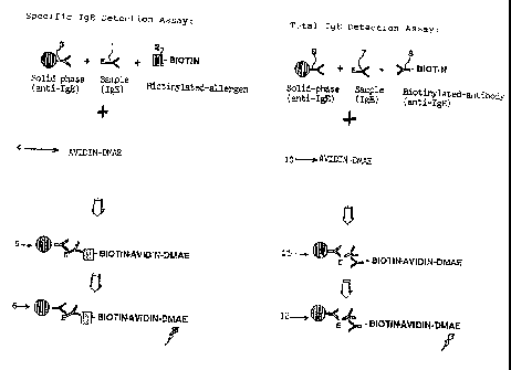
Figure 1 is a diagrammatic representation of a specific IgE assay
according to the invention and comprising a parallel total IgE
reference assay. In the figure (1) represents the specific IgE-
antibody to be detected, (2) is a specific allergen bound to biotin,
(3) is a monoclonal mouse anti-IgE bound to paramagnetic particles,
(4) is an avidin-acridinium ester and (5) represents the solid phase
labelled complex formed between (1), (2), (3) and (4) and includes
optional incubation, separation and optional washing steps, and (6)
represents a final step of initiating the chemiluminescent reaction
and measuring the light emission.
In the total IgE reference assay in Figure 1 (7) represents IgE (WHO
75/502 IU/ml), (8) is polyclonal anti-IgE bound to biotin, (9) is
monoclonal mouse anti-IgE bound to suspended paramagnetic particles,
(10) is an avidin-acridinium ester and (11) represents the solid
phase labelled complex formed between (7), (8), (9) and (10) and
includes optional incubation, separation and optional washing steps,
and (12) is the final step of initiating the chemiluminescent
reaction and measuring the light emission.
r More particularly, the immuno-assay using the method of the inven-
tion can be performed in ACS:180~fully automatic analyzer produced
by Ciba Corning Diagnostics Corp., Medfield, Mass., U.S.A.
CA 02149340 2003-11-05
9
In one preferred embodiment of the invention the immunologically
active substance to be detected is an antibody, e.g. against
penicillin or derivatives thereof, such as benzylpenicillin, peni
cilloyl, etc., and the ligand bound to biotin is a hapten, such as
penicillin or derivatives thereof.
Definitions
In the methods of the invention the antibody to be detected is
a specific immunoglobulin, preferably a specific IgA, IgD, IgE, IgG,
IgM, and isotypes thereof, and more preferably a specific IgE, or a
class of antibodies, such as immunoglobulins, preferably selected
from the group consisting of total IgA, total IgD, total IgE, total
IgG, total IgM and isotypes thereof, most preferably total IgE.
By sample is meant any liquid or liquefied sample, including solu-
tions, emulsions, dispersions and suspensions.
The ligand antigen, antibody or hapten bound to biotin can be any
immunologically active substance, such as an allergen, antibodies,
such as polyclonal antibodies, monoclonal antibodies including
recombinant antibodies or fragmented antibodies, preferably an
allergen and/or a polyclonal anti-immunoglobulin, such as goat
anti-human polyclonal serum spplied by Ventrex Laboratories, Inc.,
Portland, Maine, Catalog No. 77660. In the reference immuno-assay
said antibody is preferably directed against the constant portion of
the class of antibodies to be measured, i.e. an antibody directed
against the IgE-antibodies.
gy biological fluid is meant any clinical sample, such as blood,
plasma, serum, urine or saliva, which also includes any biological
fluid which is excreted, secreted or transported internally in an
organism.
gy paramagnetic particles (PMP) is meant particles which can be dis-
persed or suspended in a liquid medium. Throughout the examples are
used BioMag~ particles (iron oxide particles coated with amine
terminated groups) sold by advanced Magnetics Inc., Cambridge,
Massachusetts. The antibodies coupled to PMP are preferably directed
CA 02149340 2003-11-05
against the constant portion of the antibodies to be detected or
measured and may be polyclonal or monoclonal antibodies including
recombinant or fragmented antibodies, preferably a monoclonal
antibody, MAb A 5697-IA3(920325) supplied by BioInvent International
5 AB, Lund, Sweden.
The chemiluminescent acridinium compound is preferably N-hydroxy-
succinimide dimethylacridinium ester covalently bound to avidin or
streptavidin (avidin-DMAE). Avidin and OMAE are coupled according to
10 the methods of Weeks et al., Clin. Chem. 29/8, 1474-1479 (1983).
Other luminescent labelling compounds that can be bound to avidin or
streptavidin may be used in the method of the invention. E.g.
luminol, lucigenin or lophine.
preparation of biotinvlated antibodies
Biotin~rlated anti-I4E and Phleum oratense:
Goat anti-human polyclonal serum (llentrex Laboratories, Inc. MA,
USA) is purified by affinity chromatography on a CNBr-activated
sepharose~ 4B (Pharmacia, Uppsala, Sweden) with myeloma IgE (OEM
concepts, USA) as a ligand. The anti-IgE is biotinylated with the
ratio mol biotin: mol anti-IgE = 41:1.
g ~1 of Biotin (Biotin amidocaproate N-hydroxysuccinimide ester
(Sigma) 25 mg/ml in Dimethylformamide (Merck) is added to 0.4 ml of
anti-IgE 4.5 mg/ml in 0.1 M NaHC03 (Merck). The reagents are
incubated in an "end over end" mixer for 2 hours at 25'C. 0.9 ml
lysin (Sigma) solution 20 mg/ml NaHC03 is added. The solution is
filtered and the biotinylated antibody is purified by size chro-
matography on s uperdex~75 Hiload 16/60 (Pharmacia, Uppsala, Sweden).
The pool ed fracti ons are di 1 uted in phosphate buffered sal ine PBS,
pH 7.2, containing 0.1 % human serum albumin (Sigma) 0.1 % NaN3
(Sigma).
The Phleum pratense extract, (ALK Laboratories A/S, H~rsholm,
Denmark) is biotinylated in the molar ratio of 10:1 0.65 ml of
biotin 10 mg/ml is added to 0.43 ml of 10 mg/ml Phleum pratense in
0.1 M NaHC03. The reagents are incubated for 2 hours at 25'C in an
i
CA 02149340 2003-11-05
11
"end over end" mixer, after the incubation 40 ~cl lysin (Sigma)
solution 50 mg/ml is added. The solution is filtered and the
biotinylated antibody is purified from excess of biotin by size
exclusion chromatography on superdex 75 Hiload 16/60 (Pharmacia).
The fractions containing the allergens are pooled. The biotinylated
Phleum pratense is diluted with PBS pH 7.2, containing 0.1 % human
serum albumin (Sigma) and 0.1 % NaN3 (Sigma).
Prgparation of streg_tavidin-acridinium ester label
Streptavidin was conjugated with OMAE-NHS, [2',6'-dimethyl-4'-(N-
succinimidyloxycarbonyl)phenyl-10-methylacridinium-9-carboxylate
methosulphate~ using the methods of Weeks et al., Clin.Chem. 29/8,
1474-1479 (1983).
Preparation of Streptavidin-acridinium ester label:
0.96 mg N-hydroxysuccinimide dimethylacridinium ester DMAE (Ciba
Corning Diagnostics Corp., Medfield, MA, USA) is diluted in 1.92 ml
Dimethylformamide. 250 ~1 of this solution is pipetted to 2.5 ml 1
mg/ml streptavidin (Sigma) in 0.1 M sodium dihydrogenphosphate, 0.15
M NaCI pH 8.15.
The air above the solution in the vial is exchanged with nitrogen,
(AGA). The reagents are incubated for 30 min at 25°C under stirring,
after incubation 2250 ul 10 mg/ml lysin 0.1 M sodium dihydrogen-
phosphate (Merck), 0.15 M NaCI is (Merck) pH 8.15 is added. To
remove unbound DMAE the solution is loaded on a PD-10 column
(Pharmacia, Uppsala, Sweden). The eluate is collected and purified
by ultrafiltration using a cellulose Minitan-S~filter sheet 10.000
NMWL (Millipore). The filtration is performed with 1.5 1 phosphate
buffered saline, PBS pH 7.2. The retentate (retanate) is concentrat-
ed to 25 ml and 25 m1 PBS pH 7.2 containing 0.5 % HSA (Sigma) and
0.1 %a NaN3 (Sigma) is added.
The invention will be described in detail in the following examples.
WO 94/11734 ~ ~ ~ PCT/DK93/0037"
12
Immobilisation of antibody to parama4netic particles
6.5 g paramagnetics particles {Ciba Corning Diagnostics Corp., MA,
USA) are washed in 650 ml methanol (Merck) 3 times using magnetic
separation. A wash with 650 ml 0.01 M acetate buffer pH 5.5 is
performed twice. The particles are activated in 6.25 % glutar-
aldehyde (Merck), 0.01 M acetate buffer pH 5.5 for 3 hours at 25°C.
The particles are washed 3 times in 650 ml 0.01 M acetate buffer pH
5.5. The particles are coupled with 1083 mg monoclonal anti-IgE
antibody (ALK Laboratories, HOrsholm, Denmark) specific against the
IgE Fc domain, for 24 hours at 25°C. The particles are washed
twice
in 0.01 M acetate buffer pH 5.5. Blocking of excess of active groups
is performed with 200 ml 10 % IgE stripped serum (ALK Laboratories,
Harsholm, Denmark) for 24 hours at 25°C. The particles are washed
in
650 ml 0.01 M phosphate buffer (Merck) followed by 3 washes in 650
ml 1 M NaCI (Merck). The particles are washed 3 times in 0.01 M
phosphate buffer. The particles are resuspended in 650 ml PBS pH
7.2, 0.1 % w/v bovine serum albumine (Sigma), 0.001 % bovine gamma
globulin (Sigma) and heat treated for 18 hours at 50°C. The par-
ticles are washed 3 times in 650 ml PBS pH 7.2, 0.1 % w/v bovine
serum albumine (Sigma), 0.001 % bovine gamma globulin (Sigma). The
particles are heat treated for 7 days at 37°C. The particles are
washed i n 0. O1 M phosphate buffer twi ce. The parti cl es are di 1 uted
to 0.5 g per 1 in PBS pH 7.2, 0.5 % human serum albumine {Sigma).
Example 1
Detection and auantification of antigen (total IgE)
Determination of total IgE antibodies, according to the invention,
was conducted on Ciba Corning ACS:180 Benchtop Immunoassay analyzer
described in Clinical Chemistry, 36/9, 1598-1602 (1990), using the
following protocol:
50 ul of sample and 50 ~l biotinylated anti-IgE are dispensed by the
sample probe into the cuvette. The cuvette reaches the first reagent
probe R1, where 100 ul paramagnetic particles with immobilised
monoclonal anti-IgE antibody (ALK Laboratories A/S, Harsholm,
Denmark) specific against the IgE Fc domain are dispensed together
REPLACEMENT SHEET
WO 94/ 11734 - ~ ~ ~ ~ PCT/DK93/00373
13
with 200 ul of streptavidin-acridinium ester label (ALK Laboratories
A/S, H~rsholm, Denmark). The cuvette moves down the track to the
magnets and wash station. Wash with 750 ul deionized water is
performed twice. After completion of the wash cycle the particles
are resuspended i n 300 ul 0. 5 g/1 H202 i n 0.1 M HN03 . The cuvette
enters the luminometer chamber and in front of the photomultiplier
300 ul 25 mM NaOH solution is added and the photons of light emitted
are measured and quantitated and expressed as relative light units
(RLU). The amount of RLU is directly proportional to the amount of
the IgE in the sample. The time from sample dispension to first
resul t i s 15 mi n and a new resul t fol 1 ows every 20 second. Resul is
were expressed as RLU experiment/RLU background, where RLU back-
ground was the chemiluminescent reaction observed in the absence of
total IgE.
Nine Total IgE standards, calibrated in Magic Lite Total IgE Kit
(ALK Laboratories A/S, HOrsholm, Denmark) against WHO 2nd IRP no
75/502 for human serum IgE, were assayed using the protocol de-
scribed above and it is shown that 0.1 IU/ml of total serum IgE
could be detected (as determined by background x 10 standard devia-
tions), see Figure 2.
Example 2
Detection and auantification of specific antibody (Specific IaE)
Determination of Phleum pratense specific IgE antibodies (timothy
grass specific IgE, according to the invention, was conducted on
Ciba Corning ACS:180 Benchtop Immunoassay analyzer described in
Clinical Chemistry, 36/9, 1598-1602 (1990), using the following
protocol:
50 ul of sample and 50 ~cl biotinylated Phleum pratense are dispensed
by the sample probe into the cuvette. The cuvette reaches the first
reagent probe R1, where 100 ~,1 paramagnet i c part i cl es wi th i mmobi -
lised monoclonal anti-IgE antibody (ALK Laboratories A/S, H~rsholm,
Denmark) specific against the IgE Fc domain are dispensed together
with 200 ul of streptavidin-acridinium ester label (ALK Laboratories
A/S, Horsholm, Denmark). The cuvette moves down the track to the
REP~ACEMENTSHEET
WO 94/11734 ~ ~~ ~ ~ ~ PCT/DK93/0037z
14
magnets and wash station. Wash with 750 ul deionized water is
performed twice. After completion of the wash cycle the particles
are resuspended i n 300 ul 0. 5 g/1 H202 i n 0.1 M HN03 . The cuvette
enters the luminometer chamber and in front of the photomultiplier
300 ~C1 25 mM NaOH solution is added and the photons of light emitted
are measured and quantitated and expressed as relative light units
(RLU). The amount of RLU is directly proportional to the amount of
the IgE in the sample. The time from sample dispension to first
result is 15 min and a new result follows every 20 second. Results
were expressed as RLU experiment/RLU background, where RLU back-
ground was the chemiluminescent reaction observed in the absence of
total IgE.
Ten Phleum pratense specific IgE standards, calibrated in Magic Lite
SQ Specific IgE Kit (ALK Laboratories A/S, HOrsholm, Denmark)
against clinically characterised Phleum pratense allergic patients
samples and expressed as SU/ml (Standardised Units), were assayed
using the protocol described above and it is shown that between 1.43
and 800 SU/ml of Phleum pratense specific IgE can be measured as in
Magic Lite SQ Specific IgE assay, see Figure 3.
Example 3
Quantification of specific IgE against WHO Total I4E reference
Quantification of specific IgE antibodies in serum samples were
performed with reference to total IgE antibody or specific IgE
antibody using the identical assay protocols as described in example
one and two, respectively.
Thirtyfive patient samples were assayed for Phleum pratense specific
IgE along with 10 Phleum pratense specific IgE standards, calibrated
in Magic Lite SQ Specific IgE Kit (ALK Laboratories A/S, HOrsholm,
Denmark) against clinically characterised Phleum pratense allergic
patient samples and expressed as SU/ml (protocol described in
example 2).
Nine standards of IgE WHO 2nd IRP No. 75/502 (National biological
standard board) were assayed in the same run against total IgE
REPLACEMENTSHEET
~WO 94/11734 ~~~~ PCT/DK93/00373
(protocol described in example 1).
Figure 4 provides a comparison of the two dose response curves of
total IgE assay and Phleum pratense specific IgE assay respectively.
5 Parallel line test showed no significant difference in slopes
between the two dose response curves, indicating that specific IgE
in patient samples could be calibrated against the WHO Total IgE
standard and expressed in IU/ml.
10 Figure 5 provides a comparison of results from the 35 patient
samples calibrated against WHO Total IgE standards (expressed in
IU/ml) or Phleum pratense specific IgE standards (expressed in
SU/ml) and it shows very good correlation between the two units.
15 The concentration (dose) of the unknown sample was calculated using
a cubic-free spline interpolation after a log vs. log transformation
of signal/background and concentration (dose), respectively.
It was calculated from the linear regression line that one SU
(Standardised Unit) corresponds to 0.14 International Unit (IU).
In conclusion allergen specific IgE can be measured using the
embodiment of the present invention and calibrated directly from the
total IgE assay of a WHO IgE calibrated standard curve.
Example 4
Total IaE method comparison
Thirtythree patients samples were measured for total serum IgE in
the Magic Lite Total IgE kit (ALK Laboratories A/S, HOrsholm,
Denmark). The assay was performed according to the manufacturer's
instruction. The same samples were measured for total serum IgE on
the ACS:180 according to the protocol described in example 1.
Figure 6 provides a scatterplot for the method comparison for
measuring total serum IgE. A correlation with r = 0.90 was found.
REPLACEMENTSHEET
~ PCT/DK93/0037'
WO 94/11734 ~ ~ ~ 9 3 ~ ~ 16
Example 5
Specific IgE method comparison
Thirtyfive patients samples were measured for Phleum pratense
specific IgE in the Magic Lite SQ Specific IgE kit (ALK Laboratories
A/S, H~rsholm, Denmark). The assay was performed according to the
manufacturer's instruction. The same samples were measured for
Phleum pratense specific IgE on the ACS:180 according to the proto
col described in example 2.
Figure 7 provides a scatterplot for the method comparison for
measuring Phleum pratense specific IgE. A correlation with r = 0.80
was found.
Example 6
Comparison of assay precision
The within-run imprecision for ACS total IgE using the protocol
described in example 1, was compared to that of Magic Lite Total
IgE. Patient samples were run in replicates of three in the assays
described in example 4. The pooled within-run coefficient of varia-
tion (CNpwr) was calculated according to Krouwer and Rabinowitz,
Clinical Chemistry, 30, 290 (1984). The following results were
obtained:
Magic Lite ACS
%CUpwr 4.69 2.75
Min %CN 0.80 0.70
Max %C11 11.70 8.40
As seen from the results, automation and minimation of operating
steps significantly improves the precision of the analysis (by
F-test: F=1.71, p=0.049).
REF'LACEMENTSHEET
°~'WO 94/11734 ~~ PCT/DK93/00373
17
Example 7
Comparison of total and J~ecific IgE in patient samples:
The samples measured for Phleum pratense specific IgE on the ACS:180
according to the protocol described in example 2 and calibrated
against total IgE reference (WHO 75/502) as described in example 3,
were also analysed for total IgE as described in example 1. The
ratio between measured specific IgE and total IgE was calculated on
each sample and expressed as % ratio (spec. IU/total IU*100).
The following results were obtained:
Phleum pratense
No Specific IaE IU/ml Total IgE IU/ml % Ratio
1 0.973 208.300 0.467
2 0.000 153.330 0.000
3 0.000 25.119 0.000
4 0.000 43.980 0.000
5 0.082 51.997 0.158
6 0.586 30.807 1.902
7 9.130 20.796 43.903
8 2.125 168.771 1.259
9 1.545 321.553 0.480
10 0.000 299.105 0.000
11 1.664 248.660 0.669
12 8.553 46.635 18.340
13 19.427 234.858 8.272
14 0.000 178.304 0.000
15 2.380 428.987 0.555
16 4.342 112.457 3.861
17 3.975 173.793 2.287
18 0.220 206.580 0.106
19 0.158 298.630 0.053
20 4.936 17.116 28.839
21 0.376 146.939 0.256
22 4.865 23.521 20.684
23 3.115 281.804 1.105
24 0.017 17.937 0.095
REPU~CEMENTSHEET'
WO 94/11734 ,~ ~ PCT/DK93/0037'
18
25 3.446 160.651 2.145
26 1.485 167.427 0.887
27 2.763 124.309 2.223
28 3.705 247.679 1.496
29 1.289 196.314 0.657
30 8.967 416.528 2.153
31 0.355 29.784 1.192
32 15.173 290.670 5.220
33 2.141 63.847 3.353
34 3.929 96.951 4.053
35 0.154 > 620 ND
Example 7 (continued)
As seen from the results the Phleum pratense specific IgE assay was
able to measure as low as 0.05 % of Phleum pratense specific IgE out
of total IgE. Low relative amounts of Phleum pratense specific IgE
indicates that other allergen specificities are present in the
samples. Up to 44 % of total IgE was found in one patient sample to
be specific against Phleum pratense. No correlation was found
between total IgE concentration and Phleum pratense specific IgE
concentration as seen in figure 8.
30
REPLACEMENTSHEET
~'WO 94/11734 ~~~ PCT/DK93/00373
lg
Example 8
Determination of serum total IgA antibodies using paramagnetic
particles and avidin-acridinium ester label.
The determination of total IgA antibodies was assayed using the
following protocol:
25 ul of patient sample or calibrator was pipetted into a 12x75 mm
test tube. To each tube 50 ul of biotinylated polyclonal anti-IgA
antibody DAKO E484 (supplied by DAKO, Glostrup, Denmark) in 0.05 M
phosphate buffer, pH 7.4, containing 0.1 % sodium azide, 0.01
Tween~ 20 and 0.1 % human serum albumin was added and reacted for 15
minutes at ambient temperature. 400 ul slurry of paramagnetic
particles with immobilised polyclonal anti-IgA antibody DAKO A 262,
(supplied by DAKO, Glostrup, Denmark) was added to each tube and
incubated for 5 minutes. After this second incubation 50 ul of
streptavidin-acridinium ester label diluted in the same buffer as
described above was added to each tube and incubated for a further 5
minutes at ambient temperature. The paramagnetic particles were
washed twice with a 0.2 M phosphate buffer, pH 7.4, containing 0.1
Tween~ 20, after separating the magnetic particles from the liquid
by a magnetic base separator and vortexing the separated particles
with the washing buffer as described above. The contents of the
tubes were finally measured in the luminometer, where light emitted
at 426 nm, was quantitated and expressed as relative light units
(RLUs).
Total IgA standards calibrated against WHO No. 67/86 for human serum
IgA DAKO X908 (supplied by DAKO, Glostrup, Denmark) were assayed
using the above described protocol.
REPLACEMENTSHEET'
WO 94/11734 ~ I ~ e~ ~ ~ PCT/DK93/0037'
Standards:
Concentration (u4/ml) RLUs
0 370937
5 0.02 417000
0.23 557563
2.32 1252260
23.2 1872357
10 It should be apparent to one having ordinary skill in the art that
many variations are possible without departing from the spirit and
scope of the invention.
20
30
REPLACEMENTSHEET