Note : Les descriptions sont présentées dans la langue officielle dans laquelle elles ont été soumises.
~ 095/0~58 C A 2 1 6 6 0 6 9 1 PCT~S94/07866
FINGE~R ~OT OX~C PROBe
Back~round of the Invention
1. Field of the Invention
This invention relates to an oximetric sensor that is
nonadhesively attached to a human digit (e.g., a finger) via
an inexpensive, disposable finger cot to analyze the blood of
a patient by calculating the concentration of blood
constituents (e.g., the saturation level of oxygen in the
patient's blood) while m~i~;zing potentially interfering
noise artifact signals.
2. Description of Related Art
Electromagnetic energy is often transmitted through or
reflected from a medium to determine the characteristics of
the medium. In the medical field, optical energy can be
transmitted or reflected through human tissue and subsequently
measured to determine information about the tissue rather than
extracting material from a patient's body for testing. This
form of noninvasive measurement can be performed quickly and
easily, and has proven to be more comfortable to the patient.
Noninvasive physiological monitoring of body functions is
often required when treating a patient. For instance, the
available supply of oxygen in the body (i.e., blood
oxygenation) is often monitored during surgery. Today this
measurement commonly is performed by a noninvasive technique
that measures the ratio of incident to transmitted (or
reflected) light through a blood-perfused portion of the body,
such as, for example, a finger, an ear lobe, or the forehead.
Transmission of optical energy a9 it passes through the
body is strongly affected by several factors. Such factors
include the thickness of the tissue through which the energy
passes, optical coupling, the optical angle, and the distance
between the detector and the source of energy (i.e., the
optical path length).
Several parts of the human body are soft and
compressible, and therefore are ideally suited to transmit
optical energy. For example, a human digit, such as the
finger, comprises skin, muscle, tissue, bone, blood, etc.
~ W095~0~58 C A 2 1 6 6 Q 6 ~ P~T~S94J07~66
--2--
Although the bone is relatively incompressible, the tissue
~urrounding the bone is easily compressed when an external
pressure is applied to the finger. Accordingly, such digits
are well suited for the transillumination or tran5reflection
of optical energy for blood monitoring purposes.
Optical probes have been used in the past for both
invasive and noninvasive applications. In the typical optical
probe, a light emitting diode (LED) iS placed on one side of
the human tissue while a photodetector is placed on the
opposite side. Such conventional optical probes are primarily
useful when a patient i8 relatively motionless and in
environments which are characterized by low ambient light.
By way of particular example, one well-known noninvasive
measuring device in which an optical probe is used in health
applications is the pulse oximeter which measures the pulse
rate and the percent of oxygen available in an arterial
vessel. Up until the early 1980s, clinicians relied upon
arterial blood gas analysis to evaluate gas exchange and
oxygen transport within the human body. Although the arterial
blood gas test gives valuablè information, it only reflects a
patient's oxygenation status for one moment in time. On the
other hand, pulse oximetry permits a continuous, noninvasive
measurement of a patient's arterial oxygen saturation status.
Oximetry is based on the principal that blood hemoglobin
absorbs red and infrared light differently when carrying
oxygen, in the form of oxyhemoglobin, than when not carrying
oxygen, in the form of reduced hemoglobin. Prior oximetric
sensors sense the absorption rate of the optical energy by the
blood hemoglobin to determine arterial blood oxygen
saturation; i.e., the amount of hemoglobin-carried oxygen in
relation to the total hemoglobin-carrying capacity.
For this reason, prior oximetric sensor commonly includes
a photodetector and a pair of LEDS which émit both red and
infrared light. The sensor is packaged in such a way that the
LEDS and photodetector are placed on opposite sides of a
vascular bed which, in the transillumination case, is usually
a finger, ear lobe, or toe. In the reflectance case, the LEDs
~0 9S/~58 C A 2 1 6 6 0 6 9 PCT~S94/07~6
--3--
and the photodetector are placed side by 8ide over a vascular
bed, usually the forehead, but 8eparated by a barrier which
blocks light from re~h;n~ the detector without first passing
through the tissue sample. When properly po8itione~, the LEDs
emit known wavelengths of both red and infrared light for
transmission or transreflection through the vascular bed for
receipt by the detector.
The photodetector produces a signal in response to
unabsorbed light passed through the vascular bed to the
detector. This signal conventionally is con~erted to digital
form and then supplied to a computer or mic o~.o~essor which
computes the ratio of red light to infrared light absorption.
The absorption data is then utilized to determine the arterial
blood oxygen saturation values which then may be displayed on
a monitor or a strip chart. Because the light that is
directed into the vascular bed also is at least partially
absorbed by the nearby tissue and bone material, the oximeter
typically utilizes the alternating bright and dim signals
caused by arterial pulsations to further clarify the presence
of both reduced hemoglobin and oxyhemoglobin, as known in the
art.
Prior pulse oximeters provide health care providers with
the ability to assess second to second changes in a patient's
arterial oxygen saturation. This enables possible
intervention before hypoxe~;a occurs. (Hypoxemia results from
lack of oxygen in the blood which can lead to brain damage or
even death.) The health care provider also is able to
evaluate the patient's response to treatment on a continuous
basis.
Initially utilized in the operating room, pulse oximetry
is becoming increasingly common in other parts of the
hospital, including emergency rooms, adult and neonatal
intensive care units, and post anesthesia care units. It is
expected that pulse oximeters will also find their way into
the general ward and even outside the hospital by medical
emergency technicians and private physicians. It is in these
new areas that the prior optical probes have proven to be
~W095/0~58 C A 2 1 6 6 0 6 q PCT~S94/07~66
--4--
inadequate due to patient movement and the relatively noisy
environments in which they are used.
One conventional optical sensor that is adhesively
attached to a patient's finger is disclosed in U.S. Patent No.
4,830,014, issued May 16, 1989, to Goodman et al. In its pre-
application configuration, the sen90r has a planar "I" shape
with an adhesive layer covering an entire side. The area of
the Rensor which is intended to cover the curved surface of
the finger is narrowed so as to provide less stability at the
fingertip. The probe includes a complex, layered structure
formed by a plurality of juxtaposed layers joined together by
interposed layers of adhesive. A first layer includes
apertures through which a light source and an optical detector
com~n;cate with each other. Another layer firmly engages
(i.e., adheres to) the patient's finger. The sensor
consequently moves with the finger. The configuration of this
sensor also dem~n~ that the light source and the detector
must be aligned precisely with the corresponding apertures to
insure that light will pass through the apertures and between
the source and the detector.`
Prior optical sensors, such as that disclosed by Goodman,
suffer from several drawbacks. For instance, such sensors
which adhesively attach to the patient's skin, are susceptible
to decoupling when the patient's finger is moved erratically.
That is, the occasional movement of the finger commonly places
the skin on one side of the finger in tension and places the
skin on the opposite side of the finger in compression. The
source and detector, which are attached to the skin on
opposites sides of the finger, thus are moved as the skin is
placed in tension or compression, and such movement can result
in misalignment between the source and the detector and/or can
change the radiation angle between the source and detector.
The movement can also change the optical path length between
the source and the detector. As a result, the misalignment
and/or increase in the path length produce motion artifact
signals, which are unpredictable and thus uncompensatable, and
which consequently causes the output signal from the detector
- wo gs/~s8 ~ A 2 1 6 6 0 6 9 PCT~S94/0786~
--5--
to be difficult to interpret and not representative of the
amount of the transilluminated or transreflected light.
Another known optical 8en80r i8 described in U.S. Patent
No. 5,125,403 issued June 30, 1992 to Culp. A woven tube
which is folded partially inside itself secures a side-folding
light source and detector structure about a patient's finger
tip. The finger engage5 the side-fOlding structure and pushes
it inside the woven tube causing the tube to begin sliding
inside out. However, the woven tube is unstable, tending to
reverse its inside out movement. Moreover, the side-folding
structure can slide off the tip of the finger thereby
requiring the entire assembly to be refolded and refitted onto
the finger. Flexing the finger can also cause disengagement,
and the woven structure does not sufficiently act to
straighten the finger after the finger has been flexed.
Summary of the Invention
The present invention relates to a low-noise oximetric
finger-cot probe. The finger-cot probe is nonadhesively
attached to a human digit (e.g., a finger) so that the digit
will remain essentially nondeformed if the patient moves his
or her finger slightly. This advantageously avoids the
shortcomings associated with conventional optical sensors that
are adhesively bonded to a finger and, as described above, are
undesirably susceptible to a displacement and a decoupling of
the optical source relative to the detector if the patient
moves his digit. By virtue of the nonadhesive coupling
between the oximetric sensor and the digit, the optical path
is preserved and the angular displacement of the detector
relative to the sensor is reduced.
In accordance with one aspect of the present invention,
there is provided a sensor probe for analyzing at least one
characteristic of tissue. The sensor probe comprises a sensor
comprising at least one source of electromagnetic energy and
at least one detector of electromagnetic energy. A carrier
supports the sensor and has a body with sufficient flexibility
to at least partially surround a portion of the tissue. The
sensor source and sensor detector are arranged on the carrier
~ W095/0~58 C A 2 1 6 6 0 6 q PCT~S94/07~6
--6--
relative to each other so as to transmit electromagnetic
energy between each other and through the tissue, with the
carrier positioned on a portion of tissue. The probe also
includes a generally elastic, dispo9able sheath, which has a
first end and a second end. The first end of the sheath is
coupled to the carrier. The second end of the sheath has a
tubular shape and is rolled upon itself. The second end is
also adapted to be unrolled towards the carrier, such that the
sheath surrounds at least a portion of the carrier with the
carrier positioned on the portion of tissue.
In preferred embodiment, the first end of the sheath is
separably attached to the carrier, preferably by a releasable
connector positioned between the carrier and the sheath. The
releasable connector desirably is a snap-connector, a hook-
and-loop connector, a button, a pin, a clip or a similar type
of connector, which quickly and easily releasably secures the
carrier to the elastic sheath.
In accordance with another aspect of the present
invention, there is a provided a sensor probe for analyzing at
least one characteristic of tissue. The sensor probe
comprises a sensor having at least one source of
electromagnetic energy and at least one detector of
electromagnetic energy. The sensor probe also includes a
generally elastic sheath having a first end and a second end.
2~5 The first end is configured to capture at least a portion of
the tissue to be analyzed. The second end has a generally
tubular shape which is rolled upon itself and is adapted to be
unrolled over the tissue. The sensor is coupled to the sheath
proximate to the first end and is arranged within the sheath
such that the sensor source and a sensor detector transmit
electromagnetic energy between each other and through the
tissue with the sheath positioned over the tissue.
In accordance with a preferred method of analyzing at
least one characteristic of tissue, a source and a detector of
electromagnetic energy are provided. The source and detector
are interconnected and are separably attached to a securement
sheath. The source of electromagnetic energy is positioned
- ~O 95/0~58 ~ A 2 1 6 6 0 6 9 PCT~94/07~6~
--7--
against the tissue, and the detector of the electromagnetic
energy i8 likewise positioned against the tissue. The sheath
iB unrolled over the tissue and the source and detector of
electromagnetic energy to surround and secure the_source and
detector to the tissue. Electromagnetic energy is transmitted
through the tissue and is received by the detector. The
detector generates a signal indicative of the tissue
characteristic that is to be analyzed.
Brief Description of the Drawings
These and other features of the invention will now be
described with reference to the drawings of preferred
erho~ nts which are intended to illustrate and not to limit
the invention, and in which:
Figures 1-3 illustrate a conventional oximeter probe;
Figure 4 is a front elevational view of a finger-cot
probe in accordance with an embodiment of the present
invention;
Figure 5 is a side elevational view of the finger-cot
probe of Figure 4 as seen in the direction of line 5-5;
Figures 6 and 7 schematically illustrate the steps by
which the finger-cot probe of Figure 4 is applied to a finger
of a patient;
Figure 8 is a side elevational view which shows the
finger-cot probe of Figure 7 fully applied to the patient's
finger;
Figure 9 shows a partial side sectional view of the
finger-cot probe of Figure 8;
Figure 10 shows a partial side sectional view of a
modified form of the finger-cot probe in accordance with
another embodiment of the present invention;
Figure 11 is a top plan view of a finger-cot probe in
accordance with a further embodiment of the present invention;
Figure 12 i8 a partial cross-sectional view of the
finger-cot probe of Figure 11 with a permanent connector taken
along line 12-12 of Figure 11;
_~095/0~58 C A 2 1 6 6 0 6 ~ PCT~S94/07866
--8--
Figures 13 and 14 substantially illustrate the steps by
which the finger-cot probe of Figure 11 is applied to a finger
of a patient;
Figure 15 is a side elevational view which_shows the
finger-cot probe of Figure 14 fully applied to the patient~s
finger; and
Figure 16 illustrates a side elevational view of the
finger-cot probe of Figure 11 with a separable cQnnector.
Detailed Descri~tion of Preferred Embodiments
Figures 1-3 illustrate a known oximeter probe 1 that is
used to monitor blood oxygenation levels in blood-perfused
tissue, such as at the tip of a finger. With reference to
Figure 1, the prior oximeter probe 1 includes a release
layer 2, which carries a removable retA;ning bandage 4
(individually shown in Figure 2) that is adhesively bonded to
the release layer 2. As seen in Figure 2, a relatively thin
adhesive hold-down surface 6 extends longitl~;n~lly down the
middle of the retaining bandage 4. A flexible web 8 covers
the hold-down surface 6. The web 8 contains a pair of axially
spaced openings 9, 10 fo~med therethrough. A clear,
transparent (e.g., thin plastic) material preferably covers
each of the openings 9, 10.
As seen in Figure 2, the retaining bandage 4 carries an
optical source 12 and an optical detector 14. The optical
source 12 conventionally is a pair of light emitting diodes
~ (LEDs). The optical source 12 and the optical detector 14 are
bonded to bandage 4 at the hold-down surface 6 and are
received within the openings 9, 10 of the web 8 when the web
8 is secured against the hold-down surface 6. Wire are
attached to each LED of the optical source 12 and to the
optical detector 14. The wires are surrounded by an outer
protective casing 16 that terminates at a conventional plug
18. The plug 18 connects to a conventional controller (not
shown), which supplies signals to and receives signals from
the optical source 12 and the optical detector 14.
The face (i.e., the medial surface) of the bandage 4 is
covered with adhesive to permit the bandage 4 to be removably
-~095/~58 C A 2 1 6 6 0 6 9 9 PCT~S94/07866
attached to a patient's finger or other digit. That is, as
best shown in Figure 3, the bandage 4 i8 tightly wrapped
around a patient's finger 20 so that the optical source 12 and
the optical detector 14 are aligned with each_ other on
opposite sides of the finger 20. In this m-nnPr, the optical
detector can receive optical signals transmitted through the
patient's finger 20 by the LEDs of the optical source 12.
After monitoring is complete, the ret~;ning h~n~Age 4 is
removed from finger 20 and discarded.
Because the bandage 4 is tightly affixed around the
patient's finger 20, the adhesively backed ret~;n;ng h~n~ge 4
is unforgiving in the event that the patient moves his finger
during testing. That is, as described above, with the finger
held in compression, even slight movement during monitoring
tends to decouple the optical source 12 and the detector 14
and causes the optical path length to vary. Such probe
movement introduces measurement error called motion artifact
errors. Thus, the signal derived from the oximeter probe 1
may be erratic and unreliable under some conditions.
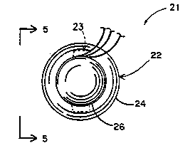
Figures 4-11 illustrate a disposable, self-adhering
finger-cot probe 21 configured in accordance with a preferred
embodiment of the present invention. The finger-cot probe 21
is designed to reduce motion artifact errors in the monitoring
signal, as well as to reduce possible noise artifact signals
from spurious light (i.e., light from sources other than the
sensor 23).
With reference to Figure 4, the present finger-cot
probe 21 principally comprises a compact finger cot 22 and a
sensor 23. The finger cot 22 is configured to be quickly and
easily attached to and Ye".oved from a patient' 9 finger or
other digit without the need of adhesives or other
uncomfortable securing means, æuch as those common to
conventional oximeter probes. It also is understood that the
present probe 21 may be used in other blood-perfused body
locations in addition to a digit.
In the pre-application configuration illustrated in
Figures 4-7, the finger cot 22 includes an open proximal
W095/0~58 C A 2 1 6 6 0 6 9 PCT~S94/0~
-10--
end 24 that is rolled upon itself and a closed distal end 26.
The cloced distal end 26 of finger cot 22 ha~ a cup-shaped
configuration to receive the tip of a patient' 8 finger 28.
The finger cot 22 desirably is formed from a thin elastic
sheath that preferably is opaque to ambient light, and more
preferably is opaque to spurious light of a wavelength within
the range detectable by the sensor 23.
With the patient's finger 28 located at the cup-shaped
distal end 26 (as illustrated in Figure 7), the rolled
10 prox;mAl end 24 is pulled prox;~lly and unrolled
longitudinally over the patient's finger 28, so as to form a
generally tubular sleeve. The sleeve of the prox;m~l end 24
desirably surrounds the circumference of the finger 28 to form
a relatively close fit without fixedly attaching the sensor 23
15 to the tissue and without significantly compressing the
tissue. Thus, the size (circumferentially and longit-l~;n~lly)
of the finger cot 22 should be configured in various sizes to
fit a variety of sizes of fingers or other digits, depending
upon the age, size, and maturity of the patient.
With reference to Figure 8, the finger cot 22 may include
a series of holes 30 through the sheathing material to
increase air flow to the finger 28 for the purpose of
maximizing comfort.
When the proximal end 28 is pulled proximally so as to be
25 fully unrolled along the patient's finger 28, the finger cot
22 terminates at a relatively thick peripheral cuff 32. The
inner diameter of the cuff 32 is slightly smaller than that of
the sleeve so as to apply sufficient pressure to enhance the
self-attachment of finger cot 22 to the finger 28 without
30 adhesive or other securing means.
With reference to Figure 9, the sensor 23 is shown with
the finger cot 22 unrolled and positioned on the tip of a
patient's finger 28. The sensor 23 includes an optical source
34 and an optical detector 36 arranged in spaced optical
35 alignment with one another at the distal end 26 of the finger
cot 22 to transilluminate the patient's finger 28 from
opposite sides. In the illustrated embodiment, the optical
~0-95,0~58 C A 2 1 6 6 0 6 9 PCT~S94/07866
-11 -
source 34 preferably is a pair of light emitting diodes
(LEDs), only one of which is shown in Figure 9.
Both the optical source 34 and the optical detector 36
are recessed within respective cavities 38, 40 fo~med in the
finger cot 22 to prevent contact between the tissue of the
patient's finger and the optical source 34 and the optical
detector 36. The patient's tissue, when slightly compressed
by the finger cot distal end 26, may be received within either
cavity 38, 40, but the tissue does not directly contact the
optical source 34 or the optical detector 36 because of the
recess depth.
An optional viscous coupling medium, such as an oil or
gel, which has an index of refraction which corresponds to
that of the patient's skin, desirably fills the cavities 38,
to couple the optical source 34 and the optical detector
36 to the patient's tissue. The optical coupling medium
rem~;nC in contact with the tissue as the patient moves his or
her finger. This ".ove...ent, however, does not move the optical
source 34 and sensor 36, and therefore the movement of the
finger does not substantially alter the optical coupling or
the path length between the source 34 and the detector 36.
As seen in Figure 9, electrically conductive wires 41, 42
are connected to the LEDs which form the optical source 34.
Another electrically conductive wire 44 is connected to the
optical detector 36. The wires 41, 42, 44 from the optical
source 34 and the detector 36 extend longitl~;n~lly through
the pro~;m~l end 24 of the finger cot 22 to be aligned side by
side one another and surrounded by an electrically insulating
outer protective casing or sleeve 46 (see Figure 8) as the
wires exit the finger cot 22. The outer sleeve 46 carries the
wires 41, 42, 44 to a suitable controller and signal
processing means (not shown), which is briefly described
below.
Figure 10 shows a finger cot 22a that is a modified form
of the finger cot 22 illustrated in Figures 4-9. For ease of
discussion, like reference numerals with an "a" suffix have
been used to indicate like components of the two embodiments.
~ W095/0~58 C A 2 1 6 6 0 6 9 PCT~S94/0~66
-12-
While the proximal and distal ends 24a, 26a of the finger cot
22a are of uniform thickness, the distal end 26a of finger cot
22a is thicker than the prox;m~l end 24a of the finger cot
22a. This variation in thickness has been found to make the
finger cot 22a easier to roll into the compact, pre-
application configuration illustrated in Figures 4 and 5.
Figures 11-15 show a finger-cot probe 50 according to a
further embodiment of the present invention. The probe 50
includes a generally planar protective backing or carrier 52
which i8 preferably formed from a flexible material (e.g.,
plastic). A finger cot 54 is attached to the backing 52 at
approximately the midpoint of the protective backing 52.
With reference to Figure 12, the finger cot 54, in its
pre-application configuration, includes an open proximal end
56 that is rolled upon itself and a closed, cup-shaped distal
end 58. As represented by phantom lines in Figures 12 and 13,
the proximal end 56 is rolled so as to lie inside the finger
cot 54.
The finger cot 54 desirably is manufactured from a thin,
elastic sheath that is generally opaque to ambient light, and
more preferably is opaque to spurious light of a wavelength
within the range detected by the sensor 57.
The finger cot 54 can be either removably or permanently
secured to the protective backing 52. Although either form of
2~5 attachment interconnects the finger cot 54 and the backing 52
equally well, it is preferred that the finger cot 54 be
separately attached to the protective backing 52 so that the
sensor 57 and backing 52 of the probe 50 can be reused, as
discussed below.
Figure 12 illustrates the finger cot 54 permanently
attached to the protective backing 52 by means of a pin 60.
The pin 60 has a narrow body, a relatively wide head 62 at one
end of the body, and a pair of flexible legs or ties 64 at the
opposite end of the body. The pin 60 extends through both the
protective backing 52 and the distal end 58 of finger cot 54.
The pin head 62 connects the finger cot 54 to one side of the
backing 52 with the flexible legs 64 projecting beyond from
C A 2 1 6 6 0 6 9 PCT~S94/07866
-13-
the opposite side of the backing 52. The legs 64 are bent
against the backing 52 and secured (e.g., sewn) to the backing
52 for reliably securing the finger cot 54 to the backing 52.
In this position, the legs 64 also serVe as a tar~et toward
5 which the patient's finger is aimed when the finger cot 54 is
applied, as shown in Figure 13 and discussed below.
Figure 16 illustrates the finger cot 54 separately
attached to the protective backing 52 by means of a releasable
connector 65. In the illustrated e~oA;~nt~ the removable
connector 65 deQirably is a col~ve~,tional snap-type connector;
however, it is understood that other types of Le,..ovable
connectors, such as, for example, buttons, hook-and-loop
fasteners (i.e., VELCRO~), pins, clips and like detachable
couplers could be used equally as well. The snap 65 includes
15 a receptacle element 67 attached to the protective backing 52
and a stud element 69 attached to the finger cot 54. Of
course, the orientation of these snap connector co..l~o~lents 67,
69 could be reversed.
The releasable attachment between the finger cot 54 and
the probe backing 52 allows the sensor portion (i.e., the
backing 52 and the optical sensor 57) of the probe 50 to be
reused, thus reducing the overall cost of the probe 50. One
sensor 57 may be used several times, where the cot 54 is
replaced after each use.
While the optical source 34 and detector 36 of the
finger-cot probe of Figures 4-10 were included as an integral
part of the finger cot 22, the finger-cot probe 50 of the
present embodiment (be~t seen in Figure 12) includes an
optical source 66 (e.g., a pair of LEDs) and an optical
detector 68 which are separated from the finger cot 54. As
seen in Figures 11 and 12, the optical source 66 and the
optical detector 68 are retained at opposite ends of the
protective backing 52. The optical source 66 and detector 68
are recessed within respective cavities 70, 72 to prevent
contact with the patient's tissue. In this manner, as
discussed above, the optical coupling and path length between
the optical source 66 and detector 68 are preserved in the
-~O9S/0~58 C A 2 1 6 6 0 6 9 PCT~S94iO7~66
-14-
event that the patient's finger is moved and/or the tissue is
compres~ed during monitoring.
With reference to Figure 12, the protective backing 52 of
probe 50 may be formed by a top layer 53 and a bottom layer 55
of flexible material to facilitate the formation of the
cavities 70 and 72. The optical source 66 and detector 68 are
carried by the bottom layer 55, while the cavities 70, 72 are
formed in the top layer 53 in axial alignment with the source
66 and the detector 68. Electrically conductive wires (not
shown) extend through the finger-cot probe 50 at the interface
between the top layer 53 and the bottom layer 55 and connect
the source 66 and the detector 68 to a conventional controller
and signal processor (not shown). As seen in Figure 15, an
electrically insulating outer protective casing or sleeve 78
desirably surrounds the wires as they exit the probe 50.
As seen in Figure 11, the sensor source 66 and the sensor
detector 68 are arranged on the protective backing 52 so as to
be an equal distance from the center or midpoint 80 of the
protective backing 52 (i.e., the point at which the finger cot
54 is attached). In the illus~rative embodiment, the distance
between the source 66 or the detector 68 and the midpoint 80
is less than the distance between the sensor source 66 or the
sensor detector 68 and a longitudinal end 81 of the protective
backing 52. Both the sensor 66 and detector 68 are exposed on
the medial side of the protective backing 52 which is
~ juxtaposed against the tissue to be analyzed, as discussed
- below.
Figures 13-15 illustrate the steps for applying the
finger-cot probe 50 to a patient's finger 74 from the pre-
application rolled configuration, shown in Figure 13, to theunrolled, extended configuration, shown in Figure 15. The
patient's finger tip 79 is first placed at the midpoint 80 of
the backing 52. For this purpose, the backing 52 preferably
includes a target located at the midpoint 80. The target may
be the extended legs 64 of the pin 60 or may be indicia marked
on the medial side of the backing 52 as well. In the
illustrated embodiment, the patient's finger tip 79 is placed
- W095~0~5& C A 2 1 6 6 0 6 9 P~T~S~4iO78~6
on the target formed at the i~tersection of the flexible legs
64 of the pin 60 opposite the unrolled distal end of finger
cot 54. A small amount of adhesive can be applied at the
midpoint 80 (e.g., applied to the legs 64) to hold_the finger
74 against the midpoint 80 during application of the finger
cot 54. The unrolled pro~;r~l end 56 of finger cot 54 is then
pulled toward the finger 74 and unrolled over the cup-shaped
distal end 58 of the finger cot 54. The distal end 58 is
inverted in this process to,surround the patient's finger tip
79. The continued rearward ..o~e-..ent of the pro~ l end 56
unrolls the sleeve over the protective backing 52. The sleeve
urges the opposite medial sides of the backing 52 toward each
other and against the respective sides of the finger 74.
In the fully unrolled and applied condition, as
illustrated in Figure 15, the protective backing S2 is bent
and retained around the finger 74 in a manner that the optical
source 66 and the optical detector 68 are held in spaced
optical alignment with each other on opposite sides of the
finger 74 so that the finger may be transilluminated. The
fully unrolled finger cot 54 terminates at a relatively thick
peripheral cuff 76, which applies sufficient pressure to hold
the finger cot 54 to the finger 74.
The optical detectors 36, 68 used with the above-
described finger-cot probes 21, 50 are responsive to light
absorption from the transillumination of the patient's muscle
tissue. The output signals provided by the detector 36, 68
can be processed for the purpose of enabling health care
providers to analyze the patient's blood by noninvasively
calculating the concentration of blood constituents. The
output signals from the detectors 36, 68 also may be converted
before processing, as known in the art.
In the illustrated embodiments, the output signals
derived from the optical detectors 36, 68 can be used to
provide a reliable indication of the percentage saturation of
oxygen (i.e., the oxygenation level) within the patient's
blood. For this purpose, a pulse oximeter (not shown)
energizes the optical source 66 to emit two signals that have
W095l0~58 C A 2 1 6 6 0 6 ~ PCT~S94/078~6
-16-
different wavelengths, one of which is typically red and the
other of which is typically infrared. The two signals
alternately pass through the patient' 8 fingertip 79. The
signals are measured by the optical detector 68 which in turn
produces two output signals. The pulse oximeter processes the
two output signals from the detector 68 to determine the
amount of oxygen available to the body. This information is
evaluated to derive the saturation of oxygenated hemoglobin in
the blood comprising both oxygenated and deoxygenated
hemoglobin.
One pulse oximeter, which is especially suitable for use
with the present finger-cot probes 21, 50, is described in co-
pending U.S. Patent Application Serial No. 07/672,890, filed
March 21, 1991, and assigned to the Assignee of this patent
application. The teachings of Patent Application Serial No.
672,890 relating to pulse oximetry are herein incorporated by
reference. It is to be understood, however, that this
oximeter is given for purposes of example only, and other
types of pulse oximeters may be used as well for processing
the output signals from the optical detectors.
Once monitoring is complete or is no longer required, a
nurse or like health-care provider removes the probe 50. The
nurse rolls the cuff 76 towards the end 79 of the finger 74 to
expose the protective backing 52. The nurse desirably rolls
the finger cot 54 completely off the end 79 of the patient's
~ finger 74 to expose the interconnection between the finger cot
54 and the backing 52. Where the cot 54 and backing 52 are
releasably attached, the nurse may unsnap or otherwise
disconnect the finger cot 54 from the backing 52. The finger
cot 54 can then be disposed, and the sensor can be sterilized
and later reused with a new finger cot 54.
It will be apparent that while preferred embodiments of
the invention have been shown and described, various
modifications and changes may be made without departing from
the true spirit and scope of the invention. For example,
although the finger-cot probes herein have been described as
having particular use with a patient's finger, it is expressly
_~075/~58 C A 2 1 6 6 0 6 9 PCT~S94iO7866
. -17-
understood that the teachings of this invention are also
applicable to any other human digit or suitable palpable
tissue area. Additionally, while the optical detectors herein
have been described as being responsive to the
transillumination of human tissue, it also is understood that
the optical detectors may be suitably located to be responsive
to transreflectance, as well. Accordingly, the scope of the
invention is intended to be defined only by the claims that
follow.