Note : Les descriptions sont présentées dans la langue officielle dans laquelle elles ont été soumises.
WO 95/24500 r~ 154
2~
INHIBITION OF PROTEASE ACTIVITY OF
HUMAN WHOLE BLOOD CELL LYSATES
Field of the Invention
The present invention relates generally to
inhibition of protease activity in biological fluids,
and more specif ically to a method of inhibiting
proteolytic degradation of Mx protein in human whole
blood cell lysates, and employing an Mx protein asE;ay as
the method that is indicative of the efficacy of
interf eron therapy .
B~ r ~1 of the Invention
Proteolytic degradation i8 a naturally occurring
process in all biological kin~l, . Proteolytic
degradation also complicates scientific investigation if
one wants to examine the undegraded level of proteins.
~etDrmin~tion of intra~ llAr protein or membrane
proteins is particularly complicated by proteolytic
degradation, as the cell lysing process also releases
proteases. A variety of protea-~e inhibitors exist for
inhibition of proteolytic degradation, and such
conventional inhibitors are known to those of ordinary
skill in the art. However, when the effectiveness of
known protease inhibitors is not sufficient to halt the
proteolytic degradation of a protein of interest, or
addition of these inhibitors only accelerated
proteolytic degradation, one would have to find an
W095l24s00 r~ . 154
21~4~5~ ~
alternative way to arrest this problem.
An example of the type of investigation that is
complicated by proteolytic degradation is the accurate
r'-~tPr7~7;nAtion of protein in biological fluid. Such
protein determination may also be useful to assess the
cl ;n;cAl relevance of therapy via determination of the
protein specif ically induced by the species of interest .
For example, it is important to evaluate the
clinical ef f icacy of interf eron therapy, which is both
costly and increasingly popular in the treatment of such
conditions as hemangiomas in children, genetically
pr^~-7~posed multiple sclerosis, autoimmune d;~ZP~ R,
certain types of cancer, a~nd AIDS. Assaying the
circulating level of interferon is technically
difficult. However, by assaying an intrAc~ 71 Ar
protein called Mx protein induced specifically by
interferon, the efficacy of interferon therapy may be
s~d. In a paper entitled, "A Whole Blood
T ~A~ Ay for the Interferon-Tnd~lc;hle Human Mx
Protein", by Towbin, et al., Journal of Interferon
Research, 12, 67 (1992), the authors describe an assay
pr~ceduLe for Mx protein in whole blood cell lysates
using an enzyme ; - - C~Ay.
Although strides in lnterf eron research in general
and Mx protein investigation in particular have been
made, it remains a goal to determine the u.. _ ; ~ed
level of Mx protein by minimizing proteolytic
degradation of Mx protein in evaluating the new
application of interferon therapy.
Accordingly, it is an object of the present
invention to provide a method of inhibiting proteolytic
degradation of an intracellular protein, i.e., .~x
WO95t24500 r. l,. `t~_154
21842~5~
protein in cell lysates. It is still another object of
the invention to provide an artif icial matrix solution
whereby an intracellular protein can be kept stable
against proteolytic degradation at a t~ c.Lu.~ at or
below 4 C f or at least three weeks .
of the Invention
The foregoing and other objects and advantages of
the invention are achieved by providing a method of
inhibiting proteolytic degradation of a thPr~ y-stable
intrac~ l Ar protein in cell lysates. The method
involves forming a solution containing one or more
denaturing agents and unknown proteases that degrade the
intr~ r protein, and heating the solution at a
t~ ~ ~ILUL_ and for a period of time sufficient to
denature the protease.
The solution may be defined as detergent lysed
whole blood cells. The solution may be defined in part
by a synthetic matriY mimicking blood cell lysates as
well. When the solution contains the intracellular
protein such as in whole blood lysates, the heating step
is carried out at conditions which do not destroy it.
When the solution is free of intracellular protein, such
as in a synthetic matrix simulating cell lysates,
harsher conditions may be applied until all of the
protease activity is deciLL~,y-~d.
The present invention also provides an indirect
method of ~lPt~r"l;nin7 interferon in patient blood. The
method involves heating a sample in the pre6ence of
denaturing agents, in order to denature 1 or more
unknown proteases from cell lysates that degrade the
intr~ r protein of 1nterest , e . g ., Mx protein .
Wo 95/24500 P~ 154
218~231
Heat i8 applied at a level and for a period of time
suf f icient to denature the proteases, but not to
denature the intracellular protein. The intrAr~ ll Ar
protein is then detormi nPA .
~et-~-rm;nAtion may be made by way of an assay, in a
manner to detect the presence of the protein induced by
interferon, thus indirectly A~et~rm;n;ng the biological
effectiveness of the interferon therapy. Such an assay
may involve the steps of providing a binding partner of
the intracellular protein to a solid phase capture
antibody, allowing the binding partner to capture the
intracellular protein by contacting the solid phase with
the solution, and coupling a second binding partner of
the intrAr~ lAr protein to the intrArplllllAr protein.
The second binding partner carries a rh~m; lllm;nPccc-nt
label, which may be detected by a lllm;n~ ter. The
courl;ng steps may be _ ' ;nPA~ in any order.
The present invention also provides an artif icial
matrix which is made to be protease-free.
IntrAr~lllllAr Mx protein remains stable in this
~rtificial protein solution at a temperature of 4C for
at least three weeks. According to one aspect of the
invention, the solution ;nrlllA-~c whole blood cell
lysates. According to another aspect, the solution
;nrlllA~ a synthetic matrix m;m;rl-;ng whole blood cell
lysates .
Other advantages, novel features and objects of the
invention will become ~ aLtllL from the following
A~tA~le~A~ description of the invention, in .~u~jull.Lion
3 0 with the z. ~ - nying ~ igures .
W0 95124500 1 .~ 54
218~51
8rief Descri7~tion of the Drawinqs
In the drawings:
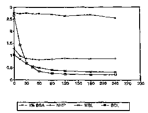
Fig. 1 i6 a graphic illustration of comparative
degradation of Mx protein in normal human plasma, whole
blood lysate, packed blood cell lysate, and in
protease-free bovine serum albumin, in units of O.D. at
405 nm vs length of incubation at 37C in minutes;
Fig. 2 is a ctandard curve for Mx antigen in a
chemilllmi n~cc~nt; - - y according to the present
invention, derived from solutions in which proteolytic
degradation of Mx had been inhibited in accordance with
a method of the present invention, in solutions where
the hematocrit is 15, 30, 45 and 70% in units of
~h~ n~7c of relative light units (7~LU) vs. Mx
cv.,. ~ L ~tion in units of ng/ml;
Fig. 3 is a graphic illustration of elimination of
proteolytic degradation of Mx protein in synthetic
matrices in accvLdc~Dce with the present invention, via
heat inactivation after heating in 2M urea and 0.1% SDS,
in synthetic matrices having hemotocrits of 15, 30, 45
~nd 7096, in units of thollCAnr7c of relative light unit6
tRLU) V5. time (in minutes) at 37C;
Fig. 4 is a graphic illustration of elimination of
proteolytic degradation of Mx protein in whole blood
lysates in accordance with the present invention, via
heating in urea at 56C for 30 minutes, in units of
thr~7lcAnr7c of relative light units (~LU) vs. time (in
minutes) at 37C; and
w0 ss/24soo r_l,~ l54
2i8~2~1 ~
Fig. 5 i8 a graphic illustration of a typical
do~c e.~ ,sive induction of Mx protein after interferon
~dministration to three human patients, determined in
;d,ll~ce with an assay of the present invention, using
8 x 10⁶ units/day of IFN (B/D), showing the ré~ e on
various days in units of l~x cull. e,.~ ion (in ng/ml) in
whole blood.
Detailed Descri~tion of tlle Preferred E ' ';-
The present inventioll provides a method of
inhibiting proteolytic degradation of a t-h~rr= 1 1 y-stable
intrAc~ 1 Ar protein by treating cell lysates with heat
and 1 or more denaturing agents at a t~ ~UL~ and for
a period of time sufficiellt to del,a~uLe the protease.
As protease activity is inhibited accnr~in~ to the
method of the invention, an assay to determine the
~Lè8ell~ ê and/or concentra~ion of the intracellular
protein may be reliably peL ru. ' . Provision of an
assay sample that is stable against proteolytic
degradation is advantageous when the sample must be
stored for any period of time prior to performance of
the assay. Additionally, an artificial mixture of
protein solution stable against proteolytic degradation
of the protein of interest may be provided and used as a
diluent for an antigen standard, controls, calibrators,
or the 1 ike .
Inhibition of the proteolytic degradation of any of
a wide variety of thermally stable proteins is provided
in accordance with the invention. As used herein, the
term "thermally-stable" is meant to define the stability
of the protein of interest at a t~ UL~ and for a
period of time n~ y to degrade the protease
W0 951~4500 P~ 154
21842~1
responsible for degradation of the protein.
ThPrr~l ly-stable protein6 include, but are not limited
to, some membrane proteins (e.g., car~--inr ' yullic
antigen) and intr~ r proteins, which include
nuclear proteins (e.g., murine Mx protein) and
cytoplasmic proteins te.g., human Mx protein, heat-shock
proteins and cytoskeletal protein6).
According to one aspect of the invention, a method
of inhibiting proteolytic degradation of an
intrlc~ll-ll~r protein (e.g., Mx protein) induced by
interferon i5 provided. Such a method makes feasible
the reliable, repro~ ih~ ~ determination of Mx protein,
thus dPt~rrninin~ the ~1 inic:~l efficacy of interferon
therapy. Approximately 30 different proteins are known
to be induced by interferon. However, only
2,5-oligo-(A) ' synthetase, p68 kinase, and Mx protein
are known to mediate anti-viral actions of interferon,
and determination of one or more of these proteins in
accordance with the invention is thus highly relevant to
evaluation of interferon therapy.
Determination of Mx protein is particularly
preferred for the following reasons: Mx is promptly
induced (2 hrs. ) after interferon treatment, and reaches
maximum levels in a relatively short period of time
(approximately 36 hours). ~ r induction of Mx
protein is not subject to feedback inhibition even at
high doses of interferon therapy. Additionally, the
biological half-life of Mx protein is relatively long
(Tl~2 is 3.5-5 days)- Thus 20-30% of the initial Mx
protein level remains even at 2 weeks following the
cessation of interf eron therapy . Thus, due to its long
half-life, Mx protein i~ a good indicator of interferon
W095l24500 r~l,~; 154
218~2~1
effectiveness. Fur~h~ ~e, the fact that it is easily
detectable make6 it a rapidly ;nAucihlp~ sensitive and
reliable indicator of interferon action in a wide range
of interf eron doses .
According to the method of the invention, a
solution containing a denaturing salt, detergents, and
proteases that degrade(s) the intracellular protein is
heated at a t~ a-u~e and for a period of time
auf f icient to denature the protease . The 601ution may
be formed by lysing cells, for example human whole blood
cells or cultured cells.
The solution also may be formed by creating an
artif icial matrix that mimics blood . Many artif icial
matrices mimicking whole blood are suitable for use in
accordance with the present invention. Preferably, an
artif icial matrix formulated in accordance with the
present invention and, --^A of protease-free bovine
serum albumin and crystalline bovine hemoglobin is
employed .
A wide variety of denaturing agents are known to
those of ordinary skill in the art, and may be used
according to the method of the invention, ;nr~l-A;n~, but
not limited to, urea and gll:ni~inP hydrochloride, which
are pref erred denaturing agents . Proteases to be
inhibited in accoLdcl~lce with the present invention
include virtually all those known to exist in white
blood cells, ;n lllA;n~ Cathepsin G, elastase,
metalloproteases, etc.
When the method of the invention has been carried
out, that is, when a solution has been treated so as to
denature a protease that degrades an intracellular
- protein, that solution th n may be spiked with the
WO 9S124500 r ~ . IS4
21842~1
intrAr~ r protein without risk of proteolytic
degradation. Such a solution may serve as a diluent for
an antigen standard in an assay, and may contain whole
blood cell lysates, or a synthetic matrix mimicking
whole blood cell lysates. According to a preferred
of the present invention, such a solution
remains stable at a t~ ~uLè of 4C for at least 3
weeks .
According to another: 'i- t, the solution may
serve as a standard in an assay, or may comprise a
sample in an assay, for example, a human whole blood
sample. When the solution contains whole blood cells or
cultured cells, a lysing agent is advantageously
i nrl~ cl in the solution prior to heating the solution
in the presence of the den~uLing agents. Thus, cells
may be lysed and the protease denatured in a single
step.
~ variety of lysing agents are suitable for use in
accordance with the present invention, including but not
limited to non-ionic detergents such as ' i ~p~rSe and
polydisperse, h~ - J ?0~ and hetélo~el.~uus
poly~ ye~lylenes. Preferred lysing agents include
Tergitol NP-40 (available from Union Carbide) or Triton
X-100 (available from Rohm and Haas), which should be
added in an amount such that its ~ e--LLation, when the
sample is heated in the presence of other denaturing
agents (such as urea and ~l~ni~lin-- hydrochloride),
should be sufficient to della-uLe the protease.
Although it is not important whether the cells are
lysed before or at the same time the protein is
solublized, it is important that the non-ionic detergent
used to lyse the cells be inrl~ in the denaturing
.
WO95/24500 1. 1~ ~ 154
2i842~1 ~
medium, since the non-ionic detergent aids the
d~ L~L ing process. In the case where lysing is not
required (e.g., when a synthetic matrix is used), the
non-ionic detergent still should be added along with 1
or more other denaturants (e.g., urea or g~ n;~inP
hydrochloride) and the anionic detergent (e.g., SDS) to
assure that denaturation occurs . ~rPn~l 1 n~ on the cell
involved, even other lysing agents may be z.l.~L~Liate.
For example, in the case of red blood cells, water is
sufficient for lysing the cell. However, when the cell
is lysed using an agent aside from a nonionic detergent,
the nonionic detelgel~t must still be used for denaturing
the protease.
Although SDS combined with heat has been used in
the past to mask the charge of the native conf iguration
of proteins, thus frequently resulting in their
denaturation (see Laemmli, Nature 227:680 (1970)), the
use of SDS, del~atuL-Int (e.g., urea) and heat in a
controlled fashion in the instant invention results only
in the destruction of proteases, without denaturing the
protein of interest. The solution is heated in the
presence of a denaturing agent at a t~ -- CltUL~ and for
a period of time suf f icient to denature the protease .
The t~ C~LUL~ and time of heating 6hould be SPIPrtp~
as to sufficiently dendLuL~ the protease, and when the
solution contains the intrA~Pl lulAr protein, the
temperature and time should be selected so as not to
denature the intrAePll~llAr protein. A temperature of no
less than 50C should be selected, and the solution
should be heated for at least 60 seconds. If the
intr~rPlllllAr protein is present in the solution, the
solution should be heated at a temperature of from about
WO 95124500 r~ 154
218~2~1
50C to about 60C for a period of time of 15-30
minutes. Ir the solution contains only the artificial
matrix (i.e., contains protease c~nt~minAtion but does
not yet contain the protein of interest), harsher
conditions may be employed before the protein of
interest is added to the matrix. For example, such a
solution may be heated at a t~ tUL ~ of from about
50C to about 100C for a period of time of from about 1
minute to 1 hour or more, preferably at a t~ C~LUL~ of
about 56C for about 1 hour (see Manwaring, W.H. (1906)
on the destruction of complement by heat, TR. Chicago
Path Soc. 6:425).
When a solution contains the intracp~ r protein,
the solution conditions should be kept within a range
compatible with survival of such a protein.
Specif ically, the pH of the solution should be kept
within a range of 7.0-8.0, and the ionic ~LLe~ Lll of the
solution should be kept at a levQl not more than about 4
M.
As noted above, a solution in accordance with the
present invention that contains a thPrr-l ly-stable
intrA~Pll~ r protein (e.g., Mx protein) and that is
free of a protease that degrades the Mx protein
facilitates a reliable and reproducible assay to
~lptprminp the intracPl l~ r Mx protein. As used herein,
the term, ~ PtPrminP" is meant to define detection of
the intracellular protein at the limit of the assay, or
determination of the ~ tion in solution of the
intracellular protein. Many typ~s of assays are known
in the art which may be modified so as to be employed in
a determination in accordance with the present
invention. General assay types include, for example,
11
wo 95/24500 P~ll l 154
2~ ~
direct, indirect, competitive and sandwich-type
het~:~;ocJP~ c or h~ assays such as those
described in U. S. Patent No. 5, 252, 459, issued October
12, 1993 to Tarcha et. al. and incorporated herein by
5 ref erence .
When human blood is to be assayed for Mx protein in
accordance with a particularly preferred ~mhorl;- L of
the present invention, an assay method may be carried
out as follows: A solution containing the proteins
derived from the whole blood cells is formed by, as
described above, '-;n;n~ the lysing agent, 1 or more
denaturing agents (preferably urea) plus sodium dodecyl
sulfate (SDS), the detergent fielected to delldLuLa the
protease and solllhi 1 i 7e the Mx protein. FUrth~ ~, it
is important that the protease be sufficiently dilute so
that the denaturants are effective. The solution is
then heated at a ~ ilLUL~ of from about 50C to about
60OC for a period of time of about 15-30 min, and Mx
protein is then det~rmi n~d.
Variations of the present invention are possible
for a variety of proteins so long as the t~ ~tu~ ~ at
which the proteases are destroyed is ~t~rminP~l and
found to be lower than the t~ ~Lur ~ at which the
analyte is denaLu-~=d. Furthermore, the proper lysing
agent, generally a non-ionic detergent, and a proper
solublizing/denaturing agent, generally an ionic
surfactant, such as the anionic surfactant sodium
dodecyl sulfate, and the d~.aLuLc~ salts (e.g., urea or
gll~n;-l;nc~) must be used. If the sample o~tained is
already lysed, there is ~o need to include the non-ionic
(lysing) agent in the system.
Fu~ther variations o the present invention are
wo ssl24soo r ~,l/LI~ 154
21842~i1
possible. For example, the sample suspected of
containing interferon may be a whole blood sample,
packed blood cells, tissue cultured cells, a solution
containing lysed whole blood cells, synthetic matrices
to which Mx protein i5 added to simulate whole blood
lysates or the like. Other variations will become
L~IIL to those with ordinary skill in the art.
The following examples are ;ntr~nrir~d to illustrate
the benef its of the present invention, but do not
exemplify the full scope of the invention. For example,
although a specific denaturing agent, solubilizing
detergent and lysing agent are exemplified, a variety of
such agents may be employed. While the determination of
Mx protein and CuLL~ ;nrJ pI~aLc.tion of standard and
control solutions containing Mx protein are exemplified,
a variety of thPrrol ly-stable proteins, ;n~ ;n~ but
not limited to those induced by interferon or other
cytokines or other biological r~D~o~.se modif iers, are
understood to be within the scope of the present
invention. These and other modifications and their
equivalents are understood to be within the scope of the
present invention.
~amules
Naterials and Methods
The protease inhibitors phenylmethylsulfonyl
fluoride (PMSF), aprotonin, antipain, ~.l-y L~ltin,
leupeptin, pepstatin A, tosyl-lysine chloromethyl ketone
tTLCK), tosyl-phenylAlAn;ne chloL~ yl ketone (TPCR),
epsilon-amino ~ Loic acid (EAQ), elastinal, and E-64
were purchased from Sigma ~'hr~;cAl Co. (St. Louis, MO) .
13
WO 95/24500 ~ C4
21~4251
A non-ionic detergent, NP-40 (used to solubilize
leukocytes), 2 - ua~1 oethanol (2-ME), protease-free
bovine serum albumin (BSA-PF), radioi - R5~A~y
(RIA)-gradQ BSA (BSA-RIA) and crystalline bovine
hemoglobin (bHB) were also purchased from sigma Chemical
Co. Sodium dodecyl sulfate (SDS) was purchased from
BioRad Laboratories (Hercules, CA). Some proteases,
i.e. elastases (porcine pàlluL~as and human leukocytes),
cathepsin G (human leukocytes) were also Sigma
rhPmirAlR. Crystalline trypsin was obtained from
Worthington BiorhPmicAlR (Freehold, NJ), and PEFAbloc~,
an analog of phenylsulfonyl fluoride, was obtained from
Pentapharm AG (Basle, Switzerland). All other chemicals
were reagent grade rhPmirAlR from MAllinkrodt (2aris,
KY) . DEAE ~PrhA~PY A25 alld 12 . 5% Phast gel were
obtained from Pharmacia Biotech Inc. (Piscataway, NJ).
Goat serum from Ventrex Laboratory (Portland, ME) was
heat-inactivated and f iltered through 0 . 2 um M; 11 i r~re
filter prior to use. Immobilized Protein-A Affinity
PaklM was purchased from Pierce rhPmirAl~: Co. (Rockford,
IL) and used as described by the manufacturer.
~-1 le 1
Investi~ation of Proteolytic Deqradation of MY
Protein
An investigation was made of degradation of Mx
protein in normal human plasma (NHP), whole blood
lysates (WBL), packed blood cell lysates ( in the absence
of plasma; BCL), in synthetic matrices mimirking whole
blood lysates, and in protease-free controls.
Mx protein in cultur d cell lines (i.e. WISH, CH0,
WO 9S124500 1 ~ .'1 . 154
~ 218~2~1
3T3) was induced with interferon (B/D) (Ciba-Geigy,
Basle, Switzerland), and the cells were lysed and stored
frozen at -80C until used. An ELISA assay d LL.lted
that ~ lo~J~ uc Mx protein present in frozen cell
lyDates exhibited a much lower quantity of
_~active Nx protein after freeze-thaw than an
original fresh sample. In C~IILLC~DL, ~ inAnt Nx
protein ~Luduced in E. Coli, purified to ~ , -;ty and
stored at -80C until used retained 100% of its initial
immunoreactivity upon repeated freezing and thawing.
A known quantity of purif ied rNx protein was spiked
into two different BSA ~.e~,lL~tiOns, i.e. BSA-PF
(protease-free) and BSA-RIA, as well as into lysed whole
blood freshly drawn from a normal healthy volunteer.
Both the whole blood lysate and two dif f erent BSA
preparations contained a lysing agent, specifically
2% tv/v) NP-40 detergent as described by Towbin, et al.,
referenced above. These preparations were further
diluted with a medium containing a denaturing agent,
specifically 2N urea, and a solllhil;~;nq detergent,
specifically 0.1% SDS and a buffer salt, i.e., 50 mM
Tris-HCl (pH 8.0). A final protein c~ tllLL-,tion was
adjusted to 1%. Nx protein was spiked into a synthetic
matrix I ~ ~ ~ of BSA-PF and crystalline bHB .
These samples were incubated at 37C for up to 120
min. Aliquots of samples were removed perio~;c~l ly and
the amount of Nx protein L. ;n;nq in the solution was
correlated with a drop in signal (RLU's) over time as
detc~m;n~ in a chemill~m;n~cc~nt; --~ID'?y
Nx protein spiked into the BSA-PF underwent minimal
degradation during this incubation period. In contrast,
the SSA-IIIA r~pldly d~gr ded VX protein with1= the fir~t
WO 9!i/24500 E.,~ '4
218~251 ~
30 minutes and Mx protein in whole blood lysate was
cont;n~lol~ly degraded Lll-uu~l~ouL a 2-hour incubation
period. Nx protein spiked into a synthetic matrix
- ~e~ of BSA-PF and crystalline bHB wa~; also
5 degraded.
Since Nx protein was m;n;r~l ly degraded in BSA-PF,
we Sllrm; ~ed that the crystalline bE~B must have been the
source of this protease activity. Indeed, synthetic
matrices with increasing cu~lc~llLL~ltions of hemoglobin
exhibited a greater degree of proteolytic degradation of
Nx. Mx protein was degraded faster in packed blood cell
lysates (in the absence of plasma), than in whole blood
lysates .
Fig 1 graphically illustrates results of this
assay, showing significant diminution of Mx protein in
normal human plasma tNHP~, whole blood lysates (WBL) and
in packed blood cell lysates (BCL), compared to a
control (1% BSA). It wa6 clear from this investigation
that Mx protein is subject to proteolytic degradation in
a variety of biolical fluids, importantly blood cell
lysates . I'hese results indicated a def inite need to
eliminate the protease activity in the synthetic
matrices as well as in whole blood lysates before one
could reliably and rc ~uluducibly cletPrm; rP the quantity
of Mx protein in rl;n;nAl samples.
E~aml~le 2
PrPnAration of Whole Blood LYsates
Whole blood lysates from normal healthy volunteers
were prepared by adding 2 % (v/v, final cull~:llL.c.tion)
of NP-40 detergent to freshly drawn blood, collected in
16
WO95/24500 r. ~ . 154
218~2~1
EDTA-or heparin-containing tubes and served as untreated
controls. ~l inic~l samples from r.l;nic~l trials of
Interferon (B/D) were also ~L~=~a~ed in the same way as
the normal control blood lysates and kept frozen at
-80C until used.
Example 3
Formulation of svnthetic matrices for Mx l~Iotein
A series of synthetic matrices which simulate the
whole blood lysates of individuals with various
hematocrits were formed. These synthetic matrices were
,-~ l of BSA-PF and bHB in PBS as follows:
~- ~o~rits
~ 30.0% 45.0% 70.0%
bHB 5g% lO.Og% 15.0g% 23.0g%
BSA 7g% 5 . 5g% 4 . 5g% 2 . 5g%
The purpose of these synthetic matrices was to
investigate the potential effects of variable hemoglobin
content on the signal readout of an Mx assay and to
define the most suitable hemoglobin content to formulate
a synthetic matrix.
r le 4
Development of Assav for Mx Protein Determination
1. Mnr~nclnn:~ 1 an~; ho~ to Mx protein
Two separate monoclonal ant;ho~ , one directed
17
WO 9~/24500 P~ 154
218~
to the C- t~inAl (clone 1302.5.32) and the other to
the N-t~rm;n~l (clone 1302.34.16.2.44) portion of M~c
protein were utilized as capture and detector ant;hoflies
in a sandwich-type; ccay. The cell lines that
produced these - --lnn 1l antihofl;~c were identified as
Hybridoma Mx 1302.5.32 and Hybridoma Mx 1302.34.16.2.44.
These cell lines were depo~ited ln the American Type
Culture Collection (ATCC) Patent Depository (12301
Parklawn Dr., Rockville, Maryland 20852, USA) and given
ATCC numbers ATCC HB-11836 (for Hybridoma Nx 1302.5.32)
and ATCC HB-11837 (for Hybridoma Mx 1302.34.16.2.44).
The deposit was made under the Budapest Treaty. These
antibodies were purified from the mouse ascites fluids
using protein-A Sepharose media and proved to be >95%
pure by densitometric Sr~nninq of the ~`- CC;f~ Blue
stained SDS-PAGE gel (20). Clone 1302.5.32 monoclonal
antibody, directed to the C terminal of Mx protein, was
conjugated to paramagnetic particles (PMP) using the
glutaraldehyde activation method of Whitehead et al. as
tlicc1oc-~fl, by U.S. Patent 4,554,088. The PMP-conjugated
antibody was EUcr~ fl~d at 10 mg/ml in PMP wash buffer,
which contained 0.25% BSA (protease-free), 0.7% bovine
gamma globulin (BGG, Pentex, Miles Scientific,
Naperville, IL), and 0.1% sodium azide in phosphate
buffered saline (PBS) and used as solid-phase capture
antibody. Clone 1302.34.16.2.44 r- Cl~nql antibody,
directed to the N-terminal o~ ~5x protein, was labeled
with acridinium ester using the
N-l~ydlu~y~ 'c-in;~;fl~-activated dimethyl acridinium ester
(DMAE-NHS, Ciba-Corning Diagnostics Corp., Walpole, MA)
at a molar ratio of DNAE: antibody = 20: 1 at room
t~ uLe for 30 min. with o~ a,.~ stirring. The
WO 95~14500 r~ 154
218~2~1
free DMAE and the DMAE-labeled antibody were separated
by chromatography on a DEAE-S~rhA~l~Y A25 column in PBS.
One ml fractions were collected and the labeled antibody
fractions were monitored using an ML2~-I or II
l~ r (Ciba-Corning Diagnostics Corp. Oberlin,
OH). Fractions containing the DMAE-labeled antibody
were pooled, diluted to a final CVI~.G.ll.LatiOn of 10l2
relative l~lm; n~c~ units (RLU) /ml in PBS containing 1%
BSA-PF, 2% NP-40 and 0.1~6 sodium azide. Both antibody
pLG~aLIltions were stored at 4C until used.
2. Development of a r`h~m;lllm;n~cc~Pnt assay
Purified L~ -;n~nt Mx-protein (rMx) derived
from the inclusion bodies of E. Coli (See Horisberger,
et al. "cDNA Cloning and Assignment to e11L~ ~ 21 of
IFI-78-k Gene, The Human Equivalent of Murine Mx Gene",
Somatic Cell & Molecular Genetics, 14, 123 (1988) ) was
used as an antigen ~L~I.d~ . The protein content was
-~rm;n~fl both by a BioRad protein assay using a BSA
standard and by quantitative Western blotting of a 2-D
gel as described by Towbin et al. referenced above. The
quantity of Mx protein was conf irmed in a modif ied
version of an ELISA assay originally published by
Towbin, et al. for whole blood lysates. The modified
version utilized a larger sample volume (50ul vs. 20ul
sample) and larger amounts of primary (50ul vs. 40ul)
and secnn~l~ry antibodies (100ul vs. 50ul). The purified
rMA protein served as the antigen standard in the
modif ied ELISA assay. For the chemil~m; n--Rc--nt
Ay of Mx protein, all samples or synthetic
19
WO 9S124500
2184231 ~1
matrices containing a known quantity of Mx protein were
incubated in 12 x 75 mm plastic tubes simult;~nP~lcl y
with DMAE-labeled detector antibody and PMP-conjugated
capture antibody. The incubation period varied from 30
min. to 120 min. at 37C in a water bath. At the end of
each incubation period, the solid pha~-c buu.,d immune
complex was separated with a m-gnot; 7eA separator rack,
Magic RackT~ (Ciba-Corning Diagno6tics Corp., E.
Walpole, MA~ for 3 min. at room t~ u..:. The
unbound antigen or antibo~y was discarded by decanting.
The separated pellets were then rPcllorpnAe~A~ in 1 ml of
deionized water using a Multi-Tube Vortexer (Model 4010,
Corning, NY) and PMP pellets were separated, and the
unbound material was removed as above. These pellets
were washed once more with 1 ml of A~Qj~n;~eA water and
finally rPsllcppnAp-A~ in 0.1 ml of deionized water before
counting in an MLA-I or -II 1 llm; - ~ pr. For the
automated assays, the ACS:180TM (Ciba-Corning
Diagnostics Corp. Oberlin, OH) wa6 used and the data was
analyzed using mathematical algorithms generated by a
statistical program.
3. Preparation of detector antibody with DMAE
label
Using the D~5AE-li~h 1 ;n~ E~L;>ceduL~ described in
the Materials and Methods section, we obtained
DMAE-labeled antibody with a specif ic activity of 7 x
10ll relative luminesce~,~e units (RLUs) per mg of
detector antibody (clone 1302.34.16.2.44), with
lllm;nPcc~,nre determined by a lll-;r Pr and protein
cu~ QI,l.L~tion APtPrm;nPd by a BioRad protein assay. The
WO 95124500 A ~,1/~3
218~2~1
APt~Ctl~r antibody was puriried with the
protein-A-Sepharose media as suggested by the
manufacturer. The DMAE-labeled antibody reacted with
the solid pha3~ bu~.l.d MY protein in a dose-~ , L
manner, similar to the biotinylated antibody used in
ELISA. This result indicates that DMAE-lAhPl ;ng did not
destroy the immunQreactivity of detector antibody to MY
antigen.
4. Preparation of PMP-bound antibody
The capture antibody (clone 1302.5.32) was
conjugated to PMP at a coupling efficiency of 74%,
resulting in 150 mg antibody bound per gm of P~qP. The
PMP-cc ~ Ay~ted antibody reacted in a dose-dprpn~lent
manner with a separate epitope of rMx, which is not
occupied by DMAE-labeled antibody.
5. DevPl :, L of l hPmi 1l7minPccDnt assay for Mx
2 0 protein
A. Effect of the hjorhPmical nature and total
protein cc,l.cc.-LL-tion of matrices on chemilllm;nPFcPnt
signal output
Since whole blood lysates consist mainly of
hemoglobin and serum albumin, the effect of these two
proteins on ~hPmilllminPccPnt signal output was PY~min~d.
The effect of the total protein cul.~el~LL~tion of the
synthetic matrix on ~hPmilllm~nDscpnt signal was also
PY~minPc~. Hemoglobin at the same 1 g~ tw/v)
col.. ~..LLation, exhibited only 1/10 of the
WO 9Sl24500 P~ .54
218~2~ --
rhPmi ll-m;nP~:cPnt signal output ~d to that of 1%
BSA. On the other hand, 2 g% of total protein (1:10
~ lu~ n of goat serum + 15g~ human h ~ hin)
generated only 112 o~ the signal output generated with
1% total protein. Thus, the h;o~hPmir:~l nature of
protein, (i.e. hemoglobin vs. BSA) as well as the
cu,~ L~Ition of total protein both influence the signal
output of the rhPm;ll-mln~cPnt assay.
Fig. 2 grilrhi cs~l ly illustrates that, upon
dilution of the matrices up to 20-fold with a buffer
(e.g., 50 mM Tris-HCl (pl{ 8.0))containing 2M urea + 0.1%
S~S, ~11 four matrices which L~ G~.~L hematocrits
between 15~ and 70% generated similar signal output .
The protease inactivation process works best at this
dilution ~8 well. Therefore this dilution ~LUCedULe was
incvr~uL c.ted as a part of the standard sample
preparation .
B. Effect of detergent ~ UlI~ t:llLL~tiOn on the
level of non-specific binding and signal output
In spite of the 2% (v/v) NP-40 detergent present
in the whole blood lysates or synthetic matrices, the
final concentration of detergent in the as6ay mixture
was low, as solutions were diluted 20-fold with a media
containing 2M urea (~ LUL ing agent) and 0.1% SDS
1llhil~zin~ detergent). Therefore, several
~;ull~ L~tiOns of NP-40 (i.e. 0.5, 1 and 2 %) were
tested in the media containing the detector antibody to
determine whether the co~l. el-LLation of detergent (0.2~)
would be suf f icient to block the non-specif ic binding of
the DMAE-labeled antibody to the solid phase (PMP). We
22
W0 95/24500 2 1 ~ 4 2 ~ 154
found that au..ce.lL.c.tion of NP-40 at 2% (v/v) in the
media containing the acridinium ester labeled antibody
gave the best 6ignal/noise ratio. Below this level of
detergent (0.525% NP-40 in the assay mixture), the level
of 1.~,.. Or,ecific binding was high, particularly at the
lower range of Nx protein aol~. el~L ~tions (below
approximately 4 ng/ml), while the signal output was
lower if the detergent CUII~C IILL ~.tion was above this
range. We also found that including 50 mN 2-NE in the
10 assay mixture elevated the level of nu.. ,~ec;f;r- binding
without anh~n~; n7 the solubility of Nx protein.
Therefore, 2-NE ;n~ rl~d in the original buffer cocktail
to enhance the solubility of Nx protins was deleted in
our denaturing media, hence in the assay mixture.
C. Effect of incubation time on assay
sensitivity level
Since the level of Mx protein in normal healthy
volunteers was at the detection limit of the instant
assay, we ~YAm;n~d whether the sensitivity limit of the
assay could be extended by longer incubation length. As
the length of incubation was increased from 30 min. to 2
hours, the absolute signal output was higher. However,
it did not extend the sensitivity limit nor improve the
precision of the assay at the lower end of Nx antigen
u~ ..LLc.tions. Therefore, a 30 minute incubation
length was chosen.
6. Dilution and ~ecuv~. ~ of Nx protein
In order to ensure that the Mx protein as~ay
23
WO9S/24500 r~,l". c'l IS4
218~251
~I vduced a linear dose-responsive curve in a wide range
of Mx protein vvl.a_.,L-c.tion, Mx protein was serially
diluted in the same assay media and assayed at seven
different Mx avllct llLL~Itions. The average recu~eLy of Nx
protein tested on 3 separate ACS:180 insLL, ~s gave an
average recvve:Ly of 95.9%, with a sensitivity limit of 1
ng/ml, indicating that the assay performance is
cu~ ellLL c.tion-; n~ L .
r le 5
Inh; hition of ProteolYtic Dearadation of a
ThPrr~l lY-Stable profP;n
A known quantity of rP. '; n~nt Mx protein was
spiked into whole blood lysates or synthetic matrices
and incubated at 37C. Aliquots of samples were taken
out perio~ Al ly and kept on ice until assayed for the
residual Mx protein.
Removal of protease activity in the whole blood
lysates or synthetic matrices was PYAmin~l after first
diluting the cell lysates or synthetic matrices with
various volume ratios of a solution containing a
dt.l~ll.uLing agent and a solllhil;~in~ detergent,
specifically 2N urea + O.l(w/v)~6 SDS in 50mN Tris-HCl
buffer solution (pH 8 . 0) . The diluted mixtures were
then subjected to heat treatment at 56C for 30 min. or
90C for 60 sec. in a water bath. Effectiveness of heat
L.~a, L in destroying the protease activity of the
synthetic matrices or whole blood lysates was PyAminpd
after spiking with a known quantity of Nx protein into
the heat-treated media and observing the changes of Nx
protein level upon further incubation at 37C for up to
24
Wo 95~24soo 2 1 8 4 2 ~ ,~ 154
12 0 min .
A buffered solution containing 2N urea + 0.1 ~
SDS + 50 mM Tris-HCl (pH 8 . 0) Was prepared, followed by
heat treatment to investigate the effect on protease
activity. Synthetic matrices or whole blood lysates
were diluted up to 20 fold tv/v) with the above solution
and subjected to heat LLeai L at 90C for 60 sec. or
nt 56C for 30 min. Following the heat LL~ai L,
solutions were further incubated at 37C for 60 min. and
the residual protease activity Was ~c~sed with
aliguots of samples taken out during this incubation
period .
As shown in Fig. 3, Mx protein spiked into the 4
different synthetic matrices, which had been
heat-inactivated in accordance with the present
invention, r~ i n~cl stable for at least 1 hour at 37C,
indicating that the protease activity of the cell
lysates was virtually abolished. Essentially identical
results were obtained by heating at 90C for 60 sec.
As shown in Fig. 4, Mx protein spiked into 6
dif~erent indiViduals~ whole blood ly6ates also ~l ;n~c~
stable at least for 1 hour at 37C following the
heat-inactivation ~JL OCeduL e in accordance with the
present invention.
The inhibition met~od o~ the present invention
was also tested With several commercially available
proteases . i . e. Cathepsin G and elastase from human
leukocytes, and trypsin and elastase type IV from
porcine pancreas. It was determined that the method of
the present invention is also effective in eliminating
the enzymatic activity of the purif ied proteases up to
an equimolar ratio of en yme tE) to sub8trate ts)-
Wo95/24500 1~~ 154
21842~1 0
In order to assess the stability of Mx protein
contained in frozen whole blood lysates, a known
quantity of purif ied rMx ~rotein was spiked into 4
different whole blood lys~tes derived from normal
healthy volunteers and stored at 4C, -20C and -80C.
levels of Mx protein in these whole blood
lysates had been previously det~rm;n~l and found to be
negliqible. An aliquot of stored sample was taken out
every week and immediately diluted with a 20x volume of
2M urea + 0.1% SDS in 50 mM ~riC HCl buffer (pH 8.0) and
heated to 56C for 30 min. in order to m;n;m; 7e further
degradation of Mx protein during assay ~lOc~-lule. Mx
protein kept in whole blood lysates underwent an
appreciable degree of autolysis even at -80C and more
y~ u~ced denL,u.;Lion at 4C (25% destruction in 1
week). In contrast, Mx protein kept in heat-treated
lysates in accordance with the present invention
~, ;n~4 stable both at -80C and at 4C for at least 3
weeks, further .' LL~ting the effectiveness of this
simple ~LO~ ~dUL~ in halting the proteolytic degradation
of Mx protein in cell lysates.
Whole blood lysates were prepared with freshly
drawn blood from normal healthy volunteers and served as
untreated normal controls. Ninety eight samples of
frozen whole blood lysates derived from a ~1 ;n;c~l trial
of interferon (B/D) were tested for Mx protein using the
manual assay. A total of 26 patients with various types
of malignancies had been treated with interferon (B/D)
doses from 2x 10⁶ to 64 x 106 units/day for days 1, 2, 3
3 0 and 7, or 8 or 9 . Group6 of three patients were treated
with each do6e of interferon (B/D) at 2, 4, 8 ,16, 32, 64 and
25 million units per day.
26
WO 95114500 1~ 154
218~2~1
Fig. 5 shows a typical doL~ L Mx
induction L.:-l.ul.se for three patients who were treated
with IFN (B/D) at 8 x lo6 units/day. ~he data was
collected according to the method of the invention.
RYAm~le 6
-ri80n between r-ml~l vs. aut~ cc~v
The maximum incubation time on our automated
assay on the ACS:180 is only 7.5 min., while the length
of incubation in the manual assay is adjustable. To
compare the manual V8. automated assay perfnrr-nr~c~ we
chose to incubate the assay mixture f or 3 0 min . at 3 7C
in the manual assay. The results ~ LL~ted that the
manual assay values are barely higher than those of the
automated assay, considering the longer incubation-time
of the manual assay. The two assays exhibited an
~Yr~ nt linear correlation (R = 0.987).
E le 7
e E~ le: Conventional Protease Tnhlh~tors
At least 14 dif ferent protease inhibitors listed
in the Materials and Methods section including an
elastase inhibitor, elastinal, and cysteine-protease
inhibitor, E-64, failed to block the protease activity
of either the whole blood lysates or the synthetic
matrices. In fact, inclusion of these ~rotease
inhibitors in whole blood lysate exacerbated proteolytic
degradation of Mx protein. Use of a high cull.~l:llLL~tion
of chaotropic salts (i.e. 3M sodium thiocyanate, or
poti~-i=m :hlor1de~, cl-n t=ring ~lt- ~ h 1: 811 ure- or
Wo 95/24~00 . ~1/1 ; ? 154
218~2~1 ~
6M ~ ;n~-Hcl, or C.~ JOZIULe to extreme pHs (pH2 or ll)
all failed to zlrrest the proteolytic degradation o~ Mx
protein .
Those skilled in the art will readily appreciate
that all parameters listed herein are meant to be
ry and actual parameters will depend on the
specif ic application f or which the sealing and grounding
O.LL~ilf~. ~5 are being used. It i8~ therefore, to be
understood that the foregoing '--';r ~5 are presented
by way of example only and that, within the scope of the
Al~ nf~ l claims and e~uivalents thereto, the invention
may be practiced otherwise than as specif ically
described .
28