Note : Les descriptions sont présentées dans la langue officielle dans laquelle elles ont été soumises.
CA 02186749 1996-09-27
'WO 95lZ6164 PGT/US95I03770
218~~49
1
APPARATTJS, INSTRUMENTATION AND METHOD FOR SPINAL FIXATION
BACKGROUND OF THE INVENTION
Field o~ the Inver,'tion
This invention relates to surgical interbody fixation
devices and in particular to a surgically implantable
device for the stabilization of adjacent vertebrae of the
human spine undergoing spinal arthrodesis and for the
prevention of the dislodgement of spinal fusion implants
used in the fusion process.
nAC~r,'_ot,'_on of the Related Art
When a segment of the human spine degenerates, or
otherwise becomes diseased, it may become necessary to
surgically remove the affected disc of that segment, and to
replace it with bone for the purpose of obtaining a spinal
fusion by which to restore more normal, pre-morbid, spatial
relations, and to provide for enhanced stability across
that segment. Performing such surgery of the spine from an
anterior (front) approach offers the great advantage of
avoiding the spinal cord, dural sac, and nerve roots.
Unfortunately, in entering the disc space anteriorly a very
important band-like structure called the anterior
longitudinal ligament, is violated. This structure
physiologically acts as a significant restraint resisting
the anterior displacement of the disc itself and acting as
a tension band binding the front portions of the vertebrae
so as to limit spinal hyperextension.
Historically, various devices have been utilized in an
attempt to compensate for the loss of this important
stabilizing structure. These devices have assumed the form
of blocks, bars, cables, or
sum'(~~
CA 02186749 1996-09-27
WO 95/26164 , PCT/US95/03770
2
some combination thereof, and are bound to the vertebrae by
screws, staples, bolts, or some combination thereof. The
earliest teachings are of a metal plate attached to
adjacent vertebrae with wood-type screws. Dwyer teaches
the use of a staple-screw combination. Brantigan U.S.
Patent No. 4,743,256 issued on May 10, 1988, teaches the
use of a block inserted to replace the disc, affixed to a
plate then screwed to the vertebrae above and below.
Raezian U.S. Patent No. 4,401,112 issued on August 30,
1993, teaches the use of a turnbuckle affixed to an
elongated staple such that at least one entire vertebral
body is removed, the turnbuckle portion is placed within
the spine, and the staple extends both above and below the
turnbuckle and engages the adjacent vertebrae to the one
removed.
Unfortunately, both staples and screws have quite
predictably demonstrated the propensity to back out from
the vertebrae. This is quite understandable as any motion,
either micro or macro, tends to stress the interface of the
metallic implant to the bone, and in doing so causes the
bone to relieve the high stress upon it by resorbing and
moving away from the metal. This entropic change is
universally from the more tightened and thus well-fixated
state, to the less tightened and less fixated state. For
a staple, this is specifically from the more compressed and
engaged state, to the less compressed and disengaged state.
Similarly, screws in such a dynamic system loosen and back
out.
ttNES~ffr(~LE2B)
CA 02186749 2005-07-28
78406-5
3
The potential consequences of such loosening and
consequent backing out of the hardware from the anterior
aspect of the vertebral column may easily be catastrophic.
Because of the proximity of the great vessels, aortic
erosions and perforations of the vena cava and iliac vessels
have usually occurred with unfortunate regularity and have
usually resulted in death.
Therefore, the need exists for a device which is
effective in restoring stability to a segment of the spine
such as, but not limited to, the anterior aspect of the
human spine and which will without danger remain permanently
fixated once applied.
SUMMARY OF THE INVENTION
According to the present invention, there is
provided a mufti-segmental spinal alignment apparatus for
linking segments of the spine, comprising: a first spinal
implant adapted to be surgically implanted at least in part
within a first disc space between two adjacent vertebrae in
a segment of the spine, said first spinal implant being
adapted to contact both of the vertebrae adjacent to the
first disc space when the disc space has been restored to
approximate a normal height for the disc space, said first
spinal implant having an end configured to receive a
connector; a second spinal implant adapted to be surgically
implanted at least in part within a second disc space
between two adjacent vertebrae in another segment of the
spine, said second spinal implant being adapted to contact
both of the vertebrae adjacent to the second disc space when
the disc space has been restored to approximate a normal
height for the disc space; and a connector attached to said
first and second spinal implants for connecting said first
and second spinal implants.
CA 02186749 2005-07-28
78406-5
4
Also according to the present invention, there is
provided an apparatus for linking multiple spinal implants,
comprising: a first spinal implant adapted to be surgically
implanted at least in part within a disc space between two
adjacent vertebrae in a segment of the spine, said first
spinal implant being adapted to contact both of the
vertebrae adjacent to the disc space; a second spinal
implant adapted to be surgically implanted at least in part
within the same disc space in which said first spinal
implant is to be implanted, said second spinal implant being
adapted to contact both of the vertebrae adjacent to the
disc space; and a connector attached to said first and
second spinal implants for connecting said first and second
spinal implants.
According to the present invention, there is
further provided a spinal fixation device for stabilizing a
portion of a human spine for use in combination with an
interbody spinal fusion implant adapted to be placed at
least in part across a disc space between two adjacent
vertebral bodies, said spinal fixation device comprising:
at least a first projection member capable of being inserted
into the vertebral body of a first of two adjacent
vertebrae, at least a second projection member capable of
being inserted into the vertebral body of a second of the
two adjacent vertebrae; a top member of sufficient length to
span the disc space between the two adjacent vertebral
bodies but not greater than the distance along a spinal
segment defined by the two adjacent vertebral bodies and the
disc space, said top member having means for engaging said
projection members, and a bottom surface for contacting the
adjacent vertebrae, said bottom surface having means for
interdigitating said top member with the spinal implant,
said interdigitating means located between said first and
CA 02186749 2005-07-28
78406-5
second projection members; and means for coupling said top
member to a spinal implant adapted to be implanted at least
in part within the disc space between the two adjacent
vertebral bodies.
5 According to the present invention, there is
further provided an apparatus comprising: an interbody
spinal fusion implant for surgical implantation within a
disc space between two adjacent vertebral bodies in a
segment of a human spine having a longitudinal axis, said
implant including upper and lower portions for contacting
each of the adjacent vertebral bodies when positioned
therein, each of said upper and lower portions having at
least one opening adapted to communicate with one of the
adjacent vertebral bodies, said openings of said upper and
lower portions being in communication with one another and
adapted for permitting for the growth of bone from adjacent
vertebral body to adjacent vertebral body through said
implant, a hollow interior for holding bone growth promoting
material, said hollow interior being in communication with
at least one opening in each of said upper and lower
portions, said implant having an insertion end for entry
into the spine, a trailing end opposite said insertion end,
and a mid-longitudinal axis passing through said implant
from said insertion end to said trailing end; and two
opposed bone screws adapted for placement one each into each
of the adjacent vertebral bodies adjacent the disc space to
be fused and into which said implant is adapted to be
positioned, each of said two opposed bone screws having a
proximal end, a distal end, and a threaded shaft, said two
opposed bone screws being connected to said implant
proximate said trailing end so that a substantial and
continuous length of said threaded shaft of each of said two
opposed bone screws in a direction from said distal end
' CA 02186749 2005-07-28
78406-5
6
toward said proximal end of said two opposed bone screws are
spaced from said implant, respectively, said mid-
longitudinal axis and said proximal ends of said two opposed
bone screws being in a plane and when in use the plane being
aligned with the longitudinal axis of the spine.
According to the present invention, there is
further provided an apparatus comprising: an interbody
spinal fusion implant for surgical implantation within a
disc space between two adjacent vertebral bodies in a
segment of a human spine, said implant including upper and
lower portions for contacting each of the adjacent vertebral
bodies when positioned therein, each of said upper and lower
portions having at least one opening adapted to communicate
with one of the adjacent vertebral bodies, said openings of
said upper and lower portions being in communication with
one another and adapted for permitting for the growth of
bone from adjacent vertebral body to adjacent vertebral body
through said implant, a hollow interior for holding bone
growth promoting material, said hollow interior being in
communication with at least one opening in each of said
upper and lower portions, said implant having an insertion
end for entry into the spine and a trailing end; said
trailing end having a rear wall between said upper and lower
portions, said rear wall being integrally formed with said
upper and lower portions of said implant; and bone
morphogenetic protein for promoting bone growth contained
within said hollow interior.
Embodiments of the present invention are directed
to a spinal fixation device for stabilizing a segment of the
human spine and for preventing the dislodgement of
intervertebral spinal fusion implants, which remains
permanently fixated to the spine once applied. The spinal
fixation device of embodiments of the present invention
CA 02186749 2005-07-28
78406-5
7
comprises a staple member made of a material appropriate for
human surgical implantation and which is of sufficient
length to span the disc space between two adjacent
vertebrae. The staple member engages, via essentially
perpendicular extending projections, the vertebrae adjacent
to that disc space. The projections are sharpened and
pointed so as to facilitate their insertion into the
vertebrae and are segmented or ratcheted to prevent the
staple member from disengaging and backing out once
inserted.
In the preferred embodiment of the spinal fixation
device of the present invention, a portion of the staple
member interdigitates with an already implanted
intervertebral spinal fusion implant and the staple member
is bound to the spinal fusion implant by a locking mechanism
such as a screw with a locking thread pattern. The
anchoring of the staple member via a locking mechanism to a
spinal fusion implant protects the patient from the danger
of the staple member itself disengaging and backing out.
Further, if the spinal fusion implant is externally
threaded, such as the spinal fusion implant taught by
Michelson, U.S. Patent No. 5,015,247 issued on May 14, 1991,
then the staple member could only back out if the spinal
fusion implant were free to rotate. However, the rotation
of the spinal fusion implant in this instance is blocked by
its connection to the staple member which is fixated across
the disc space in such a way as to be incapable of rotation.
Thus, the staple member is made safe against dislodgement by
attachment to the spinal fusion implant and the stability of
the spinal fusion implant is assured as it is also
stabilized by the staple member and each works in connection
with the other to remove the only remaining degree of
freedom that would allow for the disengagement of either.
CA 02186749 2005-07-28
78406-5
8
The spinal fixation device of embodiments of the
present invention is broadly applicable to the anterior,
posterior and lateral aspects of the spinal column, be it
the cervical, thoracic or lumbar area. In particular, the
use of a staple member spanning the disc space and engaging
the adjacent vertebrae which is applied to the anterior
aspect of the spine is of great utility in restraining those
vertebral bodies from moving apart as the spine is extended
and thus is effective in replacing the anterior longitudinal
ligament of the patient.
The spinal fixation device of embodiments of the
present invention provides the advantage of facilitating
cross vertebral bony bridging (fusion via immobilization)
which when achieved relieves all of the forces on the
inserted spinal fusion implants. The spinal fixation device
of the present invention may be coated with materials to
promote bone fusion and thus promote the incorporation and
ultimate entombment of the spinal fixation device into the
bone fusion mass. The use of a bone fusion promoting
material results in a speedier vertebra to vertebra fusion
as bone may grow along the coated spinal fixation device
bridging the two vertebrae so that the spinal fixation
device acts as a trellis and supplies essential chemical
elements to facilitate the bone fusion process.
Another advantage provided by the spinal fixation
device of embodiments of the present invention is that as it
is inserted it compresses the adjacent vertebrae together,
thus increasing the compressive load on the spinal fusion
implants or implants within the disc space, such compression
being beneficial to fusion and further stabilizing the
spinal fusion implants.
CA 02186749 2005-07-28
78406-5
8a
A further advantage of the spinal fixation device
of embodiments of the present invention is that it may be
used as an anchor such that a multiplicity of spinal
fixation devices may then be interconnected via a cable,
rod, bar, or plate, so as to achieve or maintain a multi-
segmental spinal alignment.
Alternatively, the spinal fixation device of
embodiments of the present invention could be made of
resorbable materials, such as bio-compatible resorbable
plastics, that resorb at an appropriate rate such that once
the spinal fixation device is no longer needed (i.e. when
spinal fusion is complete) the body would resorb the spinal
fixation device. The spinal fixation device could be only
in part resorbable such that the projections of the staple
member would be non-resorbable and would remain incarcerated
in the vertebrae and sealed off once the resorbable portion
of the staple is resorbed by the body.
As a further alternative, the spinal fixation
device of embodiments of the present invention could be made
wholly or in part of ceramic and more particularly made of
or coated with a ceramic such as hydroxyapatite that would
actively participate in the fusion process.
Embodiments of the present invention may provide a
spinal fixation device having a staple member spanning the
disc space and engaging two adjacent vertebrae of the spine
to restrain the vertebrae from moving apart as the spine is
extended.
Embodiments of the present invention may provide a
spinal fixation device that is effective in replacing the
function of the anterior longitudinal ligament of a patient.
CA 02186749 2005-07-28
78406-5
8b
Embodiments of the present invention may provide a
means for protecting the patient from the danger of the
spinal fixation device itself disengaging and backing out by
its being anchored to an intervertebral spinal fusion
implant.
Embodiments of the present invention may provide a
spinal fixation device that blocks the rotation of an
intervertebral spinal fusion implant by its connection to
the staple member which is fixated across the disc space in
such a way as to be incapable of rotation thereby preventing
the spinal fusion implant from backing out.
Embodiments of the present invention may provide a
spinal fixation device that is broadly applicable to the
anterior aspect of the spinal column, be it the cervical,
thoracic or lumbar area.
Embodiments of the present invention may provide a
spinal fixation device which may be applied longitudinally
at any point about the circumference of the anterior aspect
of the spine.
Embodiments of the present invention may provide a
spinal fixation device that stabilizes a surgically
implanted spinal fusion implant and works in connection with
the spinal fusion implant to prevent disengagement of
either.
Embodiments of the present invention may provide a
spinal fixation device that achieves cross vertebral bony
bridging (fusion) which ultimately relieves all of the
forces on inter-vertebral spinal fusion implants inserted
within the disc space between two adjacent vertebrae, and
provides for a permanently good result.
CA 02186749 2005-07-28
78406-5
8c
Embodiments of the present invention may provide a
spinal fixation device that serves as an anchor, such that a
multiplicity of these anchors may then be interconnected via
a cable, rod, bar, or plate, so as to achieve or maintain a
multi-segmental spinal alignment.
Embodiments of the present invention may provide a
spinal fixation device that directly participates in the
bony bridging of two adjacent vertebrae and participates in
the spinal fusion process across those vertebrae.
BRIEF DESCRIPTION OF THE DRAWINGS
Examples of embodiments of the present invention
will now be described with reference to the accompanying
drawings, in which:
Figure 1 is a perspective side view of a segment
of the spinal column having two spinal fusion implants shown
partially in hidden line inserted across the disc space
between two adjacent vertebrae with each spinal fusion
CA 02186749 1996-09-27
R'O 95/2b164 218 b ~ ~ ~ PCT/US95/03770
9
implant having a spinal fixation device of the present
invention shown partially in hidden line secured thereto,
spanning across the disc space and inserted into the
vertebrae.
Figure 2 is a perspective side view of a segment of
the spinal column having two spinal fusion implants
inserted across the disc space between two adjacent
vertebrae.
Figure 3 is an elevational side view of a cylindrical
to threaded spinal fusion implant.
Figure 4 is an end view of the cylindrical threaded
spinal fusion implant along lines 4--4 of Figure 3.
Figure 5 is a perspective side view of a segment of
the spinal column having two non-threaded spinal fusion
implants with external ratchetings, shown in hidden line,
inserted across the disc space between two adjacent
vertebrae with each spinal fusion implant having a spinal
fixation device of the present invention, shown partially
in hidden line, coupled thereto, spanning across the disc
space and inserted into the vertebrae.
Figure 6 is a perspective side view of a segment of
the spinal column having two spinal fusion implants having
truncated sides with external ratchetings shown in hidden
line inserted across the disc space between two adjacent
vertebrae with each spinal fusion implant having a spinal
fixation device of the present invention shown partially in
hidden line coupled thereto, spanning across the disc space
and inserted into the vertebrae.
su~r~~~
CA 02186749 1996-09-27
WO 95/16164 ~ PCTIUS95I03770
Figure 7 is a perspective side view of a segment of
the spinal column having two spinal fusion implants having
a knurled external surface shown in hidden line inserted
across the disc space between two adjacent vertebrae with
5 each spinal fusion implant having a spinal fixation device
of the present invention shown partially in hidden line
coupled thereto, spanning across the disc space and
inserted into the vertebrae.
Figure 8 is a tap plan view of the spinal fixation
10 device of the present invention.
Figure 9 is a side view of the spinal fixation device
of the present invention along lines 9--9 of Figure 8.
Figure 10 is a cross sectional view taken along lines
10--10 of Figure 8 showing the top member of the spinal
fixation device of the present invention.
Figure 11 is an enlarged fragmentary perspective side
view of a projection of the spinal fixation device of the
present invention taken along line 11 of Figure 9.
Figure 12 is a cross sectional view of the spinal
fixation device of the present invention inserted into the
vertebrae and secured to the spinal fusion implant with the
arrows showing the forces exerted, the rotational axis and
the longitudinal axis of the spinal fusion implant.
Figure 13A is a cross sectional view along line 13--13
of Figure 9 of the preferred embodiment of the projections
of the present invention.
Figures 13B, 13C, 13D, 13E, and 13F are cross
sectional views taken along line 13--13 of Figure 9 showing
CA 02186749 1996-09-27
WO 95/261b4 218 6 ? 4 9 PCT/U895/03770
11
alternative embodiments of the projections of the spinal
fixation device of the present invention.
Figure 14 is an enlarged elevational side view of the
locking screw used to secure the spinal fixation device of
the present invention to a spinal fusion implant.
Figure 15A is a cross sectional view of a securing
means for locking the locking screw of the present
invention.
Figure 158 is a cross sectional view of a first
alternative embodiment of the securing means for locking
the locking screw of the present invention.
Figure 15C is a cross sectional view of a second
alternative embodiment of the securing means for locking
the locking screw of the present invention.
Figure 16A is a perspective side view of the
instrumentation used for driving the spinal fixation device
of the present invention into the vertebrae.
Figure 16B is G perspective side view of a first
alternative embodiment of the instrumentation used for
driving the spinal fixation device of the present invention
into the vertebrae.
Figure 17A is a perspective side view of an alignment
rod used to align the spinal fixation device of the present
invention.
Figure 178 is a perspective side view of an
alternative embodiment of the alignment rod having splines
used to align the spinal fixation device of the present
invention.
1101FS~~.E%~
CA 02186749 1996-09-27
WO 95126164 PGT/US95I03770
218bi'°9
12
Figure 18 is a front perspective view of the drill
template instrument.
Figure 19 is a perspective side view of the alignment
rod attached to a spinal fusion implant inserted in the
disc space between two adjacent vertebrae.
Figure 20 illustrates the step of drilling guide holes
in the vertebrae adjacent to the spinal fusion implant with
the drill template instrument of Figure 18,
Figure 21 illustrates a step of the method of
IO inserting the spinal fixation device of the present
invention with the alignment rod attached to the spinal
fusion implant and the spinal fixation device placed on the
driver instrumentation.
Figure 22 illustrates a step of the short method of
inserting the spinal fixation device of the present
invention with the driver instrument engaging the splined
alignment rod and a hammer for applying an impaction force
and driving the driver instrument.
Figure 22A is an enlarged fragmentary view of a
projection being inserted into an insertion hole drilled
within a vertebra shown in cross section taken along line
22A of Figure 21.
Figure 23 illustrates another step of the method of
inserting the spinal fixation device of the present
invention in which the spinal fixation device has been
driven into the vertebrae and the driver instrumentation
has been removed.
Figure 24 illustrates another step of the method of
CA 02186749 1996-09-27
rcrnQS9sro3~~o
.. wo ~sma 2 ~ $ 6 ! ~ '~
13
inserting the spinal fixation device of the present
invention with the splined alignment rod being removed from
the spinal fusion implant and the locking screw being
inserted and secured the spinal fixation device to the
spinal fusion implant.
Figure 25 is a top plan view of a first alternative
embodiment of the spinal fixation device of the present
invention.
Figure 26 is a top plan view of a second alternative
embodiment of the spinal fixation device of the present
invention.
Figure 27 is a perspective side view of a third
alternative embodiment of the spinal fixation device of the
present invention coupled to two spinal fusion implants and
inserted in adjacent vertebrae of the spinal column.
Figure 28 is a top plan view of a fourth alternative
embodiment of the spinal fixation device of the present
invention inserted into the vertebrae of the spinal column
having a spinal fusion implant inserted in the disc space.
Figure 29 is a top plan view of a fifth alternative
embodiment of the spinal fixation device of the present
invention inserted into the vertebrae of the spinal column
having a spinal fusion implant inserted in the disc space.
Figure 30 is a perspective bottom view of the fourth
alternative embodiment of the spinal fixation device of the
present invention.
Figure 31 is a cross sectional view along lines 31--31
of Figure 29 showing the fifth alternative embodiment of
s~rrorE sue' (~.E~s)
CA 02186749 1996-09-27
WO 95/26164 PCT/US95I03770
2186749
14
the spinal fixation device of the present invention
inserted into the adjacent vertebrae and coupled to a
spinal fusion implant.
Figure 32 is a cross sectional view along lines 32--32
of Figure 29 showing the projections of the fifth
alternative embodiment of the present invention with
respect to a spinal fusion implant inserted within the disc
space.
Figure 33 is a cross sectional view of a spinal
fixation device of the present invention engaging two
adjacent vertebrae and being attached to a spinal fusion
implant, shown being used as an anchor for a multi-
segmental spinal alignment means.
Figure 34 is an enlarged elevational side view of
a threaded post used to connect the spinal fixation device
of the present invention to a multi-segmental spinal
alignment means.
Figure 35 is an exploded perspective view of a
sixth alternative embodiment of the spinal fixation device
of the present invention having independent projection
members that are screws.
DETAILED DESCRIPTION OF T'HE DRAWINGS
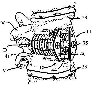
Referring to Figure 1 and 2, two identical spinal
fixation devices of the present invention, each being
generally referred to by the numerals 10 and 11,
respectively, are shown inserted into two vertebrae V
adjacent to a disc D of a segment of the human spine. Each
ssc~
CA 02186749 1996-09-27
"'~ ~1~ 218 5 i 4 v PCT~S95I03770
spinal fixation device 10 and 11 is shown coupled to
identical spinal fusion implants 40 and 41 that have been
surgically implanted in the disc space between adjacent
vertebrae V. In this manner, the spinal fixation devices
5 10 and 11 stabilize a segment of the spine, prevent the
dislodgement of the spinal fusion implant 40, and remain
permanently fixated to the spine once applied. The spinal
fixation devices 10 and 11 are identical such that the
description of one is equally applicable to the other.
l0 Thus, the description that follows will be directed to
spinal fixation device 10.
Referring to Figures 3-4, the spinal fusion implant 40
such as, but not limited to, the spinal fusion implant
described by Michelson, U.S. Patent No. 5,015,247 issued on
15 May 14, 1991, is shown. The spinal fusion implant 40 is
cylindrical in shape and has external threads 42 at its
outer perimeter for engaging the bone of the vertebrae V
adjacent to the disc D. The spinal fusion implant 40 has
an insertion end 43 having a depression 44 and a threaded
aperture 45 for engaging a portion of the spinal fixation
device 10 and also for engaging a portion of an instrument
used to insert the spinal fixation device 10 into the
vertebrae V. Referring to Figures 5-7, it is appreciated
that the spinal fixation devices 10 and il of the present
invention are not limited in use with a threaded spinal
fusion implant 40 and 41, but may be used with different
types of spinal fusion implants. For example, the spinal
fixation devices to and 11 may be coupled to spinal fusion
~~~t
CA 02186749 1996-09-27
wo 9sn6isa 218 6 7 4 9 pcT~s9sro3~~o
16
implants 40a and 41a, respectively, each having external
ratchetings 42a instead of external threads 42 as shown in
Figure 5. Alternatively, the spinal fixation devices l0
and 11 may be coupled to spinal fusion implants 40b and
41b, respectively, each having a partially cylindrical
shape with at least one truncated side 47 as shown in
Figure 6. As a further alternative, the spinal fixation
devices l0 and 11 may be coupled to spinal fusion implants
4oc and 41c, respectively, each having a knurled external
surface 48 as shown in Figure 7. It is also appreciated
that the spinal f fixation devices may be used with a variety
of other bone fusion implants without departing from the
scope of the present invention.
Referring to Figures 8-9, in the preferred embodiment,
the spinal fixation device l0 of the present invention
comprises a staple member 12 having a substantially planar
top member 14 which is of sufficient length to span one
intervertebral disc D and to engage, via a plurality of
essentially perpendicular extending projections 16 and 17,
the vertebrae V adjacent to that disc D. The top member 14
has a central opening 18 within a concentric, countersunk
recess 19 for receiving therethrough a screw or similar
coupling means for coupling the spinal fixation device l0
to the spinal fusion implant 40. The top member 14 has an
upper surface 20 having a pair of openings 22a and 22b for
receiving the posts 88a and 88b of a driving instrument 80
which is described in greater detail below in reference to
Figures 16A and 16B. Referring to Figure 10, a cross
CA 02186749 1996-09-27
WO 95126164 ~ PCT/US95/03770
17
sectional view of the top member 14 is shown. In the
preferred embodiment, the tap member :~4 is generally
triangularly shaped and is radiused along curved side 24
and straight side 26. The curved side 24 of the top member
14 is radiused at its upper edge 25 and at the upper edge
27 of straight side 26 to conform to the external curvature
of the vertebrae V. In this manner, smooth surfaces are
created at the upper edges 25 and 27 of the top member 14
that are contoured to the shape of the external curvature
l0 of the vertebrae V when the staple member 12 is in place.
The smooth contoured surface of the upper edges 25 and 27
of the top member 14 prevent aortic erosions and
perforations of the vessels proximate the vertebral column
such as the vena cava and the iliac vessels which might
otherwise result from friction. In the preferred
embodiment of the spinal fixation device 10, the top member
14 has a width ranging from 6.0 mm to 28.0 mm, with 10.0 mm
being the preferred width, and having a thickness in the
range of 2.0 mm to 4.0 mm, with 3.0 mm being the preferred
thickness. The staple member 12 is made of material
appropriate for human surgical implantation including all
surgically appropriate metals such as but not limited to,
titanium, titanium alloy, chrome molybidium alloys,
stainless steel; or non-metallic materials including
permanent or resorbable substances or composites, carbon
fiber materials, resins, plastics, ceramics or others.
Further, the staple member 12 of the present invention
may be treated with, or even composed of, materials known
S~1i101ESi~T(AI~E26~
CA 02186749 1996-09-27
WO 95/26164 PCT/I1S95/03770
21 X6749
18
to participate in or promote in the fusion process or bone
growth. The spinal fixation device 10 may be coated with
materials to promote bone fusion and thus promote the
incorporation and ultimate entombment of the spinal
fixation device 10 into the bone fusion mass. The use of
a bone fusion promoting material such as, but not limited
to hydroxyapatite, hydroxyapatite tricalcium phosphate or
bone morphogenic protein, results in a speedier vertebra V
to vertebra V fusion as bone may grow along the coated
spinal fixation device 10 bridging the two vertebrae V so
that the spinal fixation device 10 acts as a trellis and
supplies essential chemical elements to facilitate the bone
fusion process.
Referring again to Figure 9, the projections 16 and 17
are positioned at opposite ends of the top member 14 and
depend downwardly and extend perpendicularly from the
bottom surface 30 of the top member 14. The projections 16
and 17 each terminate in a distal end 32 that is pointed
and sharpened to facilitate the insertion of the
projections 16 and 17 into the vertebrae V.
The staple member 12 is most effective when the
interprojection distance I between projections 16 and 17 is
at least 4.0 mm and preferably 6.0 mm greater than the
diameter of the particular spinal fusion implant 40 for
which the spinal fixation device 10 is being used so that
at least 2.0 mm and preferably 3.0 mm of bone from the
vertebrae V will be present between the spinal fusion
implant 40 and each of the projections 16 and 17.
swc~~~
CA 02186749 1996-09-27
WO 9sn6~~a 218 6 ;' ~. 9 ~'GT/US95103770
19
Typically, intervertebral spinal fusion implants have a
diameter that ranges from 12.0 mm to 28.0 mm, therefore,
the interprojection distance I typically will range from
18.0 mm to 34.0 mm for most applications.
In the preferred embodiment, the projections 16 and 17
comprise a series of segmented and ratcheted portions 34.
The segmented and ratcheted portions 34 provide for a 'one
way" insertion of the staple member 12 to prevent the
backing-out of the projections 16 and 17 once they are
inserted into the bone of the vertebrae V. In the
preferred embodiment, each segmented and ratcheted portion
34 of the projections 16 and 17 is conical in shape and the
diameter of each segmented and ratcheted portion 34
increases in the direction from the distal end 32 toward
the top member 14 so that the projections 16 and 17
resemble a stack of cones. The segmented and ratcheted
portions 34 are spaced approximately 2.0 mm to 4.0 mm
apart, with 3.0 mm being the preferred distance between
each segmented and ratcheted portion 34. Referring to
Figure il-12, in the preferred embodiment of the spinal
fixation device 10, in order to further facilitate the
insertion of the projections 16 and 17 into the vertebrae
V, the distal end 32 of each projection 16 has an
eccentric, incline-planed inner surface 36 as shown in
Figure 11. The eccentric, incline-planed inner surface 36
of each of the projections 16 and 17 create a force F which
pushes the bone of the vertebrae V toward the spinal fusion
implant 40 as the staple member 12 is inserted into each of
CA 02186749 1996-09-27
WO 95/26164 218 6 7 4 9 pCT~S95/03770
the vertebrae V as shown in Figure 12.
Referring to Figures 13A-13F, in the preferred
embodiment of the spinal fixation device 10, the
projections 16 and 17 are cylindrical in shape having a
5 circular cross section as shown for projection 16 in Figure
13A. Alternatively, the projection 16a may have a
triangular cross section as shown in Figure 13B; the
projection 16b may have a square cross section as shown in
Figure 13C; the projection 16c may have a rectangular cross
10 section as shown in Figure 13D: the projection 16d may have
a trapezoidal cross section as shown in Figure 13E: or the
projection 16e may have a cross section with a
configuration as shown in Figure 13F.
In the preferred embodiment, the projections 16 and 17
15 each have a diameter of approximately 2.0 mm to 4.0 mm,
with 3.o mm being the preferred diameter at the widest
point. The projection 16 and 17 each have a length ranging
from 16.0 mm to 28.0 mm, with 22.0 mm being the preferred
length when the spinal fixation device l0 is implanted in
20 the direction of the anterior aspect of the vertebra V to
the posterior aspect of the vertebrae V. Alternatively, it
is appreciated that the projections 16 and 17 each could
have a longer length depending on the diameter of the
vertebrae V in which the projections 16 and 17 are
implanted.
Referring again to Figure 9, the top member 14 of the
staple member 12 has a central bar 35 extending from the
center of its bottom surface 30, for interdigitating and
r~)
CA 02186749 1996-09-27
WO 95126164 218 6 ~ ~ ~ PCT/US951Q3770
21
mating to an already implanted intervertebral spinal fusion
implant 40. In the preferred embodiment, the central bar
35 has a thickness in the range of 0.5 mm to 1.5 mm, with
0.5 mm being the preferred thickness.
Referring to Figure 1, the central bar 35 is
configured so that it complements and engages the
depression 44 at the insertion end 43 of the spinal fusion
implant 40. once engaged to the depression 44, the bar 35
interdigitates with the depression 44 of the spinal fusion
l0 implant 40 to lock and prevent the rotation of the spinal
fusion implant 40.
Referring to Figure 14, in the preferred embodiment,
the staple member 12 is secured to the spinal fusion
implant 40 by a screw 60 having threaded end 61 with a
locking thread pattern 62 and screw head 64. The locking
thread pattern 62 has a reduced pitch at the bottom of the
threaded end 61 such that the screw 60 is self-locking.
However, it is appreciated that the threaded pattern 62 may
be any of the means for locking a screw well known by those
skilled in the art.
Referring to Figures 2 and 8, the threaded end 61
of the screw 60 passes through the central opening 18 of
the top member 14 and the threaded pattern 62 threads into
the threaded aperture 45 of the spinal fusion implant 40.
The screw head 64 fits within the countersunk recess 19 of
the top member 14 such that the screw head 64 is at or
below the plane of the upper surface 20 of the top member
14. In the preferred embodiment, the central opening 18
(~A.E26)
CA 02186749 1996-09-27
WO 95/26164 / PCT/US95/03770
22
has a diameter ranging from 4.5 mm to 5.5 mm, with 5.0 mm
being the preferred diameter. The countersunk recess 19
has a diameter in the range of 6.0 mm to 8.0 mm with 7.0 mm
being the preferred diameter.
Referring to Figures 15A, 158, and 15C, an enlarged
cross sectional view of three different embodiments of a
securing means 65 for locking the screw 60 once it is
threaded to the spinal fusion implant 40 are shown. In
Figure 15A, the securing means 65 comprises a notch 66 in
the surface 20 of the top member 14 which is preferably
made of metal. Once the screw 60 is threaded and securely
tightened to the spinal fusion implant 40, a chisel C is
used to bend a portion 67 of the top member 14 into the
central opening 18 and against the screw head 64 so as to
prevent the outward excursion and any unwanted loosening of
the screw 60.
In Figure 15B, a second embodiment of the securing
means 65a is shown comprising a central score 66a
concentric with the central opening 18. A screw 60a having
a slot 61a in the screw head 64a is threaded and securely
tightened to the spinal fusion implant 40. An instrument
T is partially inserted into slot 61a after which an
impaction force Fs is applied to the instrument T to spread
apart the screw head 64a in the direction of the arrows A
so that the screw head 64a becomes deformed from the
impaction force F~ and fits within the central score 66a.
Once the screw head 64a is in the central score 66a, the
outward excursion of the screw 60a is prevented by the top
ser~aO
CA 02186749 1996-09-27
WO 95/26164 PCT/US95/03770
2185~'~y
23
lip 68 of the central score 66a.
In Figure 15C, a third embodiment of the securing
means 65b is shown comprising a screw 60b having a screw
head 64b with a slightly flanged portion 69b near the top
!5 and a slot 61b. The central opening ~.8 has along its
circumference a recess 66b for receiving the flanged
portion 69b of the screw head 64b. The securing means 65b
relies on the natural resiliency of the metal screw head
64b such that when the screw 60b is being driven by a screw
driver, the screw head 64b flexes in the direction of the
arrows B. In this manner, the flanged portion 69b of the
screw head 64b slides along the interior of the central
opening 18 so that the screw head 64b is below the top lip
68b of the recess 66b. Once the screw driver is removed
1°i from the screw 60b, the screw head 64b returns to its
natural state in the direction opposite to the arrows B so
that the flanged portion 69b is within the recess 66b. The
outward excursion of the screw 60 is thus prevented by the
top lip 68b which blocks the screw head 64b by catching the
flanged portion 69b.
Figures 16A-18 show the instrumentation used for
installing the spinal fixation device 10. Referring to
Figure 16A, a driving instrument 80 used for inserting the
spinal fixation device 10 into the vertebrae V is shown
having a hollow tubular shaft 82 which terminates at one
end to a bottom flat member 84 and terminates to a top flat
member 86 at the other end. The bottom flat member 84 is
preferably configured so that it conforms to the shape of
s~~3ttt~'(26~
CA 02186749 1996-09-27 ~~~~~ J ~ 5 ~ 0 3 7 7 0
IPEA/US 01 A P R 1996
24
the top member 14 of the staple member 12 and has a central hollow
portion 89 for receiving the alignment rod 70.
The driving instrument 80 has a pair of short posts 88a and
88b extending from the bottom flat member 84. The post's 88a and 88b are
oriented on the bottom flat member 84 so as to correspond to the position
of the openings 22a and 22b in the upper surface 20 of the top member 14
of the staple member 12. Each of the posts 88a and 88b fit into each of the
openings 22a and 22b and keep the staple member 12 aligned on the bottom
flat member 84 of the driving instrument 80. It is appreciated that the
openings 22a and 22b in the top member 14 may be depressions within the
surface 20 of the top member 14 or may be holes that pass through the top
member 14. In the preferred embodiment, the openings 22a and 22b gave
a diameter ranging from 1.5 m to 3.5 mm, with 2.5 mm being the preferred
diameter.
Referring to Figure 16B, an alternative embodiment of the
driving instrument 80" which is used for inserting into the vertebrae V the
spinal fixation device 210, described in detail below in reference to Figure
26, is shown having a hollow tubular shaft 82' which terminates at one end
to a bottom flat member 84' and terminates to a top flat member 86' at the
other end. The bottom flat member 84' is rectangular in shape so that it
conforms to the shape of the top member 214 of the spinal fixation device
210 and has a central hollow portion 89 for receiving the alignment rod 70.
The driving instrument 80' has a set of short posts 88'a, 88'b,
88'c and 88'd extending from the bottom flat member 84'. The posts 88'a-
88'd are oriented on the bottom flat member 84' so as to correspond to the
AMENDED SHEET
CA 02186749 1996-09-27
PCT/US95~03774
21 ~ 6 ;' "~ ~;~ iPEAIUS~ 1 A P R 1996
position of the openings 222a-222d of the spinal fixation device 210 and
keep the spinal fixation device 210 aligned on the bottom :lat member 84'
of the driving instruments 80'.
5 Referring to Figure 17A, an alignment rod '70 comprising a
cylindrical shaft 72 having a smooth exterior surface 73 and a threaded end
74 may be threadably attached to the threaded aperture 45 of the spinal
fusion implant 40 is shown. The alignment rod 70 fits through the central
opening 18 of the spinal fixation device 10 and is used to properly align the
10 projections 16 and 17 on each side of the spinal fusion implant 40 prior to
engaging the vertebrae V. Further, the alignment rod 70 also serves as a
guide post for the drilling template instrument 50 described in greater detail
below.
Referring to Figure 17B, as an alternative embodiment of the
1 S alignment rod 70, a splined alignment rod 70' that has a finely splined
surface 72' along its longitudinal axis and a threaded end 74' that may be
attached to the threaded aperture 45 of the spinal fusion implant is shown.
Referring to figure 18, a drilling template instrument 50 for
creating a pair of insertion holes 53a and 53b in each of the vertebrae V for
20 receiving each of the projection 16 and 17 respectively is shown. The
drilling template instrument 50 has a template 52 with a central aperture
54 therethrough and guide passagea 55 and 56 for guiding a drill bit 51 of
a drilling too. Attached to the template 52 is a handle 58 which angles
away from the template 52 so as not to
25 obstruct the line of sight of the surgeon and to allow easy access to the
template 52 and easy access to the guide holes 55 and 56 for the drill bit
ty~itNDED SHEE1
CA 02186749 1996-09-27
PCT/US 9 5 / 0 3 7.1 0
218 6 ;~ ~ '~ IPEMUS 01 AP R 1996
26
51. Extending from the center of the bottom surface of the template 52 is
a central member 59 (similar in structure and function to the central bar 35)
for mating to an already implanted intervertebral spinal fusion implant 40.
The central member 59 interdigitates with the depression 42 of the spinal
fusion implant 40 so that the template 52 is properly oriented about the
spinal fusion implant 40 and the guide holes 55 and 56 are properly
oriented with respect to the vertebrae V adjacent to the spinal fusion
implant 40. The alignment rod 70 serves as a guide post for the drill
template instrument 50 as it fits through the central aperture 54 of the
template 52 and aligns the template 52 with respect to the spinal fusion
implant 40 and insures that it is coaxial. The central aperture 54 of the
drilling template instrument 50 is smooth so that if it is placed over a
splined alignment rod 70' the drilling template instrument 50 may be easily
rotated about the splined alignment rod 70' into position such that the
central member 59 is able to mate and interdigitate with the depression 44
of the spinal fusion implant 40.
Refernng to figures 19-24, the spinal fixation device 10 of the
present invention is inserted in the following manner: At least one spinal
fusion implant 40 is surgically implanted so that it is substantially within
the disc space between two adjacent vertebrae V and engages at
i _.f
AM",(. ~ ,.
CA 02186749 1996-09-27
-~ i
WO 95/26164 ~ ~ ~ T ' PCT/US95l03770
27
least a portion of each of the two adjacent vertebrae V.
Once the spinal fusion implant 4o is in place, the
alignment rod 70 is attached to the threaded aperture 45 of
the spinal fusion implant 40. The alignment rod 70 serves
as a guide post for the drilling template instrument 50 as
it fits through the central aperture 54 of the template 52
and aligns the template 52 coaxially with respect to the
spinal fusion implant 40. Referring to Figure 20, once
the template 52 is properly aligned and the drilling
template instrument 50 is seated so that the central member
59 interdigitates with the spinal fusion implant 40, the
insertion holes 53a and 53b are drilled in each of the
adjacent vertebrae V with a drilling instrument having a
drill bit 51 with a diameter that is substantially smaller
than the diameter of each the projections 16 and 17 of the
staple member 12.
Once the drilling of the insertion holes 53a and 53b
is completed, the drill template instrument 50 is removed
from the spinal fusion implant 40 and from the alignment
rod 70. The alignment rod 70 is left in place attached to
the threaded aperture 45 of the spinal fusion implant 40.
Referring to Figure 21, the staple member 12 is placed
onto the driving instrument 80 used for driving and fixing
the staple member 12 into the vertebrae V so that the
bottom flat member 84 and the posts 88a and 88b are aligned
with the top member 14 and the depressions 22a and 22b of
the top member 14. The alignment rod 7o serves as a guide
~~t~s~
CA 02186749 1996-09-27
PCT/l; S 9 ~ 5 / 0 3 7 7 0
21 ~ 6 ' :~ ~~ r N EMUS ~ 1 N rJ t~ 1996
28
post of the staple member 12 as its fits through the central opening 18 of
the staple member 12 and aligns the staple member 12 .,~axial~y with
respect to the spinal fusion implant 40.
Referring to Figure 22, once the staple member 12 is
properly placed onto the bottom flat member 84 of the driving instrument
80, the staple member 12 and the driving instrument 80 are aligned with
respect to the alignment rod 70 so that the alignment rod 70 passes through
the central opening 18 of the staple member 12 and is inserted into the
central hollow portion 89 of the driving instrument 80. The staple member
12 and the driving instrument 80 are then lowered along the alignment rod
70 so that the sharp distal end 3~ of each of the projections 16 and 17
comes into contact with the external surface of the vertebrae V and is
aligned with the previously drilled insertion holes :53a and 53b.
As shown in Figure 22A, it is preferred that the insertion
holes 53a and 53b be drilled so that when the projections 16 and 17 are
inserted into the holes 53a and 53b, the incline planed inner surface 36 of
each of the projections 16 and 17 contacts the inner wall W of the insertion
holes 53a and 53b that is closest to the spinal fusion implant 40. In this
manner a compression force F is created as each of the projections 16 and
17 of the staple member 12 is inserted into insertion holes 53a and 53b,
respectively, compressing the bone of the vertebrae V toward the spinal .
fusion implant 40.
Referring to Figure 23, the staple member is then
a~~:~tiJJED SHF~T
CA 02186749 1996-09-27
WO 95/26164 PCTlUS95/03770
29
driven into the vertebrae V by applying a high impaction
force to the driving instrument 80 with a hammer H or other
impacting means against the top flat member 86 of the
driving instrument 80. The staple member 12 is driven into
the vertebrae V such that the projections 16 and 17 are
moved forward into the insertion holes 53a and 53b,
respectively, until the bottom surface 30 of the top member
14 of the staple member 12 comes to rest against the
surface of the vertebrae V.
l0 Referring to Figures 23-24, the driving instrument 80
is lifted away from the alignment rod 70 so that the
alignment rod 70 is no longer within the central hollow
portion 89 of the driving instrument 80. The alignment rod
70 is unthreaded from the threaded aperture 45 and is
removed from the spinal fusion implant 40. The staple
member 12 is secured to the spinal fusion implant 40 with
the locking screw 60 which has a threaded pattern 62 with
a reduced pitch. The reduced pitch of the locking screw 60
locks the locking screw 60 to the spinal fusion implant 40
with minimal turning of the locking screw 60 and prevents
any unwanted loosening. Further, any of the three
embodiments of the securing means 65, 65a or 65b described
above in reference to Figures 15A-15C may be used to
further prevent any unwanted loosening and outward
excursion of the screw 60.
Referring back to Figure 12, once the staple member 12
is driven into the vertebrae V and is secured to the spinal
fusion implant 40, the spinal fusion implant 40 is
s~snrurE r c~~
CA 02186749 1996-09-27
WO 95126164 ~ PCT/LTS95/03770
prevented from rotating along its rotational axis R by its
connection to the staple member 12 which is fixated across
the disc space between the vertebrae V. The staple member
12 is prevented from backing out from the vertebrae V along
5 the longitudinal axis L by its connection to the spinal
fusion implant 40 and by the segmented and ratcheted
portions 34 of the projections 16 and 17. In this manner,
the staple member 12 and the spinal fusion implant 40
interact to prevent the dislodgement of each other from the
10 vertebrae V in which they are implanted. Thus, the staple
member 12 is made safe against dislodgement by attachment
to the spinal fusion implant 40 and the stability of the
spinal fusion implant 40 is assured as it is also
stabilized by the staple member 12 and each works in
15 connection with the other to remove the only remaining
degree of freedom that would allow for the disengagement of
either. In addition, the incline planed inner surface 36
at the distal end 32 of the projections 16 and 17 forces
bone toward the spinal fusion implant 40 along force lines
20 F to further secure the spinal fusion implant 40 and
further prevent the dislodgement of the spinal fusion
implant 40.
It is appreciated by those skilled in the art that
when the bone of the vertebrae V is sufficiently soft, a
25 shorter method (hereinafter referred to as the "Short
Method") of inserting the spinal fixation device 10 is
possible by omitting the steps of drilling the insertion
holes 53a and 53b prior to inserting the staple member 12
~r(~x~
CA 02186749 1996-09-27
PCT/US~5~037?0
218 6 ? ~ ~~ ~pE,p,~US 01 A P R 1996
31
into the vertebrae V.
Referring to Figure 22, in the Short Method, the splined
alignment rod 70' that is finely splined along its longitudinal axis is used
instead of the alignment rod 70. Once the splined alignment rod 70' has
S been attached to the spinal fusion implant 40, the staple member 12 may be
placed over the splined alignment rod ','0' so that the splined alignment rod
70' passes through the aperture 18 and into the central aperture 89 of the
driving instrument 80. The central aperture 89 of the driving instrument 80
is correspondingly splined to the splines of the splined alignment rod 70'
so that the staple member 12 can be aligned with respect to the spinal
implant 40. The alignment of the staple member 12 and the driving
instrument 80 is maintained as the corresponding splines of the central
aperture 89 interdigitate with the splines of the splined alignment rod 70'
and prevent the rotation of the sample member 12 about the splined
alignment rod 70'. The prevention of rotation about the splined alignment
rod 70' is especially important when the Short Method is used to insert the .
spinal fixation device 10, as no insertion holes 53a and 53b have been
drilled in the vertebrae V. The staple 12 can be driven directly into the
vertebrae V by the application of a high impaction force to the driving
instrument 80 as described above and shown in Figure 22.
Once the staple member 12 is driven into the vertebrae V, the
steps of the longer method described above are used to secure the spinal
fixation device to the spinal fusion
A~UENDED SHEET
CA 02186749 1996-09-27
W O 95/26164
-; a PCT/US95/03770
32
implant 40 are the same. The Short Method of inserting the
staple member 12 reduces the amount of time required to
insert and secure the spinal fixation device 10 of the
present invention and thus reduces the overall duration of
the spinal fixation surgical procedure.
While the present invention has been described with
respect to its preferred embodiment, it is recognized that
alternative embodiments of the present invention may be
devised without departing from the inventive concept.
For example, referring to Figure 25, a first
alternative embodiment of a spinal fixation device 110
having a staple member 112 with a top member 114 generally
in the shape of an elongated oval having two curved sides
124a and 124b is shown. In this alternative embodiment,
the curved sides 124a and 124b have upper edges 125a and
125b, respectively, that are radiused to conform to the
external curvature of the vertebrae V thereby creating
smooth contoured surfaces as described above for the spinal
fixation device 10, the preferred embodiment of the present
invention. The top member 114 has openings 122a and 122b
in the upper surface 120 of the top member 114 and has two
projections 116 and 117 depending downwardly from the
bottom surface 130 of the top member 114 at opposite ends
of the staple member 112. The projections 116 and 117 are
the same as the projections 16 described above for the
preferred embodiment.
RefErring to Figure 26, a second alternative
embodiment of the spinal fixation device 210 having a
sua~rurE r c~ ~)
CA 02186749 1996-09-27
PCT/US9 /0377 0
It'EAIUS ~ 1 A P R 199'
218674~~
33
staple member 212 is shown with a top member 214 that is generally
rectangular in shape and has an upper surface 220 with cpenings 222a,
222b, 222c, and 222d. The top member 214 has four projections 216, 217,
218, and 219 depending from its bottom surface at each of its corners. The
projections 216-217 are the same as the projections 16 and 17 described
above in the preferred embodiment. The top member 214 has four straight
sides 228a, 228b, 228c, and 228d having upper edges 225a, 225b, 225c and
225d, respectively, that are radiused to conform to the to external curvature
of the vertebrae V create a smooth surface as described above for the
preferred embodiment. The driving instrument 80' shown in Figure 16B is
used to insert the spinal fixation device 210.
Referring to Figure 2'7, a third alternative embodiment of the
spinal fixation device 310 having a staple 312 with a top member 314 that
is generally triangular is shown. The top member 314 has two projections
316 and 317 depending from the bottom surface of the top member 314
that engage the vertebrae V. Extending from the center of the bottom
surface of the top member 314 is a central member 390 which is similar to
the central bar 35 of the preferred embodiment of the spinal fixation device
10 in that the central member 390 interdigitates with the depression 44 of
the spinal fusion implant 40. However, the central bar 390 also has an
extension arm 392 that extends laterally from the top member 314 to span
the diameter of an adjacent spinal fusion implant 41. The
~ ~,PrIENDED SHEET
CA 02186749 1996-09-27 pCT/!,S 9 5
1 g ~ ~ ~ '~ ~, :IPEAIUS O 1 A P R 1996
34
extension arm 392 intedigitates with the depression 44 of the spinal
implant 41. The extension arm 392 has a central aperture 394 for receiving
a screw 60b used to couple the extension arm 392 to the spinal fusion
implant 41. In this manner, a single spinal fixation device 310 is capable
S of interdigitate with two adjacent spinal fusion implants 40 and 41 to loc!c
and prevent the rotation and any excursion of the spinal fusion implants 40
and 41. The fixation of two spinal fusion implants 40 and 41 is possible
while leaving no protruding metal, such as the top member 314, on the side
of the spine where the vessels are located in close approximation to the
vertebrae as is the case with the L4 and LS vertebrae where the vessels are
located over the left side of those vertebrae. It is appreciated that any of
the
securing means 65-65b, described above may be used to lock the screw 60b
to the extension arm 392.
Referring to Figure ?8, a fourth alternative embodiment of
1 S the spinal fixation device 410 having a staple member 412 with a top
member 414 that is generally triangular in shape is shown in the installed
position. The top member 414 is wider and larger than top member 14 as
it is used with an implant 440 having a large diameter in the range of 22.0
mm to 28.0 mm. The top member 414 needs to the wider when used with
implant 440 in order to provide a central bar 435 of sufficient length to
interdigitate and mate with the depression 444 of the implant 440 in order
to prevent its rotation. Further, the top member 414 is
tapered at portion 416 so as not to cause
APA~N!~EO SHEET
CA 02186749 1996-09-27
"~4. WU 95/26164 218 5 ~ ~ 9 PCT/US95/03770
erosion or pressure against the vessels that may be present
in the area of the spine adjacent to the portion 416 of the
top member 414.
Referring to Figures 29-32, a fifth alternative
5 embodiment of the spinal fixation device 510 with a staple
member 512 having a generally rectangular top member 514 is
shown. The staple member 512 is similar in structure to
the staple 212 described above except that the top member
514 has multipronged projection blades 516 and 517
10 depending from its lower surface 530 as shown in Figure 30.
The multipronged projection blades 516 and 517 have the
same function and similar structure as the projections 16
and 17 described above and include segmented and ratcheted
portions 534 which are similar in design are function to
15 segmented and ratcheted portions 34. The muitipronged
blade projections 516 and 517 affer the added advantage of
increasing the strength and stability of the staple member
514 once it is inserted into the bone of the vertebrae V
providing a greater area of engagement of the staple member
20 512 to the vertebrae V.
The lower surface 530 has knobs 532 and 534 extending
therefrom for engaging and interdigitating with a spinal
implant 540 having an insertion end 541 with openings 542
and 544 for receiving knobs 532 and 534 respectively.
25 Referring to Figures 31 and 32, the spinal fusion
implant 540 is shown inserted within the disc space between
two adjacent vertebrae V. The spinal implant 540 is
generally rectangular in shape. The multiprong blade
CA 02186749 1996-09-27
WO 95/26164 / PCT/US95/03770
36
projections 516 and 517 have a width that is approximately
equal or slightly less than the width of the spinal fusion
implant 540. Once inserted, the spinal fixation device 510
compresses the bone of the vertebrae V towards the spinal
fusion implant 540 as discussed above in reference to
Figure 12. The spinal fixation device 510 may be secured
to the spinal fusion implant 540 with a screw 60 as
discussed above.
The spinal fixation device 510 having a staple member
512 is the preferred embodiment of the present invention
for use with a mufti-segmental spinal alignment means 600
described in greater detail below in that the staple 512
provides a more solid anchoring means that can resist
greater torsion forces resulting from the application of
the mufti-segmental spinal alignment means 600 to align the
spine.
Alternatively, for all of the embodiments described
above, the spinal fixation device l0 of the present
invention could be made of resorbable materials, such as
bio-compatible resorbable plastics, that resorb at an
appropriate rate such that once the spinal fixation device
10 is no longer needed (i.e. when spinal fusion is
complete) the body would resorb the spinal fixation device
10. One such resorbable material is polygalactone, however
any other resorbable plastic or other material safely
usable within the human body are also within the scope of
the present invention.
Further, the spinal fixation device could be only in
St~STITUTE SHEET (RULE 28)
CA 02186749 1996-09-27
WO 95/26164 218 ~ 7 ~ ~ pCT~S95/03770
37
part resorbable such that the projections 16 and 17 of the
staple member 12 would be non-resorbable and would remain
incarcerated in the vertebrae V and sealed off once the
resorbable portion of the staple is resorbed by the body.
Referring to Figures 33 and 34, as a further
application, the spinal fixation device 510 of the present
invention may be used as an anchor for a multi-segmental
spinal alignment means 600, such that a multiplicity of
spinal fixation devices may then be interconnected via a
cable, rod, bar, or plate, so as to achieve or maintain any
desired multi-segment spinal alignment. In the preferred
embodiment, the multi-segmental spinal alignment means 600
comprises more than one spinal fixation device 510 of the
present invention placed in series along the spine such
that each spinal fixation device 510 spans one disc D and
engages two adjacent vertebrae V. The spinal fixation
device 510 is preferred over the other embodiments of the
present invention in that it has a greater area of
engagement with the vertebrae V so as to provide a solid
anchoring means for the multi-segmental spinal alignment
means 600. However, it is appreciated that other
embodiments including but not limited to those described
herein may be utilized as anchoring means for the multi-
segmental spinal alignment means 600.
When used as an anchor, each spinal fixation device
510 interdigitates with and is connected to a spinal fusion
implant 610 having an insertion end 612, an interior
chamber 614 and is inserted in the disc space between the
s~rr~rEc~p
CA 02186749 1996-09-27
WO 95/26164 ~ ~ ~ ~ _/ ~ ~ PCT/US95103770
38
two adjacent vertebrae. The spinal fusion implant 610 has
a threaded blind hole 620 for receiving a threaded post 622
therein. The blind hole 620 has a casing that is made of
strong surgically, implantable material such as, but not
limited to titanium. The casing 624 extends from the
insertion end 612 of the spinal fusion implant 610 into the
interior central chamber 614. The insertion end 612 has a
rigid construction that is capable of withstanding high
torsion forces resulting from the tensioning of the multi-
segmental spinal alignment means to align segments of the
spine. In the preferred embodiment, the insertion end 612
of the spinal fusion implant has an end portion 626 that
closes the insertion end 612. The end portion is
substantially thicker than the rest of the spinal fusion
implant 610 and in the preferred embodiment, the end
portion 626 has thickness ranging from 1.5 mm to 4.0 mm,
with 2.5 mm being the preferred thickness.
Referring to Figure 34 , the threaded post 622 has a
threaded end 628 with a locking thread pattern that is
substantially longer than the locking thread pattern 62 of
the screw 60 described above and a head portion 630 having
a hole 632 for receiving a rod 634 or a cable therethrough.
The head portion 630 has a rounded exterior surface to
prevent any damage such as aortic erosion to the vessels in
the area adjacent to the spine. In the preferred
embodiment the threaded post has a diameter ranging from
3.0 mm to 6.0 mm, with 4.5 mm being the preferred diameter
and has a length ranging from 15.0 mm to 25.0 mm, with 20.0
s~r(u)
CA 02186749 1996-09-27
WO 95/26164 ~ 1% 1 ('~ PCT/US95/03770
J i
39
mm being the preferred length. The head portion 630
extends at a height above the top member 514 of the spinal
fixation device 510 of approximately 8.0 mm to 16.0 mm,
with 12.0 being the height preferred once it is threadably
attached to the spinal fusion implant 610 such that it does
not significantly protrude from the spinal column into the
tissue and vessels adjacent thereto.
Once the threaded post 622 is attached to the spinal
fusion implant 610, the head portion 630 of each threaded
post 622 are connected to one another by the rod 634 having
a sufficient diameter to fit through the hole 632 of each
head portion 630. The rod 634 has at least a portion
thereof that is threaded so that a plurality of lock nuts
638 may be used to secure the rod 634 to the head portions
630. The lock nuts 638 may also be used as length
adjusting means to adjust the length of the rod 634 between
head portions 630 so that segmental portions of the spine
may be held closer together or held further aport for the
purposes of aligning the spine. It is appreciated that a
plurality of multi-segmental spinal alignment means 600 may
be placed in series either on one side or an opposite sides
of the spine, such that one side of the spine may be
extended while the other side may be held stationary or may
be compressed in order to achieve proper spinal alignment.
The multi-segment spinal alignment may be maintained by
keeping the rod tensioned with the lock nuts 638 or by any
other means well known by those skilled in the art. It is
also appreciated that in place of a rod 634 a cable, a
S~,c"1~~~6~
CA 02186749 1996-09-27 p~T/~lS 9 5 ~ ~ 3 7 7 0
IPEAIUS 01 A P R 1996
plate or any other means well known by those skilled in the art may be used
to interconnect the mufti-segmental spinal alignment means.
Referring to Figure 35, a sixth alternative embodiment of the
spinal fixation device of the present invention is shown and generally
5 referred to by the numeral 710. The spinal fixation device 710 comprises
a top member 714 that is similar to the top member 14 described above,
except that it does not have projections 16 and 17 extending from the
bottom surface. Like numbers are being used to designate identical
features of the top members 14 and 714.
10 In the top member 714, instead of the having projections 16
and 17, independent projection members 716 and 717 in the form of screws
are used to secure the top member 714 of the spinal fixation device 710 to
the vertebrae V of the spine. The projection screw members 716 and 717
each terminate in a sharp distal end 720 and 722 respectively, have a
15 threaded portion 723, and have screw heads 724 and 726 for engaging a
screw driver or similar driving instrument.
The top member 714 has a hole 728 on one end and a hole
730 at its other end through whicf~ each of the projection screw members
716 and 717 respectively, may pass. The projection screw members 716
20 and 717 pass through the holes 728 and 730 to engage the vertebrae V.
Each of the holes 728 and 730 has a concentric counter sunk recess 732 for
receiving and seating the screw heads 724 and 726 of the projection screw
members 716 and 717 so that screw head 724 and 726 are flush or below
the top surface 20 of the top member 714 once inserted into the vertebrae
25 V.
p~~~mnGn SHE.
CA 02186749 1996-09-27
PCT/US95~03?.? 0
18 6 7 ~ '~ IPEAIUS 01 A P R 1996
41
As the projection screw members 716 and 717 are threaded,
they can be rotationally advanced into the vertebrae instead of by way of
an impaction force such that the potential for damage to the vertebrae V is
reduced. The threads of the threaded portion 723 follow one another as the
projection screw members 716 and 717 are being screwed into the bone
such that the integrity of the vertebrae V is preserved.. Also, as the
projection screw members 716 and 717 are independent from the top
member 714, the penetration depth of the spinal fixation device 710 into the
bone of the vertebrae V may be easily altered by selecting different sized
projection screw members 716 and 717 appropriate for the particular
vertebrae being fused. Further, it is possible to configure the holes 728
and 730 in the top member 714 such the t the projection screw members 716
and 717 may be inserted into the vertebrae V from a number of different
angles relative to the top member 714.
Adjacent and proximate to each of the holes 728 and 730 are
threaded openings 740 and 742, respectively, for receiving locking screws
744 and 746 respectively. Each of the locking screws 744 and 746 have a
head portion 750 and a locking thread portion 754 for threadably and
lockably engaging the threaded openings 740 and 742. The locking
screws 744 and 746 are attached to the top
AMENDED SHEET
CA 02186749 1996-09-27
WO 95/26164 ~ ' PCT/US95/03770
42
member 714 after the projection screw members 716 and 717
have been inserted into the vertebrae V. At least a part
of the head portion 750 and 752 blocks and preferably makes
contact with the screw projections 716 and 717 to prevent
any unwanted loosening and outward excursion of the screw
projections 716 and 717.
It is appreciated that the projection members 716 and
717, instead of being threaded screws, may have a number of
other configurations such as, but not limited to, the
configurations of the projections described above for the
various embodiments of the present invention. If the
projections members 716 and 717 are ratcheted instead of
being threaded, they can be driven into the vertebrae V
with a driving instrument and impaction force as described
1_°i above for the method of the present invention.
While the present invention has been described with
respect to its preferred embodiment and a number of
alternative embodiments, it is recognized that additional
variations of the present invention may be devised without
departing from the inventive concept and scope of the
present invention.
~,~TUI~ '~;~P(9tUI.E26)