Note : Les descriptions sont présentées dans la langue officielle dans laquelle elles ont été soumises.
WO 95/30004 218g 919 PCTlUS95/05262
=
MONOCLONAL ANTIBODIES WHICH PROMOTE
CENTRAL NERVOIIS SYSTEM REMYELINATION
Background
Multiple sclerosis (MS) is a chronic, frequently
progressive, inflammatory central nervous system (CNS) -
disease characterized pathologically by primary
demyelination, usually without initial axonal injury. The
etiology and pathogenesis of MS are unknown. Several
immunological features of MS, and its moderate association
with certain major histocompatibility complex alleles, has
prompted the speculation that MS is an immune-mediated
disease (Hafler, D.A. and Weiner, H.L., Immunol. Today,
10:104-107 (1989); Compston, D.A.S., "Genetic
susceptibility to multiple sclerosis," In: McAlpine's
Multiple Sclerosis (Matthews, B. ed), pp 301-319, London:
Churchil Livingstone (1991); Olsson, T., Curr. Opin.
Neurol. Neurosurg., 5:195-202 (1992)).
An autoimmune hypothesis is supported by the
experimental autoimmune (allergic) encephalomyelitis (EAE)
model, where injection of certain myelin components into
genetically susceptible animals leads to T cell-mediated
CNS demyelination (Kabat, E.A. gt al., J. Exp. Med.,
85:117-129 (1947); Lublin, F.D., SAinaer Semin.
Immunopathol., 8:197-208 (1985)). However, specific
autoantigens and pathogenic myelin-reactive T cells have
not been definitively identified in the CNS of MS patients,
nor is MS associated with other autoimmune diseases. An
alternative hypothesis, based upon epidemiological data
(Martyn, C., "The epidemiology of multiple sclerosis. In:
McAlpine's Multiple Sclerosis, (Matthews, B. ed), pp 3-40,
London: Churchil Livingstone (1991) is that an
environmental factor, perhaps an unidentified virus,
precipitates an inflammatory response in the CNS, which
WO 95/30004 21188919 PCTIUS9S/05262
-2-
leads to either direct or indirect ("bystander") myelin
destruction, potentially with an induced autoimmune
component (Lampert, P.W., Am. J. Path. 91_176-208 (1978)).
This hypothesis is supported by evidence that several
naturally occurring viral infections, both in humans (Rice,
G.P.A., Curr. Opin. Neurol. Neurosurg., 5:188-194 (1992))
and animals (Dal Canto, M.C. and Rabinowitz, S.G., Ann.
Neurol., 11:109-127 (1982)), can cause demyelination. One
commonly utilized experimental viral model is induced by
Theiler's murine encephalomyelitis virus (TMEV) (Dal Canto,
M.C., and Lipton, H.L., Am. J. Path., 88:497-500 (1977)).
The limited efficacy of current therapies for MS and
other demyelinating diseases (Goodkin, D.E. et al., Clev.
Clin. J. Med., 59:63-74 (1992)), has stimulated interest in
novel therapies to ameliorate these diseases (Martin, R.,
et al., Ann. Rev. Immunol., 10:153-187 (_1992); Steinman,
L., Adv. Immunol., 49:357-379 (1992); Weiner, H.L., et
al., Science 259:1321-1324_(1993))., However, due to the
apparently complex etiopathogenesis of these diseases,
potentially involving both environmental and autoimmune
factors, the need still exists for an effective treatment
of these demyelinating disorders.
Summary of the Invention
The present invention relates to the promotion, or
stimulation, of remyelination of central nervous system
axons in a mammal. Specifically, the present invention
relates to methods of stimulating the remyelination of
central nervous system (CNS) axons using a monoclonal
antibody obtained from a mammal immunized with spinal cord
homogenate (SCH) from a normal mammal (i.e., uninfected
with any demyelinating disease). This monoclonal (mAb) is
referred to herein as SCH94.03, and the hybridoma producing
this monoclonal antibody has been deposited on April 28,
1994, under the terms of the Budapest Treaty, with the
WO 95130004 2 18 8 9 19 PCTIUS95105262
-3-
American Type Culture Collection (ATCC) and given ATCC
Accession No. CRL 11627. As demonstrated herein, treatment
of a mammal afflicted with a demyelinating disease using
the mAb, SCH94.03, resulted in an increase in CNS
remyelination compared to mice treated with control mAb.
The present invention also relates to methods of =
treating demyelinating diseases in mammals, such as
multiple sclerosis in humans, and viral diseases of the
central nervous system of humans and domestic animals, such
as post-infectious encephalomyelitis, or prophylactly
inhibiting,the initiation or progression of demyelination
in these disease states, using the SCH94.03 monoclonal
antibody. This invention further relates to in vitro
methods of producing, and stimulating the proliferation
of,0 glial cells, such as oligodendrocytes, and the use of
these glial cells to treat demyelinating diseases.
Brief Describtion of the Ficrures
Figure 1 is a graph depicting the dose-response
characteristics of antibody-mediated proliferation of cells
in mixed rat brainculture.
Figure 2 is a graph depicting the temporal profile of
antibody-mediated proliferation of cells in mixed rat brain
culture.
Figure 3A-3D shows light and electron micrographs of
CNS remyelination promoted by mAb SCH94.03. (A) Light
micrograph of spinal cord section from a chronically
infected SJL/J mouse treated with SCH94.03 showing CNS
remyelination. (B) Light micrograph of spinal cord section
from a chronically infected SJL/J mouse treated with a
control IgM showing extensive demyelination, and the
relative absence of remyelination. Inflammatory cells,
including macrophages with ingested myelin debris are
indicated by arrows. The asterisk indicates a
representative naked axon. (C) Light micrograph of spinal
WO 95130004 218U 719 PCT/US95/05262
=
-4-
cord section with normal myelin. (D) Electron micrograph
of spinal cord section from an animal treated with SCH94.03
showing multiple axons with abnormally thin myelin sheaths
relative to axon diameter. The star in the upper
right-hand corner indicates an axon with normal myelin
sheath thickness. Arrowheads point to astrocytic
processes, which are intimately associated with
remyelinated axons. Scale bars represent 13 m in A-C, and
2 m in D.
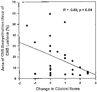
Figure 4 is a graph depicting the correlation between
the change in clinical disease and morphological
remyelination.
Figure 5 is a graph depicting the dose-response
relationship between treatment with mAb SCH94.03 and CNS
remyelination. Area of CNS remyelination (a) and
percentage of lesion area with remyelination (0) in animals
treated with various doses of mAb SCH94.03.
Figure 6 shows a Western blot nf TMEV proteins.
Lysates from infected L2 fibroblast cells were separated by
SDS-PAGE, transferred to nitrocellulose, and blotted with
SCH94.03 (lane 1), SCH94.32 (lane 2), serum from
susceptible mice chronically infected with TMEV (lane 3),
and polyclonal rabbit anti-TMEV IgG (lane 4). Molecular
weights are indicated on the left in kilodaltons (kDa).
The position and identification of the major TMEV capsid
proteins are indicated on the right.
Figure 7A-7D shows the immunostaining of cultured
glial cells andfrozen CNS tissue sections with mAb
SCH94.03. Scale bars represent 15 m.
Figure 8A-8C shows the results of SCH94.03 (Figure 8A)
and control IgMs (Figure 8D and 8C) binding to protein
antigens as determined by ELISA.
Figure 9 shows the results of SCH94.03 F(ab2)' binding
to protein antigens as determined by ELISA.
WO 95/30004 2188919 PCT/US95/05262
= -5-
Figure 10A-10C show the results of SCH94.03 (Figure
lOA) and control IgMs (Figure lOB and lOC) binding to
chemical haptens as determined by ELISA,
Figure 11 shows the alignment of the immunoglobulin
light and heavy chain variable region sequences of SCH94.03
and control IgM, CH12, and germline Ig gene segments
(SEQ ID NOS:1-11).
Detailed Description of the Invention
The present invention relates to the promotion, or
stimulation, of remyelination of central nervous system
axons in a mammal. Specifically, the present invention
relates to methods of stimulating the remyelination of
central nervous system (CNS) axons using a monoclonal
antibody obtained from a mammal immunized with spinal cord
homogenate from a normal mammal (i.e., uninfected with any
demyelinating disease). The antigen reactivity of the
monoclonal antibody, an IgM monoclonal antibody referred to
herein as SCH94.03 (also referred to herein as SCH94.32)
has been characterized as described in the present
invention using several biochemical and molecular assays,
including immunohistochemistry, immunocytochemistry,
Western blotting, solid-phase enzyme-linked immunosorbant
assays (ELISA), and Ig variable region sequencing. The
hybridoma producing monoclonal antibody SCH94.03 has been
deposited on April 28, 1994, under the terms of the
Budapest Treaty, with the American Type Culture Collection
(ATCC) and has been given ATCC Accession No. CRL 11627.
All restrictions upon the availability of the deposit
material will be irrevocably removedupon granting of the
patent.
The present invention also relates to methods of
treating demyelinating diseases in mammals, such as
multiple sclerosis in humans, and viral diseases of the
central nervous system of humans and domestic animals, such
WO 95/30004 7 r(j ! ] 8919 PCT/US95/05262
=
-6-
as post-infectious encephalomyelitis, using the SCH94.03
monoclonal antibody. Methods of prophylactic treatment
using the mAb to inhibit the initiation or progression of
demyelinating diseases are also encompassed by this
invention.
Selection of SCH mAbs to Aromote CNS remvelination
A panel of_monoclonal antibodies (mAbs) derived from
splenocytes of uninfected SJL/J mice injected with SCH was
constructed as described in detail in Example 1. After the
initial fusion and cloning, 2 of the 95 wells with viable
Ig-secreting hybridomas contained mAb with significant
binding to SCH as demonstrated by ELISA. Hybridoma cells
from these two wells, called the 79 and 94 series, were
subcloned by limiting dilution and screened again for
binding to SCH by ELISA. For the 79 series hybridomas, 14
out of 49 clones were positive by SCH ELISA, while for the
94 series, 17 out of 32 were positive for binding to SCH.
Based upon the ELISA data, two 79 series hybridomas
(SCH79.08 and SCH79.27), both of which also reacted with
myelin basic protein (MBP) by ELISA, and three 94 series
hybridomas (SCH94.03, SCH94.11, and SCH94.32), none of
which reacted with MBP, were chosen for ascites production
and j,n v'v transfer experiments.
MAbs Promote Proliferation of Glial Cells
As described in Example 2, the mAbs were tested for
their ability to promote proliferation of glial cells in
vitro. As show_nin Table 1, rat optic nerve cells grown in
the presence of mAb 94.02 or 79.27 incorporated more
['H]thymidine than controls grown in media alone or with an
isotype-matchedcontrol mAb. Data is shown from one of
five experiments which showed a similar result. '
WO 95/30004 ;j) 1889 a ~ PCTI[JS95l05262
~ L I
-7-
'i I! rl N M
O O O 0 0
p, . = .
0 0 o z ' ' = = ' i
v v v o 0
v v
m
~
Omw
y -I 41 R' r m ,i o o a m 0 0
O fd ,..~ k rq M m 0 m a ~o o .r Ln w 0
~ H N rl O rl 'i rl O ~-1 N N O ~-i
p iJ H
m
v
11 ~
G z
b7 U 'i ~-I N M d~
O O O O
.1] P.
i ~ o o i
0 R 0 0 = Z
x o v v o
v v v
v
o H
y
~ 0
-ri N .-i
U ~~~ m rl 0 M m N 0 tp rl 0 0
H .0 yj L %D r 0 N r l0 0 v 0 N 0 lO
E H m O N 'i r-1 'i N rl r-I 1i rl 'i
O v -H H U
4, 4J
.~ W
W ~
W
ro + ~ N M p ~p M tp Ol r1
0 m N 0 H r H C~ M r M
R C M 'i m N H rl N m l0 t'1 H
N
"s
~ N +i + ta +1 +1 +i +i -w +1 + ~
y +t
N ~O Ol 0 M H m H 1!f r .ry m
w .~ V' N t(1 m t0 H r 1/1 {(1 M
Cd U U l0 M M lD l0 r l0 10 m 0 N tr(1
T7 f+f N rl rl W N ri N M W G\ rl
~ O
`i
Y ~
Q~ s+ ~ M M M ~ o 0 o i o 0 o
~
0 bl
i
U
H N~ N r N r N r
N r-I M N M N
rl .U 01 O d~ O1 ~ O N H
n ~
m r ~}4 m O
l4
l+
Wc ) P~i Nu a mU0 a
SUBSTITUTE SHEET (RULE 26)
WO 95/30004 L) 9 SUqp1Q PCTIUS95/05262
] / 0
-7/1-
The dose-response characteristics of antibody-mediated
proliferation were then examined. As shown in Figure 1,
maximal stimulation with 94.03 was seen at 100 ng/ml.
Control myeloma IgMs MOPC 104E and TEPC 183 (data not shown)
also stimulated the mixed rat brain cuitures to proliferate.
However, the maximal effect was seen at a 10-fold higher
concentration than that seen with the mAbs.
The temporal profile of antibody-mediated proliferation
was also examined as shown in Figure 2. On day 8, after
culture initiation, 100 ng/ml antibody was added to the
cultures (time 0). Cells were harvested at 24 hour
SUBSTITUTE SHEET (RULE 26)
WO 95/30004 2 1 8 8 9 1 9 PCTn7S95105262
~ -8-
intervals; ['H]thymidine was present for the final 24 hours
of culture to measure the total proliferation during the
interval. The maximal_stimulation with 94.03 was seen at 72
hours after antibody addition. Similar results were
obtained with 94.32. None of the isotype control antibodies
showed any significant proliferation throughout the 120
hours of culture. These data demonstrates that both mAbs
94.32 and 94.03 induce proliferation of glial cells of mixed
rat brain culture. This proliferation is maximal at an
antibody concentration of 100 ng/ml and a culture period of
72 hours after antibody addition.
CNS Remyelination Promoted by mAbs SCH94.03 and SCH94.32
As described in Example 3, SJL/J mice chronically
infected with TMEV were treated with a total mAb dose of 0.5
mg iv or 5.0 mg ip divided into twice weekly doses for 4-5
weeks. CNS remyelination was measured by a quantitative
morphological assessment on ten spinal cord cross-sections
from each mouse. The criterion for CNS remyelination was
abnormally thin myelin sheaths relative to axonal diameter.
The data are composite of six experiments and are presented
as the mean SEM, where n indicates the number of mice.
Statistical comparisons for remyelination data were made
with the cumulative values from both IgM and buffer only
controls using a modified rank sum test. The number of
demyelinated lesions and the area of demyelination were not
significantly different between treatment groups assessed by
a one-way ANOVA. For control IgMs, we used myelomas MOPC
104E and ABPC 22 (both from Sigma), and TB5-1, an anti-
mycobacteria mAb.
SJL/J mice chronically infected with TMEV and treated
with either mAb SCH94.03 or SCH94.32 showed significantly
greater CNS remyelination than animals treated with either
isotype-matched control mAb or buffer only (Table 2).
WO 95/30004 t' ? 889 1 9 PCT/US95105262
-9-
.+ o b q +1 +i +1 -H
0 E b~" m m r N r~ in N
RC m C 'd y m M ~ w '~ =''~
N N b
0 m ~ m m
=õI ri N N N
41 O 44
On o O O O
õ~ rt 'm ~ +7 +1 ii +i
03a~
H
~ ro m
N y ~ o r
o Cm N N N N
v ~ ~ + a +1 ++
Q
~yf
o
Q~ m m r
a 7-' v N d' N N
N Q N
p U N 'i
U ; O O
i O O
ro a V V
O r N ri
p W~ O i O O
yi Q._. O O O O
-H +I 1i -H
1!1 N rl lG
A M V' ri O
y a 0 0 0 0
0
A ,d
JJ W 41 l0 M N 'i
~
y ~ O N N r
N N . '~'~ ~ ii +I
O~ m M r r1
o za C ~ C
u N N M ri
r-i >1
O M 41 Q
6 O ?1
N ~ O~1 Oi
x y U UJ
44
A H
F m Um ~i cA
co
H
WO 95130004 f 1 88 919 PCT1US95105262
-10-
Remyelination was seen with either iv or ip injections.
SCH94.03- or SCH94.32-treated animals had approximately 2-3-
fold more remyelinated lesions, and a 3-4-fold larger total
area of CNS remyelination than control animals. When a
cumulative statistical comparison was made using these two
parameters of therapeutic effectiveness, the CNS
remyelination induced by mAbs SCH94.03 and SCH94.32 was
highly significant (p < 0.005; Table 2). In a chronic
progressive disease like TMEV infection, the extent of CNS
repair is a direct function of the extent of CNS damage.
Both the number and area of CNS lesions were not different
between treatment groups, indicating similar disease
severity (Table 2). When CNS remyelination was expressed as
the percentage of lesion area showing remyelination,
approximately one-third of the cumulative demyelinated
lesion area showed CNS remyelination in mice treated with
either mAb SCH94.03 or SCH94.32 (Table 2).
Morpholocrv of CNS Remvelination
CNS remyelination was readily identified
morphologically both by light and electron microscopy
(Figure 3A-3D). Figure 3A shows a remyelinated lesion from
an animal treated with SCH94.03. The majority of axons in
the lesion show morphologic evidence of repair, with
abnormally thin myelin sheaths relative to axonal diameter
(Ludwin, S.K. "Remyelination in the central nervous system
of the mouse," In: THE PATHOLOGY OF THE MYELINATED AXON
(Adachi M, Hirano A, Aronson SM eds), pp 49-79, Tokyo:
Igaku-Shoin Ltd. (1985); Ludwin, S.K., Adv. Neurol.,
47:215-254 (1988)). For comparison, Figure 3B shows a
demyelinated lesion, with minimal remyelination, whereas
Figure 3C is an area of normal myelin, with thickly
myelinated axons. Within remyelinated lesions (Figure 3A),
there were 15.3 1.0 (mean SEM) myelinated axons per 100
m2, compared to only 1.1 0.2 myelinated axons per 100 m2
WO 95130004 2 88919 PCT1US95105262
-11- 9
in demyelinated lesions (Figure 3B). Figure 3C shows a
light micrographof spinal cord section with normal myelin.
By electron microscopy, CNS remyelination was especially
evident (Figure.3D). Almost every axon in the field has
evidence of new myelin formation, although the degree of
remyelination (ie. myelin thickness) is variable between
individual axons, suggesting different stages of the repair
process. The ratio of myelin thickness to axonal diameter
was 0.08 0.01 (mean t SEM; n = 25 axons) for remyelinated
axons compared to 0.21 t 0.01 (n = 34 axons) for normally
myelinated axons.
Correlation Between Clinical Disease and Morphological
Remvelination -
The correlation of morphological remyelination with
clinical signs of disease improvement was assessed as
described in Example 3. At each treatment injection, mice
were assessed clinically as described in Example 3. The
change in clinical score was correlated with the percentage
of lesion area showing remyelination (Figure 4).
Morphological remyelination is represented as the percentage
of lesion area showing CNS remyelination. A change in
clinical score of 0 represent stable disease over the
treatment period (4-5 weeks), whereas a positive change
indicates worsening of clinical disease, and a negative
change indicates improvement. Data represent individual
animals from all treatment groups. A positive change in
clinical score indicates worsening of disease. Using data
from all treatment groups, the change in clinical score
showed a moderate but significant negative correlation
(R=-0.40; p< 0,04) with the percentage of lesion area
showing remyelination. Although few animals actually
improved clinically (A clinical score < 0), animals with an
increase in disease severity (A clinical score > 0) tended
to have less morphological remyelination, while animals that
WO 95/30004 B B 9 19 PCTIUS95105262
~ -12-
remained stable clinically (0 clinical score = 0) showed the
most remyelination. A similar negative correlation was
obtained when the other quantitative measures of
remyelination were used (the number of remyelinated lesions
and the area of- remyelination) as shown in Table 2. These
data demonstrate that remyelination quantitated by
morphology is associated with slowing of clinical disease
progression.
Titration of mAb SCH94.03 Dose and CNS Remyelination
For the initial treatment experiments, a total mAb dose
of 25 mg/kg for iv injections and 250 mg/kg for ip injection
was empirically chosen. To assess the dose-response
characteristics, and to determine the minimal amount of mAb
needed to promote remyelination, chronically-infected mice
were treated with various ip doses of SCH94.03. Both the
number of remyelinated lesions (data not shown) and the
total area of remyelination (Figure 5) increased
significantly with larger doses of SCH94.03. Remyelination
was quantitated as described for Table 2. Data are the mean
values of 4-5 animals per mAb dose, with the final
cumulative dose indicated on the graph. SEM averaged 35% of
the mean. There was no statistical difference assessed by
one-way ANOVA in the number of demyelinated lesions or the
area of demyelination between treatment groups, indicating
similar extent of disease in all animals. The number of
demyelinated lesions and area of lesions were 33.2 7.5 and
1.25 0.43 for the 1000 g group, 31.8 8 and 1.11 0.31
for the 100 g group, 23.8 3.4 and 0.54 0.14 for the 10
g group, and 29.0 6.5 and 0.74 0.20 for the buffer only
group (represented as the 0 dose point on the graph).
Animals treated with 100 }eg control IgM (MOPC 104E) had
remyelination scores similar to control animals treated with
buffer only. The positive correlation between the dose of
mAb SCH94.03 and CNS remyelination was especially striking
WO95/30004 Z1Q Q(~ 1Q PCT/US95105262
-13-UV J / ~
when the severity of CNS disease was taken into account.
When CNS repair was expressed as the percentage of lesion
area showing remyelination, mice treated with a total dose
of 1000, 100, or 10 fcg of SCH94.03 had 6-, 5-, and 4-fold
more remyelination than control animals, respectively
(Figure 5). Mice given as little as 10 .g of SCH94.03 ip
(0.5 mg/kg) showed evidence of enhanced CNS remyelination.
These data indicated that mAb SCH94.03 and CNS remyelination
had a positive dose-response relationship, and that very
small quantities of mAb were needed to promote myelin
repair.
Antigen Specificity of SCH94 03 and SCH94 32
Although mAbs SCH94.03 and SCH94.32 were generated from
splenocytes of uninfected mice, and screened against SCH
from uninfectedmice, it was directly assessed whether
either mAb could react with TMEV capsid proteins or inhibit
viral infectivity in vitro. By Western blotting (Figure 6),
SCH94.03 and SCH94.32 did not react with any TMEV proteins
recognized by either serum from chronically infected mice or
polyclonal IgG from rabbits injected with purified TMEV
(Rodriguez, M., gi~ al., Ann. Neurol., 13:426-433 (1983)).
Western blot of lysates from control mock infected L2 cells
showed single bands with the serum from chronically infected
animals and the polyclonal rabbit anti-TMEV IgG at 32 and 43
kDa, respectively, but no reactivity with SCH94.03 or
SCH94.32.
In addition, no significant inhibition of TMEV
infectivity ig vitro with up to 5 g/ml of either SCH94.03
or SCH94.32, was observed under assay conditions where 50%
neutralization was observed with a 1:34,000 dilution of
serum from chronically infected animals. These results
indicated that the therapeutic effect of SCH94.03 and
SCH94.32 was not due to direct inhibition of the virus.
WO 95130004 , 2 , U o ~ ~ ~ PCTIUS95105262
-1(4-
O
,
To initially characterize the antigens recognized by
mAbs SCH94.03 and SCH94.32,various cell lines derived from
glial (rat C6, mouse G26-20, human U373MG and U87MG), neural
(human neuroblastoma), fibroblast (mouse L and 3T3),
epithelial (human SCC-9 carcinoma), and lymphocytic (mouse
CTLL2) origin were stained. Both mAbs stained internal
antigens of all cell lines tested, which indicated that
certain antigens recognized by these mAbs were not
restricted to unique cell types in vitro. Based on the
hypothesis that the therapeutic effect of SCH94.03 and
SCH94.32 was due to a CNS-specific interaction, the
immunostaining of cultured cells by SCH94.03 and SCH94.32
using the rat glial cell line 5.5B8 was further
investigated. This immortalized glial cell line has
phenotypic characteristics of both ac and astrocytes, with
expression of MBP and 2',3'-cyclic nucleotide
3'-phosphodiesterase (CNP), and low but detectable
expression of glial fibrillary acidic protein (GFAP) and the
lipids or proteins recognized by the mAbs A2B5 and 04
(Bozyczko, D. et al., Ann. NY Acad. Sci., 605:350-353
(1990)). SCH94.03 and SCH94.32 recognized both a surface
and cytoplasmic determinant on 5.5B8 cells. The surface
staining was most prominent on small cells which lay on top
of a layer of flat, morphologically differentiated cells
(Figure 7A). Surface staining was confirmed by flow
cytometry on live cells. When the cell membrane was
permeabilized by dehydration or brief treatment with a
non-ionic detergent to expose internal antigens, the
staining pattern was altered considerably (Figure 7B). The
cytoplasmic staining was filamentous, with a dense
perinuclear network that extended out into the cell
processes. This pattern closely resembled the staining
pattern of the intermediate filament cytoskeletal protein
vimentin. These data indicated that SCH94.03 and SCH94.32
recognized antigens that were not restricted to cells
WO 95/30004 11 8 3 919 PCT/US95105262
-15- 0
derived from thenervous system, but that they did recognize
both surface andcytoplasmic determinants on glial cells.
Immunohistochemical staining of frozen mouse, rat, and
human tissue confirmed that SCH94.03 and SCH94.32 were not
CNS-specific mAbs, but rather showed multi-organ reactivity.
Both mAbs immunostained all major organs examined, including
the brain, spinal cord, optic nerve, heart, liver, kidney,
stomach, and small intestine and skeletal muscle. However,
not all cells within an organ stained, suggesting in situ
cytological specificity. Within the CNS, SCH94.03 and
SCH94.32 stained predominantly blood vessels, ependymal
cells, and stellate-shaped cells with the morphological
features of glial cells; which were enriched in neonatal
cerebellar, periventricular, and brain stem white matter
(Figure 7C), and both neonatal and adult optic nerve.
Similar glial cells positive for SCH94.03 and SCH94.32 were
found in autopsied human brain tissue, especially at the
gray-white matter junction (Figure 7D). Identical
immunostaining results were obtained with mAb SCH94.32.
Immunostaining with a control IgM (MOPC 104E) was negative
for all samples and tissue structures which immunostained
with SCH94.03 and SCH94.32.
The identification and characterization of an entire
family of autoantibodies, referred to as "natural" or
"physiological" autoantibodies, has influenced traditional
views of autoimmunity and self-reactivity. The natural
autoantibodies that have been studied extensively are
typically IgMs, although other isotypes have been
identified, are reactive toward a wide range of antigens,
including cytoskeletal proteins, surface proteins, nucleic
acids, phospholipids, bacterial antigens such as
lipopolysaccharides, and various chemical haptens (reviewed
by Avrameas and Ternynck, Mol. Immunol., 30:1133-1142
(1993)). Natural autoantibodies share extensive idiotypic
cross-reactivity or "connectivity", which includes
WO 95/30004 889 19 PCTIUS95I05262
= -16-
expression of similar idiotypes, some of which are expressed
by pathogenic autoantibodies, as well as reactivity toward
common idiotypes expressed on other antibodies. Molecular
analysis has shown that natural autoantibodies are typically
encoded by unmutated germline immunoglobulin (Ig) genes,
with few if any somatic mutations, and therefore represent a
substantial fraction of the Ig repertoire, especially in
neonatal animals which have not had extensive exogenous
antigen exposure.
The function of natural autoantibodies remains
enigmatic. Several hypotheses have been proposed based upon
their biochemical and molecular characteristics. These
include: (1) clearance of senescent or damage tissue, (2)
providing a first line of immunological defense in the lag
period between pathogen exposure and an Ag-specific immune
response, (3) masking autoantigeus from a potentially
pathogenic autoimmune response, (4) immunomodulation,
including shaping of the neonatal immune repertoire via an
idiotypic network, and (5) participation in the positive
selection of B cells in the bone marrow, similar to the
process proposed for T cells in the thymus.
The hypothesis that antibodies SCH94.03 and SCH94.32
were natural autoantibodies was tested. To characterize the
antigen reactivities of SCH94.03 and SCH94.32, several
biochemical and molecular assays, including
immunohistochemistry and immunocytochemistry, Western
blotting, solid-phase enzyme-linked immunosorbant assays
(ELISA), and ig variable region sequencing, were used. As
described below, for all biochemical assays, SCH94.03 and
SCH94.32 were indistinguishable. In addition, SCH94.03 and
SCH94.32 had identical Ig variable region sequences, which
confirmed that they were the same mAb.
A potential mechanism whereby SCH94.03 could stimulate
remyelination in the central nervous system would be to
stimulate the proliferation and/or differentiation of cells
WO 95130004 ~ 1 88919 PCT/iJS95/05262
-17- 0
involved in myelinogenesis, primarily oligodendrocytes or
their immature precursors. Thus, it was tested whether
SCH94.03 stained the surface of various cells. Using
immortalized cells, it was determined that SCH94.03 stained
two glial cells lines, 5.5B8 (Figure 7A) and 20.2E11, but
did not stain the surface ofseveral other_glial cells lines
(l0.IA3, 20.2A40, C6, G26-20), a neuroblastoma cell line
(B104), two fibroblast lines (L2, Cos-1), or two
myoblastomas (G8, L6). Similar results were obtained with
cells isolatedfsom animal tissues and grown in culture.
SCH94.03 stained the surface of oligodendrocytes, but not
astrocytes, microglia, Schwanncells, myoblasts, or
fibroblasts.
The reactivity of SCH94.03 with proteins from glial and
lymphoid cell lines, and tissue lysates from brain, liver,
and intestine by Western blotting was also assessed.
SCH94.03 reacted with multiple bands from all cells and
tissues examined, with prominent reactivity toward bands at
50, 95, 120, and >200 kDa. The exact identity of these
protein bands has not been determined.
The reactivity of SCH94.03 with several purified
protein self-antigens by solid-phase ELISA was determined.
(Figure 8A-8C). SCH94.03 showed strong reactivity toward the
RBC antigen spectrin, but also showed consistent reactivity
toward hemoglobin, actin, tubulin, and vimentin, and
thyroglobulin, although to a lesser qualitative degree than
toward spectrin. No reactivity was observed with myosin,
transferrin, albumin, lysozyme, or myelin basic protein
under our assay conditions. Six other monoclonal or myeloma
IgM controls XXMEN-OE5 (Figure 8B), A2B5, MOPC104E, TEPC183,
01, and CH12 (Figure 8C), were also tested, and no
reactivity with any of the antigens tested was observed.
To confirm the monoclonality of,SCH94.03, 18 subclones
of SCH94.03 (9 each from SCH94.03 and SCH94.32 parents) were
tested for polyreactivity by solid-phase ELISA. All 18
WO 95130004 2-188119 PCTlUS95105262
= subclones showed identical reactivity patterns with the
panel of protein antigens as the parent SCH94.03. To
further support the conclusion that the polyreactivity of
SCH94.03 was via its Fab region, we generated F(ab)2'
fragments and assessed their reactivity with the protein
antigens by ELISA (Figure 9). SCH94.03 F(ab)2' fragments
showed similar polyreactivity as the whole IgM molecule.
A panel of chemical haptens coupled to bovine serum
albumin (BSA) was constructed and used to assess SCH94.03
reactivity by solid-phase ELISA (Figure 10A-IOC). SCH94.03
showed strong reactivity toward luorescein (FL) and
4-hydroxy-3-nitrophenyl acetic acid (NP), moderate
reactivity toward phenyloxazolone (PhOx), and weak
reactivity toward 2, 4, 6-trinitrophenyl (TNP) and
p-azophenylarsonic acid (Ars). No reactivity with p-
azophenyltrimethylammonium (TMA),
p-azophenylphosphorylcholine (PC), or the carrier protein
BSA was detected. Control IgMs (Figure lOB and lOC) showed
no significant binding to any of the haptens tested, with
the exceptions of CH12 reactivity with TMA, which has been
previously reported, and A2B5 reactivity with NP.
It was further investigated whether the ig light (L)
(SEQ ID NOS:1 and 2) and heavy (H) (SEQ ID NOS:6 and 7)
chains of SCH94.03 were encoded by germline Ig genes (Figure
11). The light chain variable (VL) and joining (J,,) region
nucleotide sequences from SCH94.03 (SEQ ID NOS:1 and 2) had
99.4% identity with the previously published sequences of
the germline Vrc10 (SEQ ID NO:4) and Jul (SEQ ID NO:5) genes,
with only two silent changes at the 3' end of both the V,,
and JL regions. The SCH94.03 VH (SEQ ID NOS:6 and 7) region
nucleotide sequence was identical to the previously
published germline VH23 (SEQ ID NO:10) sequence, the JH
region sequence differed from the published germline JH2
(SEQ ID NO:11) sequence by one nucleotide, at the 5' end of
the J region, and the diversity (D) region contained 15
WO 95130004 2, 188/ 19 PCT/US95l05262
-19- 0
contiguous nucleotides derived from the germline DFL16.1
gene. There were 8 nucleotides in the V-D junction, and 1
in the D-J junction, which did not correspond to any known
germline V or D region genes, and probably represent non-
coded (N) nucleotides inserted by the enzyme terminal
deoxynucleotide transferase during V-D-J recombination. The
only changes from the germline genes in the heavy chain of
SCH94.03 occurred at either the V-D or D-J junction, and
therefore could represent either N nucleotides or the result
of imprecise joining, rather than somatic mutations. In
addition, both the light and heavy chain variable regions of
SCH94.03 showed extensive sequence similarity with the IgM
produced by the B-cell lymphoma CH12 (SEQ ID NOS:3, 8 and 9)
(Figure 11).
SCH94.03 is a Natural Autoantibody
These preliminary antigen reactivity results suggest
that SCH94.03 is a natural autoantibody. Although this
conclusion does not readily present a mechanism as to how
SCH94.03 stimulates remyelination in the central nervous
system, it does tuggest an important physiological function
of natural autoantibodies. Autoantibodies that are produced
either during normal physiology, or in response to tissue
damage and the subsequent release of previously sequestered
antigens, might-actively participate to promote repair in
the damaged tissue. in line with previously proposed
functions of natural autoantibodies, this active
participation might be to facilitate removal of damaged
tissue, mask autoantigens thereby preventing a vigorous
pathogenic autoimmune response, modulate the immune response
which actually resulted in the tissue destruction, thereby
allowing normal endogenous tissue repair to occur, or
directly stimulate cells involved in the repair process.
Thus, as a result of the work described herein, it is
now demonstrated that an autoantibody generated and screened
WO 95/30004 ~ 18 8 919 PCT/US95105262
-20-
for its autoantigen-binding capability, also promotes CNS
remyelination. Mice chronically infected with TMEV and
treated either iv or ip with IgM mAbs from hybridomas
SCH94.03 or SCH94.32 had significantly more CNS repair than
control animals, measured by a detailed quantitative
morphological assessment of CNS remyelination. Moreover,
preliminary data suggest that the autoantibody, SCH94.03 is
also effective in promoting remyelination in mammals
afflicted with experimental autoimmune encephalomyelitis
(EAE). Thus, it is reasonable to predict that
autoantibodies, such as SCH94.03, play a critical role in
stopping an immune-mediated process of demyelination in CNS
diseases.
Two potential mechanisms can be proposed by which Abs
promote remyelination. First, Abs might inhibit some
pathogenic component of the disease process, such as virus
activity, an immune response which directly induces
demyelination, or an immune response which prevents
remyelination. If the disease outcome is based upon a
balance between tissue destruction and repair, inhibition of
pathogenic components would allow a physiological repair
response to-predominate. Experimental and clinical evidence
support this hypothesis. Spontaneous CNS remyelination is
seen in MS patients and several experimental models of CNS
demyelination as well as described herein, demonstrating
spontaneous remyelination in control mice. This indicates
that remyelination is a normal physiological response to
myelin damage. In addition, treatment of mice chronically
infected with TMEV with various immunosuppressive regiments
promotes remyelination, but does not decrease demyelination,
indicating that there is an immunological component which
inhibits remyelination. (Rodriguez, M. and Lindsley, M.D.,
Neurolocrv, 42:348-357 (1992)). Preliminary immunological
function studies have indicated that animals treated with
SCH94.03 had similar numbers of B and T (both CD4+ and CD8+)
WO 95130004 21889 1 9 PCT/US95105262
-21-
in their spleens compared to control animals, had
cells
similar in vitro splenocyte proliferative responses to
mitogens and antigens, and mounted comparable Ab responses
to both T cell-dependent and T cell-independent antigens.
The second_hypothesis is that certain Abs can actively
stimulate CNS remyelination, perhaps via stimulation of
oligodendrocyte proliferation and/or differentiation in
vivo, as has been demonstrated in vitro (Diaz, M. gt al.,
Brain Res., 154:231-239 (1978); Raine, C.S., gt al., Lab.
Invest., 38:397-403 (1979); Lehrer, G.M. et al., Brain
Res., 172:557-560 (1979); Bansal, R. gI Al., J. Neurosci.
Res., 21:260-267 (1988); Benjamins, J.A. and Dyer, C.A.,
Ann. NY Acad. Sci., 605c90-100 (1990); Dyer, C.A., Mol.
Neurobiol., 7:1-22 (1993)). MAb SCH94.03 may directly
stimulate precursor glial cells which are known to be
present at the edges of both human and experimental CNS
lesions which show active remyelination. Alternatively,
SCH94.03 may work indirectly, via activation of astrocytes
or other accessory cells, which could release factors
important for the survival or proliferation of cells in the
oligodendroglial lineage. The formation of Ab-antigen
complexes in situ with tissue components released upon
myelin destruction may also participate in Ab-mediated CNS
remyelination. Although SCH94.03 is not CNS-specific, the
recognition of.both surface and cytoplasmic antigens on
glial cells by the mAb supports an active mechanism
hypothesis. In contrast to the immunomodulatory hypothesis,
which would not necessarily require that Abs have direct
access to the CNS, the hypothesis that Abs actively
stimulate CNS remyelination implies the prerequisite of
direct access to the CNS. This is contrary to the view of
the selective permeability of the blood-brain barrier,
especially toward large molecules such as pentameric IgM.
However, during chronic inflammatory conditions such as TMEV
infection or MS, peripheral leukocytes migrate into the CNS,
WO 95/30004 ~188r17 PCTIUS95105262
-22-
indicating an alteration in the blood-brain barrier
permeability. Therefore, large proteins such as serum Ig
might also enter, via either passive diffusion through
"open" endothelium, or perhaps via an unidentified active
transport mechanism.
Treatment of Demyelinating Diseases
The results of the experiments described herein have
practical applications to multiple sclerosis (MS), EAE, and
other related central nervous system demyelinating
disorders. Rare examples ofspontaneous CNS-type
remyelination ("shadow plaques") are found in MS and
occasional peripheral nervous system (PNS)-type
remyelination is found in demyelinated spinal cord plaques
near the root entry zone. Oligodendrocytes are infrequent
at the center of the chronic plaques in MS but they appear
to proliferate at the periphery of plaques, where they are
associated with abortive remyelination. The process of
remyelination may correlate with the spontaneous remission
and improvements observed clinically in MS. These clinical
observations indicate that new myelin formation i$ possible
in MS. The remyelination that has been stimulated in mice
with TMEV-induced demyelination by using a mAb may hold
promise for therapeutic application in multiple sclerosis.
Of importance clinically is the question of whether
morphologic regeneration of thin myelin sheaths contributes
to functional recovery. Computer simulations indicate that
new myelin formation even by inappropriately thin sheaths
improves impulse conduction. Since the axon membrane of
normally myelinated fibers is highly differentiated, it is
necessary for sodium channels to be present at high density
at the node of Ranvier to propagate saltatory conduction.
Experimental evidence suggests that newly formed nodes do
develop the required high sodium channel density as
demonstrated by saxitoxin binding. Data to date suggest
WO 95130004 L~O8 9 1 9 PGT/US95105262
-23-
remyelination even by inappropriately thin myelin
that
improves conduction in a previously demyelinated axon.
Therefore, any strategy to promote this morphologic
phenomenon has the potential of producing functional
recovery.
The data presented herein demonstrates, for the first
time, that administration of a monoclonal antibody to a
mammal is capable of stimulating remyelination of central
nervous system axons 3,n vivo. Specifically, treatment of
chronically infected TMEV-infected mice with as little as 10
ug of SCH94.03 resulted in a 4- to 5-f.old increase in the
total area of CNS myelination compared to mice treated with
a control mAb.
Thus, as a result of the experiments described herein,
the method of the present invention can be used to treat
mammals, including humans and domestic animals, afflicted
with demyelinating disorders, and to stimulate remyelination
of the CNS axons. As described herein, an effective amount
of the monoclonal antibody can be administered by
intravenous (iv) or intraperitoneal (ip) injection. An
effective amount of the antibody can vary depending on the
size of the mammal being treated, the severity of the
disease, the route of administration, and the course of
treatment. For example, each dose of mAb administered can
range from approximately 0.5 mg/kg to approximately 400
mg/kg, with the preferred range from approximately 0.5 mg/kg
to approximately 250 mg/kg. It is important to note that a
dose as low as 10 gg (0.5 mg/kg) was effective in promoting
remyelination ofCNS axons in mice. The dose of mAb will
also depend on the route of administration. For example, an
iv dose administered to mice was 0.5 mg/kg, and an ip dose
was 5.0 mg/kg. The course of treatment includes the
frequency of administration of the mAb (e.g, daily, weekly,
or bi-weekly) and the duration of the treatment (e.g, four
weeks to four tnonths). Thus, for example, a larger amount
WO 95/30004 Z 18 8 919 PCT/US95105262
-24-
of mAb can be given daily for four to five weeks, as opposed
to a smaller amount of mAb given for four months.
The effectiveness of the amount of the monoclonal
antibody being administered can be assessed using any number
of clinical criteria, for example, as described in Example
3, including overall appearance of the mammal, the activity
of the mammal and the extent of paralysis of the mammal.
The effectiveness of the amount of monoclonal antibody
necessary to induce remyelination in humans can also be
assessed in a double blinded controlled trial. Patients
with fixed neurological deficits from demyelinating disease
can be treated with monoclonal antibody or controls.
Improvement in isometric muscle strength as detected by
quantitative biomechanics muscle testing could be used as
the primary therapeutic end-point.
An effective amount of the monoclonal antibody can be
combined with, or diluted with, an appropriate
pharmaceutically acceptable carrier, such as a physiological
buffer, or saline solution. Additionally, the monoclonal
antibody may be genetically altered, e.g. "humanized" by the
substitution of human antibody nucleotide sequences in non-
variable regions of the murine mAb to reduce immunogenicity.
In addition to i}x vivo methods of promoting
remyelination, ex vivo methods of stimulating remyelination
in CNS axons are also encompassed by the present invention.
For example, the monoclonal antibody may be used jn vitro to
stimulate the proliferation and/or differentiation of glial
cells, such as oligodendrocytes, as described in Example 2.
These exogenous glial cells can then be introduced into the
CNS of mammals using known techniques. Remyelination of CNS
axons would be increased by increasing the number of
endogenous glial cells present (glial cells, such as
oligodendrocytes play a critical role in the production of
myelin).
L ? 889I 7 PCT1US95/05262
W O 95/30004
-25-
In vitro methods of producing glial cells, or
stimulating the proliferation of glial cells from mixed
culture (e.g., rat optic nerve cell, or rat brain cell
cultures) are also encompassed by this invention. For
example, cells obtained from rat optic nerve, or rat brain,
containing glial_cells, are cultured as a mixed culture
under conditions sufficient to promote growth of the cells.
An effective amount of mAb capable of promoting
remyelination of CNS axons, such as SCH94.03, is then added
to the mixed culture of cells and maintained under
conditions sufficient for growth and proliferation of cells.
The mAb stimulates the proliferation of glial cells in the
mixed culture. Thus the proliferation of glial cells
cultured in thepresence of the mAb is increased, relative
to the proliferation of glial cells grown in the absence of
the mAb.
The invention will be further and more specifically
illustrated by the following Examples, which are not
intended to be limiting in any way.
Example 1: Monoclonal Antibody Production. Screening and
Purification
Animals
Spleens of two SJL/J mice (Jackson Laboratories, Bar
Harbor, ME) that had been injected twice with spinal cord
homogenate (SCH) in incomplete Freund's adjuvant were used
as the source of B cells forfusion and hybridoma
production. Splenocytes were fused with NS-i myeloma cells
using polyethylene glycol, and viable cell fusions were
selected with hypoxanthine-aminopterin-thymidine (HAT) media
and cloned by limiting dilution as described (Katzmann, J.A.
et al., Proc. Nat. Acad. Sci. USA, 78:162-166 (1981)).
CA 02188919 2005-03-29
WO 95130004 faCT/US95105262
-26-
ELISAs
Hybridoma supernatants from viable Ig-producing clones
were screened for binding to SCH by an enzyme-linked
immunosorbant assay (ELISA). The following antigens were
used for screening mAbs: SCH - (10 g) reconstituted in
carbonate-bicarbonate buffer (pH 8.53), MBP -(1 g)
dissolved in PBS, GC (1 g) dissolved in absolute alcohol,
PLP (1 g) dissolved in water. PLP was provided by Dr. W.
Macklin (UCLA) who has published a solid phase immunoassay
for PLP. For SCH, MBP or GC ELISA, Immuno II plates were
coated with prepared antigen (100 1/well) which was
incubated overnight at 4oC. The following day wells were
washed in PBS and blocked with PBS -F 1a serum for 1 hr at
room temperature. Plates were washed again in PBS and
serial dilutions of primary Ab diluted in PBS/0.1% BSA were
added and incubated at room temperature for 2 hrs. Plates
were washed in PBS/O.05o Tween and appropriate secondary Ab
conjugated to alkaline phosphatase (1:1000 in PBS 0.1 s BSA)
was added. Plates were incubated at 370C for 2 hrs, washed
in PBS 0.05o Tween, and the substrate (Sigma 104 Phosphatase
Substrate Tablet in 5 ml diethanolamine buffer) was added
for 30 min. The reaction was terminated with 50 1 of 1 N
NaOH. The plates were read on a Dynatech ELISA plate
reader.
Ascites production
The hybridomas chosen for treatment experiments were
injected into pristane-treated BALB/c mice for ascites
production. Hylridomas were also grown in RPM1-1640 media
supplemented with 10o fetal bovine serum for IgM production.
IgM mAbs were purified by either ammonium sulfate
precipitation and gel filtration on a Sephacryl S-400 HR
(Sigma) column for the initial transfer experiments, or by
affinity chromatography using goat anti-mouse IgM ( -chain
specific; Jackson Immunoresearch, West Grove, PA) coupled to
X Trade-mark
CA 02188919 2005-03-29
WO 95130004 PCT/US95/05262
-27-
Reacti-Gel 6X matrix (Pierce, Rockford, IL) for later
transfer experiments.
Example 2: In Vitro TestincT of Monoclonal Antibodies
Selection of mAbs that promote glial cell proliferation
The ability of the mAbs to promote proliferation of
glial cells in vitro was tested. Glial cells isolated from
rat brain or optic nerves were seeded in Falcorz Microtest II
plates at a concentration of 2 x 10⁴ cells per well in 0.1
ml of DME. Whole serum (SCH, IFA, MBP, GC, MBP/GC, PBS or
PLP), purified Ig or mAb, was serially diluted and 0.1 ml
aliquot was added to cells and assayed in triplicate. Three
days later 3H-thymidine was added (1 ACi/ml) and cells were
harvested after 17 hrs with an automated cell harvester
(Mash II Harvester). To document identity of cells
proliferating (i.e., , astrocytes, progenitor glial cells,
macrophages), selected cultures after exposure to
3H-thymidine, were incubated with appropriate Ab specific
for cell type followed by ABC immunoperoxidase technique.
After reaction of Hanker-Yates reagent, the slides were
immersed in Ilford K2 nuclear emulsions, exposed for 4 days
at 4oC and developed.
mAb 94.03 and 94.32 induce proliferation of mixed rat optic
nerve brain cultures
One- to two-day-old rats were killed with ether.
Through careful dissection, optic nerves were removed from
5 the optic nerve chiasm to the eye. Nerves were transferred
to centrifuge tubes containing 2 mis=of DMEM. An equal
volume of 0.25% trypsin was added and incubated to 37oC in a
water bath for 45 min. 0.2 ml of FCS was added to terminate
trypsinization. Nerves were passedthrough a sterile needle
10 and syringe (gauge no. 21) and then centrifuged at 14.00 rpm
for 10 min. The cell count was adjusted to provide
* Trade-mark
2~ 88919
WO 95/30004 PCT/US95/05262
~
-28-
concentration of 5 x 10⁵ cells/100 l of media in 24-well
trays in DMEM + 0.5~k FCS. After 12 to 16 hrs, appropriate
antibodies or growth media were added as per experimental
protocols.
Brains of 1-2 day old rats were removed and placed in
Hank's Balanced Salt Solution with 10 mM HEPES buffer
(HBSS/H), approximately 1-2 ml per brain. The brain stem,
cerebellum, and midbrain was discarded whereas the forebrain
was minced with a bent syringe. The tissue was further
disrupted by repeated passage through a 10 ml pipet and
transferred to a 50 ml conical tube. The tissue suspension
was shaken on a rotary shaker (75 rpm) for 30 min at 37oC.
Trypsin was added to a final concentration of 0.125k and the
suspension was shaken for an additional 60 min. Trypsin
digestion was stopped by adding FCS (10t). The cell
suspension was passed sequentially through 120 and 54 m
Nytex, centrifuged, resuspended in serum-free medium with
10& FCS, and filtered again through 54 m Nytex. Serum-free
media was DMEM with 3.7 g/1 sodium bicarbonate, 6.0 g/1
glucose, 2 mM L-glutamine, 0.1 nM nonessential amino acids,
5 g/ml insulin, 5 g/ml transferrin, 5 ng/ml selenite, l00
U/ml penicillin and 100 {Cg/mi streptomycin. The cells were
counted, plated onto uncoated tissue culture flasks or
plates at 5xlO cells/cm~ and cultured at 370C in 5!k COZ.
The media was changed after 72 hrs, and every 48 hrs
thereafter. On day 8 after culture initiation, the media
was aspirated and replaced by SFM with various supplements
(for example, antibody). For most experiments, the cells
were grown for an additional 48 hrs before harvesting.
Cells were pulsed with ['Hlthymidine (5 Ci/ml) for the
final 1824 hrs of culture.
Western Blot Procedure
Antigens were denatured and solubilized by heating at
100oC in sodium dodecyl sulfate (SDS) sample buffer.
WO95/30004 ~ j8 89 19 PCT/US95105262
~
-29-
Samples were electrophoresed on stacking and separating gels
containing 4.75* and 12.0W acrylamide at 200 volts. After
electrophoresis,- gels and nitrocellulose membranes were
equilibrated for,30 min in transfer buffer (25 mM Tris, 192
mM glycine, 20W methanol, pH 8.1-8.3). All steps were done
at room temperature. Gels were electroblotted for either 1
hr at 100V or overnight at 30V using the Bio-Rad Mini
Trans-blot apparatus. The nitrocellulose membrane was cut
into strips and washed, 3X TBS (100 mM NaCI, 50 mM TriG, pH
7.6) with 0.03t Tween 20. Nitrocellulose strips were
blocked (TBS with 3k non-fat milk and 0.03% Tween 20) for
2-4 hrs, washed 3X, and incubated with primary Ab or
antisera (diluted in blocking buffer) for 4 hrs or
overnight. After primary Ab incubation, strips were washed
3X, incubated with either biotin- or alkaline
phosphate-labelled secondary Ab (diluted in blocking buffer)
for 2 hrs, washed 3X, and incubated with
alkaline-phosphatase labeled-streptavidin (diluted in
blocking buffer) for 2 hrs if the biotin system is used.
Nitrocellulose strips were washed 4X (final wash in TBS
without Tween 2Q) and incubated with substrate solution
(0.165 mg/ml BCIP and 0.33 mg/ml NBT in 100 mM NaCI, 100 mM
TriG, 5 mM MgG12, pH 9.5) until sufficient color developed
(approximately 10-15 min). The reaction was stopped by
adding PBS with 5 mM EDTA.
Cell lines or mixed brain cultures were lysed in 1X SDS
reducing sample buffer (2.3% SDS, 10t 2-ME, 0.125 M Tris,
20t glycerol) and heated to 85oC for .15 min. Nucleic acids
were sheared by repeated passage of lysate through 21-
27-gauge needles. Lysate proteins were separated on a 12%
acrylamide reducing gel, transferred to nitrocellulose
membranes, and blotted with various antibodies as previously
described.
2188919
WO 95/30004 PCT/US95105262
~
-30-
Bxamnle 3: Promotion of CNS Remvelination Using a Monoclonal.
Antibody
Virus
The DA strain of TMEV was obtained from Drs. J. Lehrich
and B. Arnason after eight passages in BHK cells. The virus
was passaged an additional four times at a multiplicity of
infection of 0.1 plaque forming units (PFU) per cell.
Cell-associated virus was released by freeze-thawing the
cultures followed by sonication. The lysate was clarified
by centrifugation and stored in aliquots at -70oC. All
subsequent experiments will use passage 12 virus. This
virus isolate causes white matter pathology without
destruction of anterior horn cells.
In vitro TMEV neutralization assay
Viral plaque assays were done as previously described
(Patick, A.K., et al., J. NeuroDath. Exo. Neurol.,
50:523-537 (1991)). To assess neutralization, aliquots of
TMEV (200 PFU/ml) were incubated with various concentrations
of Ab for 1 hour at room temperature prior to plating onto
confluent L2 cells. As a positive control, we used serum
from susceptible mice chronically infected with TMEV. Under
the assay conditions described above, a serum dilution of
1:34,000 gave 50% neutralization, which corresponded to an
estimated 20 ng/ml of TMEV-specific Abs, assuming a total
serum Ig concentration of 15 mg/ml, and a TMEV-specific
fraction of 5%.
Demvelination protocol
Demyelination was induced in female SJL/J mice, ages
four to six weeks, from the Jackson Laboratory, Bar Harbor,
ME. Mice were inoculated intracerebrally with 2 x 10⁵
plaque-forming units of DA virus in a volume of 10 l. Mice ,
WO 95/30004 2 B8y 19 PCTIUS95/05262
-31-
chronically with TMEV (4 to 6 months following
infected
infection) were assigned randomly to groups of treatment.
Treatment protocol and clinical disease assessment
Chronically infected mice were given either intraperitoneal
(ip) or intravenous (iv) injections of mAb twice weekly for
4-5 weeks. At each treatment injection, mice were assessed
clinically by three criteria: appearance, activity, and
paralysis. A score for each criterion was given ranging
from 0 (no disease) to 3 (severe disease). For appearance,
1 indicated minimal change in coat, 2 indicated a moderate
change (scruffy_appearance), and 3 indicated a severe change
(incontinence and stained coat). For activity, 1 indicated
decreased spontaneous movements (minimal ataxia), 2
indicated moderate slowing (minimal spontaneous movements),
and 3 indicated severe slowing (no spontaneous movement).
For paralysis, 0.5 indicated a spastic extremity, 1
indicated a paralyzed extremity, 1.5 indicated two or more
spastic extremities, 2 indicated two paralyzed extremities
(unable to walk)_, 2.5 indicated no righting response, and 3
indicated three or four paralyzed extremities (moribund).
The total score_.for each mouse was the cumulative total from
each criterion (maximum of 9). As the clinical score was an
ordinal, but not a cardinal scale, the change in clinical
score to assess clinical disease was used. The clinical
assessment data were not disclosed until after the
morphological assessment of remyelination was completed.
Liaht and electron microaraph orenaration and assessment of
remyelination
Preparation of light and electron microscopy sections
and morphological assessment of remyelination were done.
Briefly, treated mice were anesthetized with pentobarbital
(0.2 mg ip), exsanguinated by cardiac puncture, and killed
by intracardiac perfusion with Trump's fixative (100 mM
2188919
WO 95/30004 PCTIUS95/05262
~ -32-
phosphate buffer, pH 7.2, with 4g formaldehyde and 1.5k
glutaraldehyde). The entire spinal cord was removed
carefully from the spinal canal, and sectioned into 1 mm
transverse blocks. Every third block was post-fixed in ik
osmium tetroxide and embedded in Araldite (Polysciences,
Warrington, PA). One micron sections from each block were
cut and stained with p-phenylenediamine. On each section,
remyelination was quantitated using a Zeiss interactive
digital analysis system (ZIDAS) and camera lucida attached
to a Zeiss photomicroscope (Carl Zeiss Inc., Thornwood, NY).
Abnormally thin myelin sheaths relative to axonal diameter
was used as the criterion for CNS remyelination. Ten spinal
cord sections from each mouse were examined; this
corresponded to 8-9 mm2 of white matter examined per mouse.
To avoid bias, slides were coded and quantitation was done
without knowledge of the treatment groups.
Myelin thickness and axonal diameter measurements and
cuantitation of mvelinated axons
Electron micrographs of normal and remyelinated axons
from plastic-embedded spinal cord sections were imaged with
a Hamamatsu video camera, digitized, and analyzed using an
IBAS 2000 Image Analysis System (Kontron, Munich, Germany).
The axonal cross-sectional area with and without the myelin
sheath was measured, and equivalent circle calculations were
used to determine the axonal diameter and myelin sheath
thickness. For myelinated axon quantitation, the number of
myelinated axons in lesions from plastic-embedded spinal
cord sections were counted using the analysis system
described above attached to an Axiophot microscope (Carl
Zeiss, Inc.). 17 remyelinated and 15 demyelinated lesions
in spinal cord sections from animals treated with mAb
SCH94.03, control IgM, or buffer only were analyzed. This
corresponded to 0.6 mm2 of remyelinated area and 0.8 mmZ of
' demyelinated area. The criterion for selection of a lesion
WO95130004 218 8 919 PCTITJS95/05262
-33-
demyelinated iaas the presence of substantial
as
demyelination with minimal repair, whereas remyelinated
lesions were chosen based upon the presence of almost
complete remyelination throughout the lesion.
Immunostainina
Rat 5.5B8 glial cells were grown on
poly-D/L-lysine-coated chamber slides in Dulbecco's modified
Eagle's medium (DMEM) supplemented with 1.5 g/L D-glucose,
30 nM SeOa, 15 nM triiodothyronine, 10 ng/ml biotin, 100 M
ZnC12, 50 fcg/ml gentamicin, and 10t fetal bovine serum. All
staining steps were done at room temperature. For surface
staining, slides were briefly rinsed with PBS, and cells
were lightly fixed with 1% formaldehyde in PBS for 10 min to
prevent cell detachment during subsequent staining steps.
For cytoplasmic staining, slides were rinsed twice in PBS
and either air dried for 1 hour or incubated with 0.1t
Triton X-100 in PBS for 10 min. Cells were blocked in 2t
BSA for 30 min, washed, incubated with control IgM or mAb
SCH94.03 (10 gg/ml in 1t BSA) for 1 hour, and washed
extensively with PBS. Afterfixation with 4t
paraformaldehyde for 15 min, slides were incubated with
fluorescein-labeled goat anti-mouse IgM (Jackson
Immunoresearch) for 1 hour, washed with PBS, coverslipped
with 10k MOWIOL (Hoechst) in 100 mM Tris, 25k glycerol, pH
8.5 with 25 g/ml 1,4-diazobicyclo-[2.2.2]-octane (DABCO) to
prevent fading, and allowed to set overnight in the dark.
For frozen tissue sections, fresh neonatal rat, adult mouse,
qr autopsied human cortical brain tissue was quick frozen in
isopentane chilled in liquid nitrogen prior to liquid
nitrogen storage. Frozen sections (10 m) were transferred
onto gelatinized glass microscope slides, air dried for 4-8
hours, and stored at -70 C. Prior to immunostaining, slides
were placed at room temperature overnight. The
immunoperoxidase staining protocol was similar to that
CA 02188919 2005-03-29
WO 95130004 PCTIUS95105262
-34-
described above, using the ABC immunoperoxidase reagent
(Vector Laboratories, Burlingame, CA), developed with 1.5
mg/ml Hanker-Yates reagent (p-phenylene diamine-procatechol)
in 50 mM Tris, pH 7.6 with 0.03401 H202, counterstained with
Mayer's hematoxylin, and mounted with Permount*(Fischer
Scientific, Pittsburgh, PA).
Data Analysis
A modified cumulative rank sum test (O'Brien, P.C.,
Biometrics, 40:1079-1087 (1984)) was used to compare
remyelination between treatment groups. This statistical
test takes into:account several numerically unrelated
parameters of therapeutic effectiveness, and is used
routinely for clinical trial efficacy assessment. Parallel
analyses using a standard unpaired Student's t-test to
compare individual parameters of remyelination gave
equivalent results. Comparisons of disease severity and
correlation significance were determined by a one-way
analysis of variance (ANOVA). Statistical analyses were
done with the either the SigmaStat (Jandel Scientific, San
Rafael, CA) or EXCEL (Microsoft Corporation, Redmond, WA)
software programs. Calculated values were considered
significant when p was < 0.05.
Equivalents
Those skilled in the art will recognize, or be able to
ascertain, using no more than routine experimentation, many
equivalents to the specific embodiments of the invention
~described herein. Such equivalents are intended to be
encompassed by the following claims:
* Trade-mark
WO 95/30004 PCT/US95/05262
-35-
SEQffENCE LISTING
(1) GENERAL INFORMATION: (i) APPLICANT: Mayo Foundation for Medical Education
Research
(ii) TITLE OF INVENTION: MONOCLONAL ANTIBODIES WHICH PROMOTE
CENTRAL NERVOUS SYSTEM REMYELINATION
(iii) NUMBER OF SEQUENCES: 11
(iv) CORRESPONDENCE ADDRESS:
(A) ADDRESSEE: Hamilton, Brook, Smithy & Reynolds, P.C.
(B) STREET: Two Militia Drive
(C) CXTY: Lexington
(D) STATE: Massachusetts
(E) COUNTRY: USA
(F) ZIP: 02173
(v) COMPUTER READABLE FORM:
(A) MEDIUM TYPE: Floppy disk
(B) COMPUTER: IBM PC compatible
(C) OPERATING SYSTEM: PC-DOS/MS-DOS
(D) SOFTWARE: PatentIn Release #1.0, Version #1.25
(vi) CURRENT APPLICATION DATA:
(A) APPLICATION NUMBER:
(B) FILING DATE:
(C) CLASSIFICATION:
(vii) PRIOR APPLICATION DATA:
(A) APPLICATION NUIMER: U.S. 08/236,520
(B) FILING DATE: April 29, 1994
(viii) ATTORNEY/AGENT INFORMATION:
(A) NAME: Granahan, Patricia
(B) REGISTRATION NUMBER: 27,227
(C) REFERENCE/DOCKET NUMBER: MMV92-01 PCT
(ix) TELECOMMUNICATION INFORMATION:
(A) TELEPHONE: 617-861-6240
(B) TELEFAX: 617-861-9540
(2) INFORMATION FOR SEQ ID NO:l:
(i) SEQUENCE CHARACTERISTICS:
(A) LENGTH: 393 base pairs
(B) TYPE: nucleic acid
(C) STRANDEDNESS: double
(D) TOPOLOGY: linear
(ix) FEATURE: (A) NAME/KEY: CDS
(B) LOCATION: 1..393
WO 95/30004 21889 19 PCT/US95/05262
.
-36-
(xi) SEQUENCE DESCRIPTION: SEQ ID NO:1:
ATG ATG TCC TCT GCT CAG TTC CTT GGT CTC CTG TTG CTC TGT TTT CAA 48
Met Met Ser Ser Ala Gln Phe Leu Gly Leu Leu Leu Leu Cys Phe Gln
1 5 10 15
GGT ACC AGA TGT GAT ATC CAG ATG ACA CAG ACT ACA TCC TCC CTG TCT 96
Gly Thr Arg Cys Asp Ile Gln Met Thr Gln Thr Thr Ser Ser Leu Ser
20 25 30
GCC TCT CTG GGA GAC AGA GTC ACC ATC AGT TGC AGG GCA AGT CAG GAC 144
Ala Ser Leu Gly Asp Arg Val Thr Ile Ser Cys Arg Ala Ser Gln Asp
35 40 45
ATT AGC AAT TAT TTA AAC TGG TAT CAG CAG AAA CCA GAT GGA ACT GTT 192
Ile Ser Asn Tyr Leu Asn Trp Tyr Gln Gln Lys Pro Asp Gly Thr Val
50 55 60
AAA CTC CTG ATC TAC TAC ACA TCA AGA TTA CAC TCA GGA GTC CCA TCA 240
Lys Leu Leu Ile Tyr Tyr Thr Ser Arg Leu His Ser Gly Val Pro Ser
65 70 75 80
AGG TTC AGT GGC AGT GGG TCT GGA ACA GAT TAT TCT CTC ACC ATT AGC 288
Arg Phe Ser Gly Ser Gly Ser Gly Thr Asp Tyr Ser Leu Thr Ile Ser
85 90 95
AAC CTG GAG CAA GAA GAT ATT GCC ACT TAC TTT TGC CAA CAG GGT AAT 336
Asn Leu Glu Gln Glu Asp Ile Ala Thr Tyr Phe Cys Gin Gln Gly Asn
100 105 110
ACG CTT CCG TGG ACG TTC GGT GGA GGC ACC AAG CTG GAA ATC AAA CGG 384
Thr Leu Pro Trp Thr Phe Gly Gly Gly Thr Lys Leu Glu Ile Lys Arg
115 120 125
GCT GAT GCT 393
Ala Asp Ala
130
(2) INFORMATION FOR SEQ ID NO:2:
(i) SEQUENCE CHARACTERISTICS:
(A) LENGTH: 131 amino acids
(B) TYPE: amino acid
(D) TOPOLOGY: linear
(ii) MOLECULE TYPE: protein
(xi) SEQUENCE DESCRIPTION: SEQ ID NO:2:
Met Met Ser Ser Ala Gln Phe Leu Gly Leu Leu Leu Leu Cys Phe Gln
1 5 10 15 -
Gly Thr Arg Cys Asp Ile Gln Met Thr Gln Thr Thr Ser Ser Leu Ser
20 25 30
Ala Ser Leu Gly Asp Arg Val Thr Ile Ser Cys Arg Ala Ser Gln Asp
35 40 45
Ile Ser Asn Tyr Leu Asn Trp Tyr Gln Gln Lys Pro Asp Gly Thr Val
50 55 60
WO 95/30004 218 8 919 PCT/US95/05262
~
-37-
Lys Leu Leu Ile Tyr Tyr Thr Ser Arg Leu His Ser Gly Val Pro Ser
65 70 75 80
Arg Phe Ser Gly Ser Gly Ser Gly Thr Asp Tyr Ser Leu Thr Ile Ser
85 90 95
Asn Leu Glu Gln Glu Asp Ile Ala Thr Tyr Phe Cys Gln Gln Gly Asn
100 105 110
Thr Leu Pro Trp Thr Phe Gly Gly Gly Thr Lys Leu Glu Ile Lys Arg
115 120 125
Ala Asp Ala
130
(2) INFORMATION FOR SEQ ID NO:3:
(i) SEQ'QENCE CHARACTERISTICS:
(A) LENGTH: 324 base pairs
(B) TYPE: nucleic acid
(C) STRANDEDNESS: double
(D) TOPOLOGY: linear
(xi) SEQUENCE DESCRIPTION: SEQ ID NO:3:
GATATCCAGA TGACACAGAC TACATCCTCC CTGTCTGCCT CTCTGGGAGA CAGAGTCACC 60
ATCAGTTGCA GGGCAAGTCA GGACATTAGC AATTATTTAA ACTGGTATCA GCAGAAACCA 120
GATGGAACTG TTAAACTCCT GATCTACTAC ACATCAAGAT TACACTCAGG AGTCCCATCA 180
AGGTTCAGTG GCAGTGGGTC TGGAACAGAT TATTCTCTCA CCATTAGCAA CCTGGAGCAA 240
GAAGATATTG CCACTTACTT TTGCCAACAG GGTAATACGC TTCCTCCGAC GTTCGGTGGA 300
GGCACCAAGC TGGAAATCAA ACGG 324
(2) INFORMATION FOR SEQ ID NO:4:
(i) SEQIIENCE CHARACTERISTICS:
(A) LENGTH: 285 base pairs
(B) TYPE: nucleic acid
(C) STRANDEDNESS: double
(D) TOPOLOGY: linear
(xi) SEQIIENCE DESCRIPTION: SEQ ID N0:4:
GATATCCAGA TGACACAGAC TACATCCTCC CTGTCTGCCT CTCTGGGAGA CAGAGTCACC 60
ATCAGTTGCA GGGCAAGTCA GGACATTAGC AATTATTTAA ACTGGTATCA GCAGAAACCA 120
GATGGAACTG TTAAACTCCT GATCTACTAC ACATCAAGAT TACACTCAGG AGTCCCATCA 180
AGGTTCAGTG GCAGTGGGTC TGGAACAGAT TATTCTCTCA CCATTAGCAA CCTGGAGCAA 240
GAAGATATTG CCACTTACTT TTGCCAACAG GGTAATACGC TTCCT 285
WO 95/30004 2' " " 919 PCTIUS95105262
.
-38-
.(2) INFORMATION FOR SEQ ID NO:5:
(i) SEQUENCE CHARACTERISTICS:
(A) LENGTH: 39 base pairs
(B) TYPE: nucleic acid
(C) STRANDEDNESS: double
(D) TOPOLOGY: linear
(xi) SEQUENCE DESCRIPTION: SEQ ID NO:5:
TGGACGTTCG GTGGAGGCAC CAAGCTGGAA ATCAAACGT 39
(2) INFORMATION FOR SEQ ID NO:6:
(i) SEQUENCE CHARACTERISTICS:
(A) LENGTH: 429 base pairs
(B) TYPE: nucleic acid
(C) STRANDEDNESS: double
(D) TOPOLOGY: linear
(ix) FEATURE:
(A) NAME/KEY: CDS
(B) LOCATION: 1..429 (xi) SEQUENCE DESCRIPTION: SEQ ID NO:6:
ATG GGA TGG AGC TGT ATC ATC CTC TTT TTG GTA GCA GCA GCT ACA GGT 48
Met Gly Trp Ser Cys Ile Ile Leu Phe Leu Val Ala Ala Ala Thr Gly
1 5 10 15
GTC CAC TCC CAG GTC CAA CTG CAG CAG CCT GGG ACT GAA CTG GTG AAG 96
Val His Ser Gln Val Gln Leu Gln Gln Pro Gly Thr Glu Leu Val Lys
20 25 30
CCT GGG GCT TCA GTG AAG CTG TCC TGC AAG GCT TCT GGC TAC ACC TTC 144
Pro Gly Ala Ser Val Lys Leu Ser Cys Lys Ala 8er Gly Tyr Thr Phe
35 40 45
ACC AGC TAC TGG ATG CAC TGG GTG AAG CAG AGG CCT GGA CAA GGC CTT 192
Thr Ser Tyr Trp Met His Trp Val Lys Gln Arg Pro Gly Gln Gly Leu
50 55 60
GAG TGG ATT GGA AAT ATT AAT CCT AGC AAT GGT GGT ACT AAC TAC AAT 240
Glu Trp Ile Gly Asn Ile Asn Pro Ser Asn Gly Gly Thr Asn Tyr Asn
65 70 75 80
GAG AAG TTC AAG AGC AAG GCC ACA CTG ACT GTA GAC AAA TCC TCC AGC 288
Glu Lys Phe Lys Ser Lys Ala Thr Leu Thr Val Asp Lys Ser Ser Ser
85 90 95
ACA GCC TAC ATG CAG CTC AGC AGC CTG ACA TCT GAG GAC TCT GCG GTC 336
Thr Ala Tyr Met Gln Leu Ser Ser Leu Thr Ser Glu Asp Ser Ala Val 100 105 110
TAT TAT TAT GCA AGA CGG GCC CCT TAC TAC GGT AGT AGG AAC TTT GAC 384
Tyr Tyr Tyr Ala Arg Arg Ala Pro Tyr Tyr Gly Ser Arg Asn Phe Asp
115 120 125
TAC TGG GGC CAA GGC ACC ACT CTC ACA GTC TCC TCA GAG AGT CAG 429
Tyr Trp Gly Gln Gly Thr Thr Leu Thr Val Ser Ser Glu Ser Gln
130 135 140
WO 95/30004 ? 18 8 919 PCr/US95/05262
.
-39-
(2) INFORMATION FOR SEQ ID NO:7:
(i) SEQUENCE CHARACTERISTICS:
(A) LENGTH: 143 amino acids
(B) TYPE: amino acid
(D) TOPOLOGY: linear
(ii) MOLECULE TYPE: protein
(xi) SEQUENCE DESCRIPTION: SEQ ID NO:7:
Met Gly Trp Ser Cys Ile Ile Leu Phe Leu Val Ala Ala Ala Thr Gly
1 5 10 15
Val His Ser Gln Val Gln Leu Gln Gln Pro Gly Thr Glu Leu Val Lys
20 25 30
Pro Gly Ala Ser Val Lys Leu Ser Cys Lys Ala Ser Gly Tyr Thr Phe
35 40 45
Thr Ser Tyr Trp Met His Trp Val Lys Gln Arg Pro Gly Gln Gly Leu
50 55 60
Glu Trp Ile Gly Ann Ile Asn Pro Ser Asn Gly Gly Thr Asn Tyr Asn
65 70 75 80
Glu Lys Phe Lys Ser Lys Ala Thr Leu Thr Val Asp Lys Ser Ser Ser
85 90 95
Thr Ala Tyr Met Gln Leu Ser Ser Leu Thr Ser Glu Asp Ser Ala Val
100 105 110
Tyr Tyr Tyr Ala Arg_Arg Ala Pro Tyr Tyr Gly Ser Arg Asn Phe Asp
115 120 125
Tyr Trp Gly Gln Gly Thr Thr Leu Thr Val Ser Ser Glu Ser Gln
130 135 140
(2) INFORMATION FOR SEQ ID NO:8:
(i) SEQUENCE CHARACTERISTICS:
(A) LENGTH: 366 base pairs
(B) TYPE: nucleic acid
(C) STRANDEDNESS: double
(D) TOPOLOGY: linear
(ix) FEATURE:
(A) NAME/BEY: CDS
(B) LOCATION: 1..366
(xi) SEQUENCE DESCRIPTION: SEQ ID NO:8:
CAG GTC CAA CTG CAG CAG CCT GGG ACT GAA CTG GTG AAG CCT GGG GCT 48
Gln Val Gln Leu Gln Gln Pro Gly Thr Glu Leu Val Lys Pro Gly Ala
1 5 10 15
TCA GTG AAG CTG TCC TGC AAG GCT TCT GGC TAC ACC TTC ACC AGC TAC 96
Ser Val Lys Leu Ser Cys Lys Ala Ser Gly Tyr Thr Phe Thr Ser Tyr
20 25 30
WO 95/30004 PCTlUS95105262
= -40-
TGG ATG CAC TGG GTG AAG CAG AGG CCT GGA CAA GGC CTT GAG TGG ATT 144
Trp Met His Trp Val Lys Gln Arg Pro Gly Gln Gly Leu Glu Trp Ile
35 40 45
GGA AAT ATT AAT CCT AGC AAT GGT GGT ACT AAC TAC AAT GAG AAG TTC 192
Gly Asn Ile Asn Pro Ser Asn Gly Gly Thr Asn Tyr Asn Glu Lys Phe
50 55 60
AAG AGC AAG GCC ACA CTG ACT GTA GAC AAA TCC TCC AGC ACA GCC TAC 240
Lys Ser Lys Ala Thr Leu Thr Val Asp Lys 8er Ser Ser Thr Ala Tyr
65 70 75 80
ATG CAG CTC ASC AGC CTG ACA TCT GAG GAC TCT GCG GTC TAT TAT TAT 288
Met Gln Leu Ser Ser Leu Thr Ser Glu Asp Ser Ala Val Tyr Tyr Tyr
85 90 95
GCA AGA GAT TAC TAC GGT AGT AGC TGG GGG TAC TAC TTT GAC TAC TGG 336
Ala Arg Asp Tyr Tyr Gly Ser Ser Trp Gly Tyr Tyr Phe Asp Tyr Trp
100 105 110
GGC CAA GGC ACC ACT CTC ACA GTC TCC TCA 366
Gly Gln Gly Thr Thr Leu Thr Val Ser Ser
115 120
(2) INFORMATION FOR SEQ ID NO:9:
(i) SEQUENCE CHARACTERISTICS:
(A) LENGTH: 122 amino acids
(B) TYPE: amino acid
(D) TOPOLOGY: linear
(ii) MOLECULE TYPE: protein
(xi) SEQUENCE DESCRIPTION: SEQ ID NO:9:
Gln Val Gln Leu Gln Gln Pro Gly Thr Glu Leu Val Lys Pro Gly Ala
1 5 10 15
Ser Val Lys Leu Ser Cys Lys Ala Ser Gly Tyr Thr Phe Thr Ser Tyr
20 25 30
Trp Met His Trp Val Lys Gln Arg Pro Gly Gln Gly Leu Glu Trp Ile
35 40 45
Gly Asn Ile Asn Pro Ser Asn Gly Gly Thr Asn Tyr Asn Glu Lys Phe
50 55 60
Lys Ser Lys Ala Thr Leu Thr Val Asp I,ys Ser Ser Ber Thr Ala Tyr
65 70 75 80
Met Gln Leu Ser Ser Leu Thr Ser Glu Asp Ser Ala Val Tyr Tyr Tyr
85 90 95
Ala Arg Asp Tyr Tyr Gly Ser Ser Trp Gly Tyr Tyr Phe Asp Tyr Trp
100 105 110
Gly Gln Gly Thr Thr Leu Thr Val Ser Ser
115 120
WO 95130004 2188919 PCT/US95105262
-41-
INFORMATION FOR SEQ ID NO:10:
(2)
(i) SEQUENCE CHARACTERISTICS:
(A) LENGTH: 351 base pairs
(B) TYPE: nucleic acid
(C) STRANDEDNESS: double
(D) TOPOLOGY: linear
(xi) SEQUENCE DESCRIPTION: SEQ ID NO:10:
ATGGGATGGA GCTGTATCAT CCTCTTTTTG GTAGCAGCAG CTACAGGTGT CCACTCCCAG 60
GTCCAACTGC AGCAGCCTGG GACTGAACTG GTGAAGCCTG GGGCTTCAGT GAAGCTGTCC 120
TGCAAGGCTT CTGGCTACAC CTTCACCAGC TACTGGATGC ACTGGGTGAA GCAGAGGCCT 1S0
GGACAAGGCC TTGAGTGGAT TGGAAATATT AATCCTAGCA ATGGTGGTAC TAACTACAAT 240
GAGAAGTTCA AGAGCAAGGC CACACTGACT GTAGACAAAT CCTCCAGCAC AGCCTACATG 300
CAGCTCAGCA GCCTGACATC TGAGGACTCT GCGGTCTATT ATTATGCAAG A 351
(2) INFORMATION FOR SEQ ID NO:11:
(i) SEQUENCE CHARACTERISTICS:
(A) LENGTH: 45 base pairs
(B) TYPE: nucleic acid
(C) STRANDEDNESS: double
(D) TOPOLOGY: linear
(xi) SEQUENCE DESCRIPTION: SEQ ID NO:11:
TACTTTGACT ACTGGGGCCA AGGCACCACT CTCACAGTCT CCTCA 45