Note : Les descriptions sont présentées dans la langue officielle dans laquelle elles ont été soumises.
CA 02207561 1997-06-12
DRILLING CATHETER ASSEMBLY
BACKGROUND OF THE INVENTION
1. Field of the Invention
The present invention is directed to a drilling catheter assembly for intra-osseous
S delivery of medication and is particularly suitable for delivery of dental anaesthesia.
An intermediate assembly is provided by ~e m~mlf~cturer and comes sterile with a
c~n~ ..in~l ion protective cap and, once used, the intermedi~t~ assembly may be simply
disposed of. A preferably sterilizable stainless steel adaptor engages the intermediate
assembly and interfaces it to the dental handpiece for drilling. Once drilling is
10 completed, the adaptor is removed leaving the drilling catheter component of the
intermediate assembly in place for insertion of an injection needle therethrough to
infuse the medication to the target site.
2. Related Art
There are a variety of methods currently in use for providing local anaesthetic in
15 dentistry. These methods and appaldluses however all have disadvantages, either
heing difficult for practitioners to perform or painful and unpleasant to the patient.
CA 02207561 1997-06-12
An example of a method used ~;ull~;;nlly in dentistry is the infiltration method, whereby
a local anaesthetic solution is injected into the soft tissue of gingiva. In doing so, the
solution eventually passes through the cortical plate affecting the nerve bundle entering
the tooth. Disadvantages of this method include the delay of onset of anaesthesia after
5 the injection and, in most cases, ballooning of the injected tissue. As well, there is
an extended period of time for recovery of the tissue until return to normal condition.
Another method which is cullelllly used is the regional block method whereby an
anaesthetic solution is injected locally in proximity to the nerve trunk as it enters the
bone. Disadvantages of this procedure are that it is extremely difficult to locate the
10 nerve trunk, there is discomfort to the patient and a delay for the anaesthetic to take
effect. As in the case of the infiltration method, this method necessitates a long
recovery period for tissue to return to normal.
At present, two types of a~ tus have been used to ~l rO~ intra-osseous
anaesthesia. These are surgical burs used to perforate the cortical plate and the villet
15 injectors.
The use of a surgical bur has disadvantages in that burs are ~ el~ive and they have
to be sterilized between uses or a new bur used each time. In addition, the method
is slow, requiring the ~ h~d gingiva and periosteum to be anaesth~ d before the
cortical plate is perforated. The villet injector is an appa-alus that services both as a
CA 02207~61 1997-06-12
perforator and injector. It uses specifically designed needles rotated by a conventional
dental motor. A disadvantage of this device is that the needle often becomes clogged
with pulverized bone which obstructs the passage in the needle and prevent~s injection
of the anaesthetic solution. It is generally difficult to remove the clogging material
S from the needle and often the use of a second needle is necessary. Other
disadvantages of this method include the initial capital cost of the instrument purchase,
and the cost of the needles which are somewhat expensive. In addition, the design of
the instrument makes access to various parts of the mouth difficult and sometimes
impossible.
10 Intra-osseous and targeted root-canal nerve anaesthesia have not become popular for
the reason that there has not heen a practical technique of making the injections
successfully. For example~ there has been a general belief that this method is radical
and to be restored to only if nerve block and infiltration anaesthetic do not accomplish
the desired result. However, intra-osseous and targeted injections produce positive7
15 more ~roround anaesthesia and could be made with less pain than either of the other
types according to the present invention.
Targeted . - ~sthe~i~ has several advantages over prior art nerve block or infiltration
methods. There is no feeling of numbness in the tongue7 cheek7 or lips during or after
the injection and there is no after-pain. The anaesthetic is profound and acts
20 immediately alleviating the necessity of waiting for the anaesthetic to take effect as
CA 02207~61 1997-06-12
with the nerve block and infiltration methods. Furthermore, as only a few drops of
anaesthetic are injected, there is no feeling of faintness or increa~sing of the pulse rate.
To achieve targeted anaesthesia one must gain access, if intra-osseous, to the
cancellous bone by going through the cortical layer; or to the bottom of the booth, if
S root-canal targeted an ~Sthf~ci~ is desired. Because of instant anaesthesia and profound
pulpal ~n~esth~si~, there is a much greater control over the region one wishes to
anaesthetize, resulting in a much smaller does of anaesthetic; as well as, of course,
other medication, where applicable.
United States Patent No. 5,173,050 ~Dillon) discloses a dental appa-alus for
10 perforating the cortical plate of human m~xill~ry and mandibular bones. The
appalalus of Dillon comprises a metal needle moulded into a plastic shank. The shank
is being formed with means for cooperation with a dental hand piece for transmitting
the rotational movement to the needle. The needle used for drilling is solid and has
a sharp bevelled free end. The appa.~lus described by Dillon is disposable.
15 However, the device disclosed in Dillon's patent cannot be used as a catheter for
injecting ~n:lesth~tic by inserting a hypodermic needle through the drilling needle. As
well, the device disclosed by l~illon is not provided with means for blocking entry of
bone debris into the needle passageway. In addition, the direct connection between
the hand piece and the pelroIat(~r does not provide for a safe and reliable id~ntifi~hle
CA 02207561 1997-06-12
passageway through which the ~n~esth~si;~ can be transported and is only available to
use via the fixed gingiva.
In United States Patent No. 5,261,877 (Fine et al.) there is provided an intravascular
c~th~,~er and thrombectomy procedure utili7ing the cathe~er. The catheter comprises
S a flexible jacket with a distal working head having a c~n~ ing tip rotatable at high
speeds for removing ~1I1V111I~US from the lumen of a vessel. A flexible drive assembly
extends through the jacket to rotate the tip and a plurality of infusion ports are formed
adj:lcent the distal end of the jacket, capable of delivering a fluid contrast media at
relatively high volumetric flow rates into the lumen of the vessel to locate the site of
10 the ~hrolllbus. The c~n~1i7ing tip is then rotated at high speed to homogenize and
remove the thrombus from the lumen.
United States Patent No. 3,534,476 (Winters) discloses a drilling and filling root canal
app~lus. The drilling is performed by a drill having a central bore. The depth of
the root canal is d~lellllh~ed in advance and a stop is placed on the drill to limit the
15 depth of drilling. The device is provided with a flexible rod which is pushed into the
root canal so that the drill is directed along this road to follow the contour of the canal
so that the resulting bore will have an uniform ~i~mf~tçr which is free of shoulders or
leges. The al)pa.alus disclosed by Winters is concerned with enlarging the root canal
after the nerve has been extracted. This apparatus is not used for injecting medication
20 in close proximity to a targeted area for tre~tm~nt or anaesthetic.
CA 02207561 1997-06-12
United States Patent No. 4,944,677 (A1exandre~ discloses a smooth hollow needle with
a bevelled point for dAlling a hole into the jawbone near the apex of the tooth to be
~n~estheti7ed Theredfler, the drilling device 13 removed from the jaw, and a
hypodermic needle of substantially the same gauge is inserted into the hole and
5 ~n~h~si~ is injected. Thus, there is no cath~ti7ed delivery of medication, with the
~tt~nd~nt disadvantage that the pre-drilled hole may be difficult to locate when
inserting the hypodermic needle.
One significantly older United States patent that is discussed by Alexandre (above) is
United States Patent No. 2,317,648 (Siqveland) granted in 1943. In addition to the
10 disadvantage mentioned by Alexandre, the fact that Siqveland teaches use of threaded
sleeve which penetrates the bone during drilling and is left (screwed) in the bone to
serve as a guide for insertion of the actual injection needle. I~ue to the cost of such
a device, it cannot be made disposable; but more impo~ ly, for the threaded sleeve
to be securely fastened in the bone it would have to rotate at a much slower speed than
15 the drill (as in Siqveland) or the drilling catheter (as in the present invention~.
SUMMARY OF THE INVENTION
The present invention provides a drilling clthftet assembly comprising: a hollow
drilling needle adapted to serve as a cath~ter affixed to a first flange at its non-drilling
end; a second flange adapted at one side thereof to engage said first flange and adapted
CA 02207561 1997-06-12
at its opposite side to engage a rotating shaft; and said first flange with af~lxed drilling
needle and said second flange held together in an assembled position by means an
outer protection housing open at its end near said opposite side. The assembly may,
however, be provided witbout the housing, which could, if desired, be added later.
S The dril1ing c?~het~qr assembly, said second flange having a dowel projecting from its
centre and located inside of, and concentrically with~ said hollow drilling needle.
The drilling c~theter assembly, said dowel having a ~ m~ter such that it is easily
withdrawn from said hollow drilling needle after drilling.
The drilling ca~he~qr assembly, said dowel being sufficiently long to prevent drilling
l0 debris from blocking the hollow drilling needle once the dowel has been withdrawn.
The drilling catheter assembly, said first and second flanges having a lon,~itu(1in~1
groove for receiving a prong projecting from a third flange adapted its side opposite
said prong to engage a drilling handpiece.
BRIEF DESCRIPTION OF THE DRAWINGS
15 The preferred embodiment of the present invention will now be described in
conjunction with the anneYed drawing fgures, in which:
CA 02207561 1997-06-12
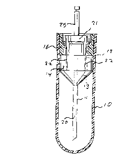
Figure 1 is an exp10ded view of the full drilling catheter assemb1y, including the
handpiece interface adaptor;
Figure 2 is a partial cross-section of the fully assembly drilling cathPter shown in
Figure l just prior to the removal of the co.~ ion protective cap; and
5 Figure 3 shows in more detail the upper left-hand corner of the assembly shown in
Figure 2.
DETAILED DESCRIPTION OF THE PREFERRED EMBODIMENTS
Referring to Figures 1, 2 and 3 of the drawings (Figure l shows an exploded view)
the full drilling catheter assembly comprises a co.~ 1ion protective cap 107 a
IO hol10w drilling cathç~er 11 having a drilling tip 12 and afflxed at its other end to an
oulward]y flaring flange 13 which te~ ates in an integral cylindrical portion 14,
which has four (but preferably at least two) equal1y spaced longitudinal grooves or slits
15 in its wall. A cylindrical flange 16, adapted to engage the top of the cylindrical
portion 14 in a fixed position by means of a protruding spur 17 and a co-operating
lS notch 18 in the wall of the portion 14, is provided with four equally spaced
longitudinal grooves or slits 19 in its wall which are aligned with the slits 15 (by
means of the spur 17 and notch 18). The flange 16 has a theleli()lll centrally
CA 02207~61 1997-06-12
protruding rod or dowel 20, which fits inside the hollow catheter 11 preferably
reaçhing down to near the bevelled drilling tip 12. The components described so far
represent the m~nuf~ct~rer assembled intermefli~te assembly, which is delivered
contained inside the cap 10 and which is held together by the slight pressure of the
S walls of the cap 10. The final part of the full assembly is disc-like flange 21, which
has projecting therefrom four (or two or three, as ~e case may be) prongs 22
distributed equidistantly along the cil~;u~llrer~nce of the flange 21 to fit into the slits
19 and 15, the prongs 22 being slightly wider at their point of ~ hm~Mt to the
circumference of the flange 21 and projecting slightly ~ulwaldly from the
10 cilcul~lrelence such that when the flange 21 is pushed down into the provided
interme~ te assembly beyond the upper edge of the cap 10, the prongs 22 push-out
the wall of the cap 10 at the points of contact 23 and, if the cap 10 had been creased
along the points of contact 23, cause the wall of the cap 10 to rupture along the
creases 23. At the same time, once the flange 21 has been pushed below the edge of
the flange 16 (as shown more clearly in Figure 3) it is retained by lip portion 24 in
the flange 16. With the flange 21 thus retained and the wall of the cap widened or
ripped along the creases 23, it is then a simple matter to pull the cap 10 off while
holding it and a shaft 25 protruding outwardly from the centre of the flange 21. The
shaft 25 is, of course, adapted to engage the dental handpiece.
20 Once the cap 10 has been removed, the dental handpiece ~not shown) engage the shaft
25 and drilling is commenced according to normal practice. When drilling is
CA 02207561 1997-06-12
completed, the cy1indrical portion 14 is pl~feldbly held between thumb and indexfinger while the handpiece is pulled away removing with it the flanges 21 and 16, with
the c~th~t~r 11 left in place. It is then a simple matter of inserting an injection needle
through the portion 14 and into the catheter l 1 to deliver the an~esthesi~ to the target
5 site. A1though the presence of the dowel 20 may not be always necessary, it is
desirable in order to prevent bone debris from blocking catheter 11.
The entire interm~li~t~ assembly may be made from a (disposable) plastic material
except, of course, for the drilling cath~ter 11 (and possibly the dowel 20). Thus, the
interme(li~te assembly comes sterilized and protected from co~ ion in a
10 protective wrap (not shown) and is disposed of after use. The only part that is
plerel~bly made from st~in1es.~ steel is the handpiece interface flange 21, which may
thus be sterilized after use.
