Note : Les descriptions sont présentées dans la langue officielle dans laquelle elles ont été soumises.
CA 02224640 1997-12-12
W O 97/00047 PCTrUS96/10582
- 1 -
SURGICAL KIT AND METHOD FOR PERFORMING LAPAROSCOPIC
URETHROPEXY, AND APPARATUS EMPLOYED IN SAME
BACKGROUND OF THE INVENTION
1. Field of the Invention
The present invention is directed towards a surgical kit and method for
performing laparoscopic urethropexy for the correction of female stress urinary
incontinence, as well as the apparatus employed in the same. More particularly,
the present invention provides apparatus for performing urethropexy through
laparoscopic techniques, thereby greatly reducing the duration, discomfort, and
recovery period of such surgeries.
2. Description of Related Art
Female stress urinary incontinence (SUI), defined as the unintentional loss
of urine, can be a socially unacceptable problem for many women. Most often,
the incontinence occurs during coughing, sneezing, or physical activity in womenafflicted with this problem. While effective surgical treatment for this condition
has existed for nearly 50 years, the procedures typically involve major abdominal
surgery with accompanying post-operative limitations lasting six to eight weeks.Because of the nature of these surgical procedures, many women simply resort
to diaper-like incontinence pads, or simply avoid any activities which result in the
unintentional loss of urine.
In the normal resting state, the external pressure exerted on the
collapsible urethra by the surrounding musculature is greater than the pressure
exerted on the bladder, and therefore continence is maintained. During moments
CA 02224640 1997-12-12
WO 97/00047 PCT~US96/10582
of coughing, sneezing, or physical activities, greater pressure will be exerted on
the dome of a filled bladder. In women not afflicted with stress incontinence, acorresponding increase in the external pressure on the urethra acts to prevent
the unwanted loss of urine from the bladder. Sufferers of SUI, however, aren't
so fortunate.
Stress incontinence is generally caused by two etiologies: a spastic
detrusor muscle; or a loss of support of the periurethral tissue at the urethra-vesicular junction ("UVJ" - the region where the urethra enters the bladder).
When the latter situation occurs, the UVJ will sag into the vagina, thereby
reducing the pressure which can be exerted on the urethra during moments of
stress. Diagnosis of any sagging of the UVJ can be easily determined by
inserting the tip of a cotton swab into the urethra until it reaches the UVJ. The
patient is then asked to bear down as if urinating, and loss of the UVJ support is
1 5 readily identified by the upward movement of the wooden end of the cotton swab.
In this test, the external urethral meatus acts as a fulcrum for the tip of the swab,
and the elevation of the opposite end indicates the downward descent of the
UVJ. U.S. Patent No. 4,07Z,144 provides an alternative device which may be
utilized to readily measure the angle of the UVJ in a similar manner.
The first urethropexy procedure for eliminating SUI caused by a sagging
urethra was developed in 1948 by Drs. Marshall, Marchetti, and Krantz, and
generally involves the fixation of the periurethral tissue at the UVJ on either side
of the urethra (MMK procedure). Fixation in the MMK procedure, also known as
urethropexy or abdominal culposuspension, is accomplished by suturing the
periurethral fascia at the UVJ on either side of the urethra to the periosteum of
the pubic bone. The procedure essentially alters the angular relationship
between the urethra and bladder by elevating the UVJ, and therefore preventing
the sagging of the UVJ when downward pressure is applied to the region by
various stresses.
_ _ _, CA 02224640 1997-12-12 - ---
- 3.1 -
The MMK procedllre has been perfected o~er the years, however the
essential principles ha~e remained the san e. In 1g55 Burch developed the
tochnique of afflxing til2 periurethral ~ascia bil~terally to Cooper's ligament,thereby resultin~ in a technically ea9ier procedure becau~e of the pr~vious
difficulties in passing a needle through ~he periosteurn of the pub;c bone.
AJthough the Burch prooedure t~as ~een performe~ lapar~seopically, the five-yearfailure rate forthe open ~3urch procedure is appr~xirnately ~0%. A laparoscoplo
Bu~h procedure is even more problematic since it is extremely difficult and time-
consuming to tie sutures laparoscopically.
Alternati~/ely, uroto~ical proc~dures such as that of Stamey, Raz and
Peyerra ha~e been develope~, however these are typically blind procedures
which reqllire the passing of long needles tkr~ugl the re~tus fascia to the
periurethral ~ascia utili~ing a cystoscope. Aithough these urol~gical proceduresa~oid the 1 0-~entimeter midline or Pfannenstiel incision and its requfred thr~e-
day or longer hospital sby, the gynecologicai procedures of MMK, Burch and
o~er~ have proven to be the most effecth~e. In fact, the scarring of the urethraand int~ior bladder as well as the scarrin~ of the periurethral tissues, aids infi~tion of all of the inYolved tissues during the MMK ancl Burch proce~ures,
thereby assisting in the preventiorl of Incontinence.
A recen~ modification to the "needle suspension" procedures ~f Stamey,
~az ~nd Peyerra is dic~losed in WO g3t107~5 for ~hich Benderev is ~e named
irtventor (hereina~er referred to as "the Bender~v prooeduren). The Benderev
pr~oedure differs from pre~ious needle suspension procedures primarily in that
the susperldin~ sutures may be attached to bone anchors secured to the upper
sur~ace o~ the pubic bone (the ~anterior-superio~' edge o~ the pubic bone, as
deflned with the pat;ent in the harizorl~l positlon). Anc~ors sinlilar to those
described herein may be employed, and a bar~ for each anchor is first created
3Cl in the pubic bone using a drill bit. A suture passer ~or di~ecting suture p~ss~e
is also provided.
h,,~E~ ~.
Substitute Sheet
~_ .CA 02224640 1997-12-12
,, . . . ~, . . . _
3.2 -
Recently, a modified \~er~ion of the MMK prQced~re has been dcvcto,~
whic~ utilizes bane anchors secured directly to the pubic bone on either side ofthe symphysis f~r fixation of th~ UVJ. The ~pparatus for perforrning this modi~i~d
MMK procedure are sald by Mitek Surgical Products, Inc. of N~wocd,
5 Massachusetts, and a nllmber of U.S. patents concem th~se products ~see, e.g.,US Patent No's 5,207,6-79, 5,217,486 and 4,899,743). In the Mitek-MMK
procedure, a P~annenstiei incision must be made in the abdomen in order to
pr~vlde access to the spac~ ~f Retzius. Tl-e spac~ of Retziu~: is in actuality a"potential" space in th~t it conta;ns va~i~u~ connective tissues and ~ats which
must be disse~ted in orderto pr~vide sufficient acc~ss to this rQgls~n. In ~act, this
connectiYe tissue, particularly the areolar adventitial tissue, generally breaks
AMENDED SHEET
Substitute Sheet
CA 02224640 1997-12-12
W O 97/00047 PCT~US96/10582
down after delivery of a child, and this breaking down of the connective tissue
often contributes to the onset of SUI in many women.
Once the space of Retzius has been dissected in the Mitek-MMK
procedure, small anchors are secured in the pubic bone on either side of the
pubic symphysis. Each of the bone anchors has a suture attached thereto, and
these sutures are threaded through the periurethral tissue on either side of theurethra. The sutures are then tied off in the abdomen so that the periurethral
tissue is pulled upward, which in turn restores the angle of the urethra at the
UVJ, thereby restoring the urethra to its proper location. While the Mitek-MMK
procedure is highly effective, it is a lengthy and complicated procedure which can
generally only be performed by highly-skilled surgeons.
The present invention offers an unique anchor design for use in a
laparoscopic urethropexy procedure, as well as an inserter for placing the anchor
within a bore created in the patient. These apparatus are particularly suited for
laparoscopic urethropexy, however the use of the devices is not so limited.
BRIEF DESCRIPTION OF THE DRAWINGS
While the specification concludes with claims particularly pointing out and
distinctly claiming the present invention, it is believed the same will be better
understood from the following description taken in conjunction with the
accompanying drawings in which:
Figure 1 is a cross-sectional view taken through the midline of a patient
who has lost support of the periurethral tissue at the UVJ, and is thereby
suffering from stress urinary incontinence;
Figure 2 is the same view as Fig. 1, however the structural defect has
been corrected using the method and apparatus of the present invention;
~.
-
CA 02224640 1997-12-12
W O 97/00047 PCT~US96/10582
- 5 -
Figure 3 is a top plan view of a bone anchor used in the method of the
present invention;
.
Figure 4 is a side plan view of the bone anchor of Fig. 3;
Figure 5 is a top plan view of a drill tamper tool of the present invention
wherein a portion of the tool has been broken-away;
Figure 6 is a side plan view of the tamper tool of Fig. 5;
1 0
Figure 7 is an end plan view of the tamper tool of Fig. 5, taken along line
7-7 thereof;
Figure 8 is a top plan view of a bone anchor insertion tool of the present
invention, wherein a portion of the tool has been broken-away;
Figure 9 is a side plan view of the insertion tool of Fig. 8;
Figure 10 is a side plan view of the insertion tool of Fig. 8 with the bone
anchor of Fig. 3 loaded thereon;
Figure 11 is a side plan view of a suture retriever of the present invention;
Figure 12 is an end plan view of the suture retriever of Fig. 11, taken along
the line 12-12 thereof;
Figure 13 is a top plan view of a suture template of the present invention;
Figure 14 is a side plan view of the template of Fig. 13;
Figure 15 is a bottom plan view of the template of Fig. 13;
,
CA 02224640 1997-12-12
W O 97/00047 PCTnUS96/10582
-- 6 --
Figure 16 is an end plan view of the template of Fig. 13, taken along line
16-16 thereof;
Figure 17 is a perspective view of the template of Fig. 13 in use during a
surgical procedure with portions of the patient's anatomy cut-away for clarity;
Figure 18 is a perspective view of an altemative embodiment of the suture
template according to the present invention;
Figure 19 is a perspective view of the insertion tool of Fig. 8 in use during
a surgical procedure with portions of the patient's anatomy cut-away for clarity;
Figure 20 is a perspective view of the surgical procedure of the present
invention wherein portions of the patient's anatomy cut-away for clarity, and
wherein the suture retriever of Fig. 11 is being employed;
Figure 21 is a perspective view of the surgical procedure of the present
invention wherein portions of the patient's anatomy cut-away for clarity, and
wherein the sutures have been retrieved from the pre-peritoneal region for tying;
Figure 22 is a perspective view of the space of Retzius, and illustrates the
proper placement of the anchors and sutures employed in the present invention;
Figure 23 is a side plan view of the anchor of the present invention in
place in the pubic bone of a patient, wherein the pubic bone is shown in cross-
section;
Figure 24 is a perspective view of the anchor of Fig. 23;
Figure 25 is a side plan view of the anchor of Fig. 23;
Figure 25a is another side plan view of the anchor of Fig. 23;
CA 02224640 1997-12-12
W 097/00047 PCTrUS96/10582
Figure 26 is a top plan view of the anchor of Fig. 23;
Figure 27 is an end plan view of the anchor of Fig. 23, viewed from the
proximal end towards the distal end;
Figure 28 is an end plan view of another embodiment of an anchor
according to the present invention, viewed from the proximal end towards the
distal end;
Figure 29 is an end plan view of another embodiment of an anchor
according to the present invention, viewed from the proximal end towards the
distal end;
Figure 30 is a cross-sectional view of the anchor of the present invention
1 5 taken along the line 30-30 of Fig. 26;
Figure 31 is a side plan view of an anchor-insertion tool of the present
invention, wherein a portion of the tool has been cut-away for clarity;
Figure 32 is a side plan view of the anchor-insertion tool of Fig. 31 with an
anchor loaded thereon, said anchor having a suture extending therefrom,
wherein a portion of the tool has been cut-away or cross-sectioned for clarity;
Figure 33 is a cross-sectional view of the loaded anchor-insertion tool of
Fig. 32 taken along line 33-33;
Figure 34 is a cross-sectional view of the loaded anchor-insertion tool of
Fig. 32 taken along line 34-34; and
~ 30 Figure 35 is a cross-sectional view of the loaded anchor-insertion tool of
Fig. 32 taken along line 35-35.
CA 02224640 1997-12-12
W O 97/00047 PCT~US96/10582
SUMMARY OF THE PREFERRED EMBODIMENTS
In accordance with one aspect of the present invention, a surgical kit for
use in a laparoscopic urethropexy procedure wherein the urethra is elevated to
the desired angle by securing a suture between an anchor which has been
laparoscopically secured to the pubic bone and the periurethral tissue adjacent
the urethra, is provided. This surgical kit comprises:
(a) at least one anchor, said anchor laparoscopically securable to a
pubic bone;
(b) an anchor-insertion tool for laparoscopically securing said anchor
to a pubic bone; and
(c) a suture retrieval tool for retrieving a suture extending from said
anchor into the vagina.
The kit further preferably comprises at least one suture, and said anchor
preferably has a passageway through which said at least one suture may
inserted. A pair of said anchors and a pair of said sutures are more preferably
provided.
The surgical kit may further comprise a surgical template for guiding said
suture through the periurethral fascia and vaginal mucosa adjacent the urethra
during said urethropexy procedure, as well as a drill tamper tool for creating abore in the pubic bone into which said anchors may be inserted.
A surgical method for performing laparoscopic urethropexy is also
provided, and this method comprises the steps of:
(a) providing at least one anchor and at least one suture;
(b) laparoscopically securing said anchor to the pubic bone of said
patient;
(c) securing said suture between said anchor and the periurethral
tissue adjacent the urethra, thereby elevating the urethra to the desired
angle.
== =
CA 02224640 1997-12-12
W O 97/00047 PCTAUS96/10582
The space of Retzius is also preferably prior to the step of securing said anchor,
and said anchor may thereafter be secured to the pubic bone adjacent the space
of Retzius. In order to secure the anchor to the pubic bone, this method furthercomprises the step of laparoscopically creating a bore in the pubic bone, and
said step of securing said anchor to the pubic bone comprises laparoscopically
inserting said anchor into said bore such that the anchor is secured within the
bore.
In this method, the anchor is provided with said suture extending
therefrom, and said step of securing said suture between said anchor and the
periurethral tissue comprises the steps of pulling said suture through the
periurethral fascia and vaginal mucosa into the vagina, and tying said suture
within the vagina. Preferably:
-a pair of anchors are provided, each of said anchors having a suture
extending therefrom;
-each anchor is secured to the pubic bone adjacent the space of Retzius;
and
-each of said sutures is secured to the periurethral tissue adjacent to the
urethra by pulling said suture through the periurethral fascia and vaginal
mucosa into the vagina, and tying said suture within the vagina.
The step of pulling said suture through the periurethral fascia and vaginal
mucosa into the vagina preferably comprises the steps of:
(a) providing a suture retriever, said retriever having a tip;
(b) inserting said tip of said suture retriever into the vagina, through the
vaginal mucosa and periurethral fascia adjacent the urethra into the
patient;
(c) grasping said suture with said retriever;
(d) pulling said tip of said retriever back into the vagina, thereby pulling
said suture into the vagina.
- =
CA 02224640 1997-12-12
W 097/00047 PCT~US96/10582
- 10 -
A suture template may also be employed in this procedure, wherein said
ten~pldle has at least one guide aperture. This template is positioned within the
vagina of the patient, such that said aperture is alignably positioned against the
vaginal mucosa adjacent the urethra; and wherein said suture is pulled through
said guide aperture of said template. The suture preferably has first and secondtails, such that each tail is pulled through the periurethral fascia and vaginalmucosa into the vagina such that said first tail is located a predetermined
distance from said second tail, and wherein said tying step comprises tying saidfirst tail to said second tail.
1 0
An anchor securable within a bore created in bone is also provided. The
anchor comprises:
(a) a body having a longitudinal axis, and proximal and distal ends;
(b) at least three wing members extending outwardly from, and
secured to the body, each wing member capable of being elastically
flexed from a normal deployed position to a compressed position, each
wing member further having an external end positioned away from the
anchor body, at least a portion of the external ends of the wing members
substantially aligned along a first imaginary plane extending through the
anchor body at an angle to a transverse cross-section through the body,
this transverse cross-section perpendicular to said longitudinal axis; and
(c) an aperture positioned adjacent the proximal end of the anchor
body.
The anchor body has upper and lower portions, and the top of the body is
defined by a "top-line" extending along the surface of the body parallel to the
longitudinal axis, such that the first imaginary plane described above intersects
this top-line at the point of intersection between the surface of the body and the
first imaginary plane which is nearest to the proximal end of the anchor body.
The angle between the first imaginary plane and the transverse cross-section is
between about 20~ and about 60~, and most preferably about 45~.
CA 02224640 1997-12-12
W O 97/00047 PCTAJS96/10582
- 11 -
The anchor body should be cylindrical, and the proximal end of the anchor
body is substantially perpendicular to its longitudinal axis to thereby provide a flat
proximal end. A tab member extends from this flat proximal end, and the
aperture described previously is preferably positioned in this tab member. The
~ 5 tab member preferably has a width less than the diameter of the anchor body to
thereby provide a means for matingly engaging the proximal end of the anchor
body with an anchor-insertion tool.
The wing members or barbs of the anchor may extend outwardly from the
anchor body along a second imaginary plane extending through the body at an
angle to a transverse cross-section through the body, wherein the transverse
cross-section perpendicular to the longitudinal axis of the body. Alternatively,each of the wing members may extend outwardly from the body at points
eq~ ist:~nt from the proximal end of the anchor body. In this case, the length of
each of the wing members may then be chosen so that at least a portion of the
external end of each wing member will be substantially aligned along the first
imaginary plane.
The anchor body further comprises a channel positioned directly beneath
each of the wing members, each channel having a length at least as great as the
portion of its corresponding wing member positioned externally of the body, so
that the wing members will be positioned at least partially within the channels
when the wing members are in their compressed position. Each of the wing
members preferably comprises a cylindrical barb which, in its deployed state,
curvilinearly extends radially and axially away from said body. The external endof each barb is also preferably tapered so as to substantially align the entirety of
each external end along said first imaginary plane. The channels are also
preferably sized to accept the entirety of each barb upon compression.
The actual barb configuration may vary considerably, however
embodiments having 3, 4 or 5 barbs are preferred. When 3 barbs are employed
one of the barbs should extend downwardly away from the lower portion of the
CA 02224640 l997-l2-l2
WO 97/00047 PCT~US96/10582
- 12 -
body, while the other two barbs extend upwardly from the upper portion of the
body. More preferably, when viewed in a plan end view from the proximal end
towards the distal end with the top-line of the anchor body defned as the 12
o'clock position, the 3 barbs are at the following positions: between about 10 and
11 o'clock; between about 1 and 2 o'clock; and about 6 o'clock. When four barbs
are employed, two should extend downwardly away from the lower portion of the
body, and two should extend upwardly away from the upper portion of the body.
More preferably, the barbs are at the following positions: between about 10 and
11 o'clock; between about 1 and 2 o'clock; between about 4 and 5 o'clock; and
between about 7 and 8 o'clock. When 5 barbs are employed, two should extend
downwardly away from the lower portion of the body, and three should extend
upwardly away from the upper portion of the body. More preferably, the barbs
are at the following positions: about 12 o'clock; between about 10 and 11 o'clock;
between about 1 and 2 o'clock; between about 4 and 5 o'clock; and between
about 7 and 8 o'clock.
According to another aspect of the present invention, an anchor-insertion
tool for laparoscopically inserting and securing the anchor described above is
provided. This anchor-insertion tool comprises an elongate, rigid member
having:
(a) an anchor-receiving tip capable of matingly engaging the anchor
described above;
(b) a cylindrical, hollow intermediate portion having one end adjacent
the, this intermediate portion having a diameter greater than the diameter
of said anchor-receiving tip;
(c) a handle attached to the intermediate portion at the end opposite
the anchor-receiving tip
(d) a p~ss~geway providing communication between the interior of the
intermediate portion and the exterior of the tool, such that a suture
extending from an anchor portioned on the anchor-receiving tip may pass
from the anchor-receiving tip, through at least a portion of the
intermediate portion, and through the passageway.
CA 02224640 1997-12-12
W 097/00047 PCTnJS96/10582
The anchor-insertion tool further comprises at least one, and preferably two
notches for holding a suture, wherein the suture may be wedged within these
notches in order to tension the suture between an anchor portioned on the
anchor-receiving tip and the notches. Both of the notches are also preferably
~ 5 positioned adjacent the passageway so that suture tails exiting the passageway
may be secured directly in the notches. Preferably the p~ss~geway extends
through the handle, and has an elongated diamond-shaped exit such that the t~,vonotches con,~ e opposing corners of the diamond-shaped exit. In this manner,
two tails of a single suture threaded through the aperture of an anchor held on
the tip of the tool can be tensioned with the two tails separated from one another
within the respective notches. This feature further helps in preventing the suture
tails from becoming entangled with one another.
The anchor-receiving tip of the insertion tool is preferably hollow, with the
interior of the tip in communication with the interior of the intermediate portion.
The tip also preferably has a slot extending across the diameter of the tip and at
least partially along the length of the tip, this slot positioned at an end of said tip
opposite said interrnediate portion. The tab member of the anchor should have
a width equivalent to or less than the width of the slot so that the tab member
may be positioned within the slot to thereby engage the anchor with the anchor-
receiving tip. The anchor body and anchor-receiving tip should have
approximately equivalent diameters, so that the end of the intermediate portion
adjacent the tip may act as a stop when the loaded tool is employed to insert the
anchor into a bore having a diameter less than the diameter of the intermediate
portion. In addition, the interior dial "eLer of the anchor-receiving tip is preferably
greater than the sum of the width of the tab member plus twice the diameter of
the suture, thereby pemmitting the tab member with the suture extending through
the aperture to be positioned within the slot. To hold the anchor in place on the
tip, a releasable adhesive may also be employed.
The handle may be threadably secured to the intermediate portion of the
tool. The handle further preferably has a top portion and a bottom portion
CA 02224640 1997-12-12
W O 97/00047 ~CTAUS96/10582
- 14 -
aligned with the slot in the anchor-insertion tip. The bottom portion may then be
made heavier than the top portion in order to facilitate proper insertion of an
anchor when the tool is employed. In addition, the intermediate portion of the
tool has a plurality of ridges extending about its circumference positioned
adjacent the handle. These ridges are sized so to interact with the operative
channel of a laparoscope to provide enhanced control during use of the tool.
The present invention also provides a loaded anchor-insertion tool
comprising, in combination, the anchor-insertion tool described above, the
anchor described above, and a suture positioned within the aperture of the
anchor. The anchor is matingly and rele~hly engaged with the anchor-
receiving tip, and the suture extends from the anchor through the interior of the
intermediate portion and through the passageway.
A suture retrieval tool for use in a laparoscopic urethropexy procedure is
also provided. This suture retrieval tool (or suture retriever) comprises:
(a) a retrieving end, said retrieving end comprising a rigid, rod-like
shaft and a sharp tip capable of penetrating periurethral tissue, said shaft
having a longitudinal axis; and
(b) a handle, said handle having a longitudinal axis;
whereby said retrieving end may be inserted into a vagina, through the vaginal
mucosa and periurethral tissue adjacent a urethra, and into the space of Retzius,
such that said retrieving end may be employed to snare a suture positioned in
the space of Retzius for retrieving said suture into the vagina. The suture
retrieval tool further preferably comprises a midshaft positioned between said
retrieving end said handle. The n ,idsl ,~n and said shaft of said retrieving end are
secured to one another in an angular relationship, and said handle and said
midshaft are secured to one another in an angular relationship, such that the
angle between said midshaft and said shaft of said retrieving end is
approximately equivalent to the angle between said handle and said midshaft,
thereby positioning the longitudinal axis of said shaft of the retrieving end
substantially parallel to the longitudinal axis of said handle. Preferably, both of
CA 02224640 l997-l2-l2
W O 97/00047 PCTAJS96/10582
- 15 -
these angles are between about 50 and about 80 degrees, so that when said
retrieving end is inserted into the space of Retzius of a patient in order to snare
a suture therein, said handle will be positioned outside of the patient's vagina in
order to facilitate manipulation of said retrieving end in the space of Retzius.
The diameter of the midshaft is also preferably greater than the diameter
of said shaft of said retrieving end, such that the end of said midshaft adjacent
said shaft of said retrieving end will act as a stop preventing the midshaft from
penetrating soft tissue when said retrieving end is inserted through the soft tissue
of a patient. In order to facilitate snaring of the suture, the retrieving end further
comprises a return leg, said return leg extending from said sharp tip parallel to,
and spaced from said rod-like shaft, such that said return leg, the underside ofsaid sharp tip, and said rod-like shaft define a U-shaped region capable of
snaring a suture therein when said U-shaped region is pulled over a suture. The
surfaces of said retum leg, the underside of said sharp tip, and said rod-like shaft
are should also be sufficiently rounded and smooth so as to prevent nicking or
fraying of a suture snared within said U-shaped region. The retrieving end
should also have a length sufficient to provide access to the space of Retzius in
a patient when said retrieving end is fully inserted into the vagina and through the
vaginal mucosa and periurethral fascia adjacent the urethra, but not so long as
to perrnit said sharp tip to penetrate beyond the space of Retzius. The retrieving
end is preferably stainless steel~ and said midshaft and said handle are
preferably plastic and may be singularly molded.
A surgical method for vaginally retrieving a suture extending from an
anchor, said anchor secured within a bore in the pubic bone of a patient, said
bore positioned adjacent the space of Retzius above the periurethral fascia, is
also provided. This method comprises the steps of:
(a) providing a suture retrieving tool, said retrieving tool comprising:
- 30 -a metal retrieving end, said retrieving end comprising a rigid, rod-
like shaft, a sharp tip capable of penetrating periurethral tissue,
said shaft having a longitudinal axis, and a return leg extending
CA 02224640 l997-l2-l2
W O 97/00047 PCTAUS96/lOS82
- 16 -
from said sharp tip parallel to, and spaced from said rod-like shaft,
such that said retum leg, the underside of said sharp tip, and said
rod-like shaft define a U-shaped region capable of snaring a suture
therein when said U-shaped region is pulled over a suture.;
-a handle, said handle having a longitudinal axis; and
-a midshaft positioned between said retrieving end and said
handle;
(b) inserting said retrieving end into the patient's vagina;
(c) forcing said sharp tip through the vaginal mucosa and periurethral
fascia adjacent the urethra at a predetermined location;
(d) pushing said retrieving end upwardly into the space of Retzius by
means of said handle so that said U-shaped region is positioned directly
above said suture;
(e) pulling said retrieving end downwardly by means of said handle so
1~ as to snare said suture within said U-shaped region;
(f) continuing to pull said retrieving end downwardly by means of said
handle until said retrieving end is completely pulled back into the vagina,
such that said suture will also be pulled into the vagina.
The U-shaped region is preferably sized so that said suture freely slides
therewithin as said retrieving end is pulled back into the vagina, thereby ensuring
that said suture is not damaged during the retrieval process. In addition, the
handle remains substantially outside of the vagina during the retrieval process,thereby facilitating manipulation of the retrieving end within the patient.
A template for guiding at least one suture through the periurethral fascia
and vaginal mucosa adjacent a patient's urethra during a urethropexy procedure
is also provided, wherein said at least one suture is attached to an anchor
secured within the body of said patient above the patient's urethra, said template
comprising:
(a) first and second wing members extending laterally from opposite
sides of the template; and
CA 02224640 l997-l2-l2
W O 97/00047 PCT~US96/lOS82
- 17 -
(b) at least one suture guide aperture positioned in each of said wing
members at a predetermined location;
said template configured to be alignable within the vagina of a patient such that
one of said wing members will be positioned adjacent either side of said urethra- 5 with said at least one guide aperture in each wing member positioned such that
a suture may be retrieved from within the patient's body through said at least one
aperture.
The template may further comprise a trough of arcuate cross-section, said
trough having a length and first and second ends, said first and second wing
members extending away from opposite sides of said trough along said length.
The second end of said trough preferably comprises an end wall, and wherein
said trough is sized such that a patient's urethra may be portioned within said
trough with said end wall adjacent to the end of the patient's urethra, to thereby
locate said apertures on either side of the patient's urethra. The template alsopreferably further comprises an alignment member extending away from the end
wall of said trough, said alignment member having a longitudinal axis, said
alignment member extending parallel to said trough with the longitudinal axis ofsaid alignment member parallel and aligned with the centerline of said trough.
The alignment member is preferably arcuate in cross-section and extends away
from said trough (Fig. 13), and said alignment member is sized such that the
alignment member may be position~d about the circumference of a catheter
inserted in a patient's urethra to thereby position said template in the desiredlocation. To facilitate such placement, the alignment member should be made
of a resilient material such as plastic. Alternatively, the alignment member mayextend from said end wall towards said first end of said trough above the interior
surface of said trough (Fig.18), and said alignment member is then insertable ina patient's urethra in order to align said template. The template also preferably
has first and second guide apertures in each of said wing members, said first and
- 30 second guide apertures in each wing member spaced from one another by a
predetermined amount. The first and second guide apertures in each wing
CA 02224640 1997-12-12
W O 97/00047 PCT~US96/10~82
- 18 -
member are aligned along an imaginary line extending perpendicularly away
from said trough.
A surgical method for performing urethropexy on a female patient is also
provided, said surgical method comprising the steps of:
(a) providing first and second anchors, each of said anchors having a
suture extending therefrom, each of said sutures having a pair of tails;
(b) securing said anchors within the abdominal cavity of said patient;
(c) positioning the surgical template within the vagina of the patient
such that one of said wing members extends laterally away from either
side of the patient's urethra adjacent the vaginal mucosa;
(d) passing a tail of one of said sutures through the periurethral fascia,
the vaginal mucosa, and one of said guide apertures into the vagina;
(e) repeating step (d) for the remaining three tails and guide apertures,
such that a single tail is pulled through each guide aperture;
(fl tying the two tails of each suture to each other within the vagina
such that said sutures will elevate the urethra to the desired angle.
The two tails of the suture extending from said first anchor are passed through
the periurethral fascia and vaginal mucosa on one side of the patient's urethra,and two tails of the suture extending from said second anchor are passed
through the periurethral fascia and vaginal mucosa on the opposite side of the
patient's urethra, such that after said tying step each suture will provide an
upward force on the tissue on opposite sides of the patient's urethra. When the
surgical template further comp,i~es a trough of arcuate cross-section, said trough
having a length and first and second ends, and wherein said wing members
extend awayfrom opposite sides of said trough along its length, said positioningstep comprises placing the template within the patient's vagina such that the
urethra will be positioned within said trough to thereby align said wing membersadjacent the vaginal mucosa on either side of the urethra. The surgical method
may further comprise the step of inserting a catheter into the patient's urethra to
the bladder, wherein said positioning step further comprises securing said
CA 02224640 1997-12-12
WO 97/00047 PCT/US96/10582
- 19-
alignment member of the template about at least a portion of the circumference
of the portion of said catheter positioned immediately outside of the urethra,
thereby securing said template in the proper location. The surgical method may
further comprise the step of determining the length of the patient's urethra, sothat the lemplale may be sized so as to correspond with the length of the urethra,
and thereby positioned within the vagina such that said end wall will abut against
the end of the patient's urethra.
The step of passing the tails of said sutures through the periurethral
fascia, the vaginal mucosa, and one of said guide apertures into the vagina
comprises:
(a) inserting a portion of a suture retriever into the vagina, through one
of said guide apertures, through the vaginal mucosa and periurethral
tissue above said aperture, and into the abdominal cavity of the patient in
the region wherein said anchors are positioned;
(b) grasping a tail of a suture with said suture retriever;
(f) pulling said tail grasped by said retriever back through the
periurethral fascia, vaginal mucosa and guide aperture by means of said
retriever so that said tail extends from said anchor into the patient's
vagina.
In yet another embodiment of the present invention, a surgical method for
performing laparoscopic urethropexy on a patient is provided. This surgical
method comprises the steps of:
(a) dissecting at least a portion of the space of Retzius in the patient;
(b) laparoscopically creating a bore in the pubic bone adjacent the
space of Retzius;
(c) providing at least one anchor, said anchor having at least one
suture extending therefrom;
- 30 (d) laparoscopically inserting an anchor in said bore such that said
anchor is thereby secured in said bore; and
CA 02224640 1997-12-12
WO 97/00047 PCT~US96/lOS82
- 20 -
(e) securing said suture to the periurethral tissue adjacent to the
urethra to thereby elevate the urethra to the desired angle.
The suture is secured to the periurethral tissue by pulling said suture through the
periurethral fascia and vaginal mucosa into the vagina, and tying said suture
within the vagina. More preferably, a pair of bores are created in the pubic bone,
with one of said bores on either side of the pubic symphysis above the
periurethral fascia. A pair of anchors are then provided, each of said anchors
having a suture extending therefrom, and one of said pair of anchors is insertedinto each of said bores (i.e., each bore has an anchor inserted therein). One ofsaid sutures is pulled through the periurethral fascia and vaginal mucosa
adjacent either side of the urethra into the vagina, and each of said sutures isthen tied within the vagina, such that one of said sutures will provide an upward
force on the periurethral tissue positioned on either side of the urethra, thereby
elevating the urethra to the desired angle. In other words, the periurethral tissue
(preferably both the periurethral fascia and vaginal mucosa) on each side of theurethra will have a suture providing an upward force thereon.
The bore creation step preferably comprises:
(a) inserting a laparoscope into the patient to provide access to and
vision of the space of Retzius;
(b) creating a crater in the pubic bone at the desired location for said
bore by means of a laser inserted through the laparoscopic channel, said
crater being sufficiently deep to provide access to the cancellous bone;
(c) providing a drill tamper tool, said drill tamper tool having a boring
tip, said boring tip having a distal end; and
(d) inserting said boring tip of said drill tamper tool through the
laparoscopic channel;
(e) urging the distal end of said boring tip into said crater to create said
bore in the cancellous bone beneath said crater.
The distal end of said boring tip is preferably sufficiently sharp to enable said tip
to create said bore in the cancellous bone by means of hand force, while being
CA 02224640 l997-l2-l2
W 097/00047 PCTrUS96/10582
- 21 -
insufficiently sharp to inadvertently pierce soft tissue or organs during the bore
creation step.
The drill tamper tool preferably comprises an elongate rigid member
- 5 having:
(a) said boring tip, the boring tip being of conical cross section, and
having flat sides which taper from said distal end of the boring tip to a
cylindrical rear portion of said boring tip, said rear portion having a
diameter which corresponds to the diameter of the bore created in the
pubic bone;
(b) a collar having a diameter greater than that of the rear portion of
said boring tip;
(c) a cylindrical intermediate portion having a diameter greater than
that of the rear portion of said boring tip, said intermediate portion
attached to said rear portion of said boring tip at said collar;
(d) a cylindrical guide portion having first and second ends, said first
end attached to the end of said intermediate portion opposite said collar,
said guide portion having a diameter greater than said intermediate
portion and slightly less than the operative channel of said laparoscope;
and
(e) a handle attached to said second end of said guide portion;
wherein said bore is cl~led by inserting said distal end of the boring tip into said
crater, and rotatingly urging said boring tip into said crater by means of said
handle. The collar acts as a stop during said bore creation step such that the
length of said bore is approximately equivalent to the length of said boring tip.
While the collar may comprise a conically-shaped portion positioned between the
rear portion of the boring tip and the intermediate portion, the collar may alsosimply comprise the end of said intermediate portion positioned adjacent the
cylindrical rear portion of said boring tip.
The dissecting step preferably comprises:
CA 02224640 1997-12-12
W O 97/00047 PCTAJS96/10582
- 22 -
(a) inserting a balloon dissection apparatus infraumbilically into the
space of Retzius;
(b) inflating the balloon of said balloon dissection apparatus in order
to separate the adventitial tissue in the space of Retzius;
(c) further dissecting the adventitial tissue using a laser, a
electrocautery device, or both a laser and a electrocautery device, as
needed, wherein the extent of dissection is observed by means of said
laparoscope.
The anchor insertion step preferably comprises:
(a) providing an anchor-insertion tool, said anchor-insertion tool
comprising a rigid, elongate member having:
-an anchor-receiving tip at one end, and
-a handle at the opposite end;
(b) loading said anchor on said anchor-receiving tip, with said suture
extending along the length of said anchor-insertion tool towards said
handle;
(c) inserting said tip of said anchor-insertion tool through the
laparoscopic channel;
(d) inserting said anchor into said bore by urging said anchor-receiving
tip, with said anchor loaded thereon, toward said bore;
(e) pulling said anchor-insertion tool away from said bore, thereby
releasing said anchor from said anchor-receiving tip.
The anchor-insertion tool of this embodiment may further comprise an elastic
band extending about its circumference. In this fashion, the anchor loading stepfurther comprises positioning said suture between said elastic band and said
anchor-insertion tool so that said suture is securely held against said insertion
tool by said elastic band. Of course the anchor-insertion tool previously
described may also be employed in this method (suture routed internally of tool).
Thus, during said step of pulling said insertion tool away from said bore, said
suture is permitted to slide between said elastic band and said insertion tool so
that said suture is tensioned between said anchor and said insertion tool to
CA 02224640 1997-12-12
W O 97/00047 PCT~US96/10582
- 23 -
thereby pull outwardly on said anchor in order to seat the anchor within said
bore.
The step of pulling said insertion tool away from said bore may also
comprise removing said insertion tool at least partially from the laparoscopic
channel, so that after said pulling step said suture is tensioned between said
anchor and said insertion tool. The suture securing step may then comprise:
(a) providing a suture retriever, said retriever having a pointed tip;
(b) inserting said tip of said suture retriever into the vagina, through the
vaginal mucosa and periurethral fascia adjacent the urethra, into the
space of Retzius;
(c) snaring said tensioned suture with said suture retriever;
(d) pulling said snared suture into the vagina by means of said suture
retriever; and
(e) tying said suture within the vagina.
Most preferably the suture has first and second tails, and the tails are pulled into
the vagina in this fashion a predetermined distance from each other. The two
tails are then tied to each other within the vagina in order to elevate the urethra
to the desired angle.
The anchor-insertion tool of this embodiment also preferably further
comprises a shouldered depression extending about the circumference of said
anchor-insertion tool, wherein said elastic band is positioned within said
depression during said anchor loading step. In addition, the insertion tool may
comprise a cylindrical intermediate portion having a distal end adjacent said
anchor-receiving tip, said intermediate portion having a diameter greater than the
diameter of said anchor-receiving tip. The diameter of said anchor-receiving tipis approximately equivalent to the diameter of said anchor; and the combined
length of said anchor and said anchor-receiving tip is approximately equivalent
- 30 to the depth of said bore. In this fashion, during the step of inserting said anchor
into said bore, said distal end of said intermediate portion of the anchor-insertion
CA 02224640 1997-12-12
WO 97/00047 PCT~US96/10582
- 24 - _
tool acts as a stop, thereby ensuring that the anchor is inserted into said boreto the maximum possible depth.
The present invention also provides a drill tamper tool for laparoscopically
creating a bore in the pubic bone of a patient during a urethropexy procedure,
said tool comprising an elongate, rigid member having:
(a) a boring tip, said boring tip having a distal end which is sufficiently
sharp to enable said tip to create said bore in cancellous bone by means
of hand force, while being insufficiently sharp to inadvertently pierce soft
tissue or organs during the bore creation step;
(b) a collar having a diameter greater than that of said boring tip;
(c) a cylindrical intermediate portion having a diameter greater than
that of said boring tip, said intermediate portion attached to said boring tip
at said collar;
(d) a cylindrical guide portion having first and second ends, said first
end attached to the end of said intermediate portion opposite said collar,
said guide portion having a diameter greater than said intermediate
portion, and insertable in the operative channel of a laparoscope; and
(e) a handle attached to said second end of said guide portion;
wherein said tool may be employed laparoscopically to create a bore in the pubicbone by inserting said distal end of the boring tip into a crater previously created
in said pubic bone, said crater being sufficiently deep to provide access to thecancellous bone, and rotatingly urging said boring tip into said crater by meansof said handle in order to create said bore, said bore having a length
corresponding to the length of said boring tip.
The boring tip of the drill tamper tool preferably has a conical cross
section, and flat sides which taper from said distal end of the boring tip to a
cylindrical rear portion of the boring tip, wherein the tool may be employed to
create a bore having a diameter which corresponds to the diameter of said rear
portion. The total length of said drill tamper tool is such that said tool may be
readily employed through a laparoscope in order to create a bore in the pubic
CA 02224640 1997-12-12
W O 97/00047 PCT~US96/10582
bone of a patient during a laparoscopic urethropexy procedure. Preferably, the
boring tip has a length of between about 1 and about 3 centimeters, the
intemmediate portion has a length of between about 2 and about 6 centimeters,
and the guide portion has a length of between about 50 and about 55
- 5 centimeters.
DETAILED DESCRIPTION OF THE INVENTION
Referring now to the drawings in detail, wherein like numerals indicate
identical elements throughout the views, Fig. 1 is a cross-sectional view taken
along the midline of a patient suffering from stress urinary incontinence. For
reference, Fig. 1 depicts bladder 1, urethra 2, urethra-vesicular junction (UVJ) 3,
periurethral tissue 4, vagina 5, uterus 6, pubic symphysis 7, and space of Retzius
8. In this patient, urethra 2 and the associated periurethral tissue 4 have sagged
into vagina 5. During periods of stress such as coughing or sneezing, pressure
will be exerted on bladder 1. Due to the collapse of urethra 2, the surrounding
musculature will be unable to provide sufficient counteractive pressure on urethra
2 to prevent loss of urine during these periods of stress. As known from the
methods of the prior art, particularly the MMK procedure, fixation of periurethral
tissue 4 at UVJ 3 on either side of urethra 2 will act to support the urethra and
prevent the sagging of urethra 2 into vagina 5. This in turn will enable the
surrounding musculature to provide sufficient pressure on urethra 2 to prevent
loss of urine during moments of stress.
Figure 2 depicts the resulting support of urethra 2 at UVJ 3 by means of
the surgical procedure of the present invention. It should first be noted that pubic
bone 12 is shown in Figure 2, and is that portion of the pubic bone Iying
immediately to the right of the pubic symphysis. As will be more fully understood
later, the anchors of the present invention are secured in the pubic bone on
- 30 either side of the pubic symphysis. A bore 13 has been produced in pubic bone
12, and anchor 9 has been secured within bore 13. It should be noted that bore
13 and anchor 9 have been enlarged for purposes of clarity. A suture 10 is
CA 02224640 1997-12-12
W O 97/00047 PCTAUS96/1058
- 26 -
secured to anchor 9, and the two tails of suture 10 extend downwardly through
the space of Retzius 8 into vagina 5. The tails of suture 10 extend into the
vagina immediately to the right of urethra 2 through periurethral tissue 4 at UVJ
3. In the vagina, the two tails of suture 10 are tied to one another such that
suture 10 provides an upward force on periurethral tissue 4 on the right side ofurethra 2 adjacent UVJ 3. An identical anchor and suture combination is secured
to the pubic bone on the left side of the pubic symphysis, and the suture entersthe vagina in a similar fashion as before in order to provide an upper force on
periurethral tissue 4 on the left side of urethra 2. In this fashion, the sutures on
either side of the urethra act to restore the angle of the urethra at the UVJ.
As will be described in further detail below, the securing of the anchors to
pubic bone 12 can be accomplished laparoscopically. Suture 10 may then be
pulled into vagina 5 through periurethral tissue 4 immediately adjacent to urethra
1 5 2. The two tails of suture 10 may then be tied to one another within vagina 5 by
hand. It has been found that the portion of suture 10 positioned within vagina 5will be epith~ d within a few days after the procedure. In this fashion, suture
10 will not cause any discomfort or irritation to the patient since suture 10 will
quickly be covered by the epithelium of vagina 5.
SURGICAL TECHNIQUE
A. PreParatory Procedures
Identification of patients suitable ~or the techniques of the present
invention may be made by any of the known techniques for identifying patients
amenable to SUI correction by MMK or similar procedures. For example, as
discl Isse~l previously a cotton swab may be inserted into the urethra until the end
of the swab reaches the UVJ. The patient is then asked to bear down and the
movement of the portion of the swab outside of the urethra is monitored. The
external urethral meatus will act as a fulcrum for the cotton swab, and a loss of
urethral support at the UVJ can be readily identified by the upward movement of
CA 02224640 1997-12-12
W O 97/00047 PCTrUS96/10582
the extemal end of the cotton swab. This indicates a downward descent of the
urethra at the UVJ, which in turn provides an indication of the structural causeof the patient's SUI. Other means known in the art, however may be employed
to confirm the diagnosis and/or to rule out other possible causes.
The preoperative preparation of the patient follows standard procedures
for laparoscopic and gynecological surgeries, however no enema is needed. The
patient is placed in the dorsal lithotomy position, and standard parentera
antibiotics are applied. Preferably, the patient is also placed under general
anesthesia in order to minimize discomfort.
A Foley catheter (16 French with 10cc balloon) is then inserted into the
urethra. The balloon of the Foley catheter is inflated, and the catheter is gently
pulled outwardly to ensure proper placement of the balloon at the juncture of the
bladder and the urethra. Proper placement of Foley catheter 14 is shown in Fig.
17 wherein a portion of vagina 5 has been cut-away for purposes of clarity.
Bladder 1 is thereafter drained in the usual fashion using the Foley so that thebladder will become deflated. As will be understood below, maintaining the
bladder in a deflated state greatly simplifies the procedure of the present
invention. In addition, when the template of the present invention is employed,
it is preferable that Foley catheter 14 be positioned in the manner shown in Fig.
17 for reasons which will be described further herein.
It is also desirable to measure the length of the patient's urethra in order
to ensure proper placement of the supporting sutures, particularly when the
template of the present invention is employed. If the sutures are placed too
close to the bladder, there is a considerable risk that the suture retrieving tool will
puncture the bladder. Likewise, if the sutures are placed too far from the UVJ,
then proper support of the urethra will not be accomplished. The length of
- 30 urethra 2 may be readily measured by means of any suitable apparatus which
may be inserted into the urethra, as long as the surgeon can be certain that oneend of the device is positioned at the juncture of the bladder and the urethra (i.e.,
CA 02224640 1997-12-12
W O 97/00047 PCTAUS96/10582
- 28 -
the UVJ). The simplest means of obtaining this measurement is to provide
graduations along at least a portion of the length of Foley catheter 14, as shown
in Fig. 17. In this fashion, when the balloon of the Foley is properly inflated within
the bladder and the catheter pulled outwardly to ensure proper seating of the
balloon at the juncture of the bladder and the urethra, the length of the urethra
can be determined using the gr~du~fiQns which will be readily visible immediately
adjacent the end of urethra 2. While the average urethra is 3 cm in length, thiscan often vary between about 2.7 and about 3.3 cm. As will be more fully
understood below, the suture template employed in the method of the present
invention can thus be manufactured in different sizes to accommodate the
differing urethra lengths. A minimum of two sizes for the template may be
provided, and more preferably at least three different sizes. Alternatively, thegraduations may be employed to facilitate proper placement of a single-sized
template.
1 5
After the placement of Foley catheter 14 and drainage of bladder 1, an
infraumbilical incision is made in the patient in order to provide access to the pre-
peritoneal region (the area between the abdominal wall and the peritoneum), and
more particularly space of Retzius 8. Surgical dissection of space of Retzius 8
iS necessary in order to provide visual access to the pubic bone for placement
of the bone anchors. Thus, dissection is performed below the fascia, thereby
eliminating the adventitial or supportive connective tissue in space of Retzius 8.
Although dissection of the connective tissue in space of Retzius 8 can be
accomplished in the typical fashion through a laparoscope, Applicant has found
that a balloon dissection procedure is simpler and more effective.
Balloon dissection can be accomplished using the SPACEMAKER
surgical balloon dissector manufactured by General Surgical Innovations of
Portola Valley, California, or an equivalent device. This device has a guide rodto which a small balloon is attached. The guide rod is inserted into the
infraumbilical incision until the tip of the rod reaches the pubic symphysis in the
space of Retzius (i.e., between the symphysis and the bladder). The balloon is
CA 02224640 1997-12-12
W O 97/00047 PCT~US96/10582
- 29 -
then inflated in space of Retzius 8 by filling the balloon with approximately 300cc
of saline solution or other suitable fluid, thereby further deflating bladder 1 and
separating the surrounding connective tissue in order to provide sufficient roomin space of Retzius 8 for the fixation procedure of the present invention. The
- 5 balloon is then aspirated and removed from the pre-peritoneal region.
Although the SPACEMAKER device has an integral trocar which may
normally be left in the infraumbilical incision for placement of the laparoscope,
the only size currently available is too small for the procedure of the present
invention. Obviously a properly sized integral trocar could remain in the patient
after removal of the deflated balloon. Alternatively, and as presently preferred,
the SPACEMAKER device is removed in its entirety, and a larger 12mm trocar
is inserted into the infraumbilical incision. A 12mm WOLF operating/laser
laparoscope (preferably with a WOLF 50/50 beam splitter camera) is inserted
into the trocar. The pre-peritoneal region is then insufflated, preferably with C ~2
at a pressure between about 10 and about 30 mm Hg, thereby further expanding
the space of Retzius and providing excellent laparoscopic vision in this region.
Although the balloon dissection procedure is highly effective, further
dissection of the space of Retzius is typically necessary in order to provide the
necessary access to the pubic bone and the periurethral tissue. Although this
may be accomplished by means of a CO2 laser or a electrocautery device
through the laparoscope already inserted, it is presently preferred that an
additional 5mm trocar be inserted in the midline suprapubically. A
2~ iy~Liolllsuctionlbovie device (such as that manufactured by US Surgical) is then
inserted into the space of Retzius through the smaller trocar. This device will not
only assist in further dissection of the space of Retzius, but will also provide the
necessary irrigation and suction while the other instruments necessary for
pe, r~"",i, ,9 the present procedure are employed through the infraumbilical trocar.
- 30 The result of further dissection is that vision far superior to the standard MMK or
Burch procedures employing a full abdominal incision will be provided, since it
is difficult in these procedures for the surgeon to see the underside of the pubic
CA 02224640 1997-12-12
W O 97/00047 PCTfUS96/10582
- 30 -
bone where the anchors must be placed without the surgeon placing his or her
head on the stomach of the patient. In this fashion, unobstructed laparoscopic
access to the pubic bone and the periurethral tissue necessary for performing the
procedure of the present invention is provided.
B. Creation of Bore in Pubic Bone
It should initially be noted that the procedure of the present invention may
be employed with any of a variety of bone anchors, provided that the anchor can
be readily secured to the pubic bone and a suture can be attached thereto. It ispresently preferred, however, that the MITEK bone anchors known to those
skilled in the art be employed for this purpose. As discussed more fully herein,these anchors are secured in place by pressing them into properly-sized bores
created in the pubic bone.
1 5
The MITEK-MMK and related procedures require the use of either a
mechanical drill or hand-operated awl in order to provide the bore for insertionof a bone anchor (such as those manufactured by Mitek Surgical Products, Inc.)
into the pubic bone. While these devices may be readily employed with large
abdominal incisions, they cannot be used through a laparoscope for a number
of reasons Most importantly, these tools must be sufficiently sharp to enable
the surgeon to penetrate the hard outer layers of the pubic bone (periosteum andcortical bone). Since the field of vision through a laparoscope may be limited at
times, however, it is very risky to employ such sharp implements as there is a
tremendous risk of puncturing the bladder or other soft tissue in the operative
area. In addition, as best shown in Fig. 12, pubic bone 12 falls away from the
laparoscope at an angle of approximately 45~. The angularity of pubic bone 12
therefore provides vision and operative difficulties which are overcome by the
apparatus and methods of the present invention. Simply drilling into pubic bone
12 using prior art apparatus through the laparoscope is not advisable because
the drill or awl tip will tend to slide downwardly during the drilling operationbecause of the manner in which pubic bone 12 angles downwardly away from
CA 02224640 1997-12-12
WO 97/00047 PCT~US96/10582
the laparoscope. While the drill tip may eventually penetrate the hard outer
periosteum of the bone, the drill may enter at an improper location or angle dueto downward slippage of the drill tip. Ideally one would like to produce a bore in
pubic bone 12 which is at an angle of approximately 45-degrees to the surface
~ 5 of the bone into which the bore is produced. This angle may, however, be
between about 20 and about 60-degrees, to thereby provide sufficient support
for the bone anchor to be placed in the bore thus produced.
Applicant has developed a novel method and apparatus for creating the
required bores in the pubic bone through a laparoscope. The method and
apparatus avoid the use of any sharp tips, while still enabling the surgeon to
properly place the bores in the pubic bone without a risk of misalignment duringthe bore creation process. In order to produce the bore without a need for a
sharp instrument, a laser is first employed to produce a cone-shaped crater in
the pubic bone at the desired bore location. The crater is produced in the
periosteum and cortical bone, thereby providing access to the soft cancellous,
or trabecular, bone. In this fashion, a drill tamper tool (to be described further
herein) may then be çmployed to create the properly-sized bore.
In orderto create the starter "crater" in the pubic bone, a CO2 laser (such
as a SHARPLAN 20 watt) is inserted through the laparoscopic channel, and is
employed to create a cone-shaped crater slightly larger than the diameter of thelaser beam in the pubic bone on either side of the pubic symphysis. The
diameter of the laser beam is preferably about 2mm, and therefore the cone-
shaped crater created in the pubic bone is slightly larger than 2mm in diameter.The crater should be sufficiently deep to reach the cancellous bone. A crater isestablished on either side of the pubic symphysis directly above and
approximately 1cm lateral to the periurethral fascia at the UVJ. Fig. 21 depictsthe space of Retzius after creation of bores 9 and 17 in pubic bone 12 on eitherside of pubic symphysis 7. The desired placement location can be readily
detemmined through means of the optics of the laparoscope, as proper dissection
of space of Retzius 8 will provide suffcient vision for proper identification of the
CA 02224640 l997-l2-l2
W O 97/00047 PCTAUS96/10582
- 32 -
appropriate structures in the patient. If needed, the surgeon may use one or
more fingers to press upwardly on the periurethral tissue on either side of the
urethra within the vagina in order to properly position the two starter craters in
the pubic bone.
Once access to the cancellous bone has been provided by the cone-
shaped craters created on either side of the pubic symphysis using the laser, the
bore for insertion of the anchor may be readily created using the drill tamper tool
of the present invention. Since the cancellous bone is significantly softer thanthe periosteum or cortical bone, it is not necessary that a sharp awl or drill bit be
used to create the bore. Rather, a bluntly pointed drill tamper tool may be used,
wherein the end of the tamper tool is not suffficiently sharp to puncture the
bladder or other soft tissue under normal use. This provides a significant
advantage in that damage to the bladder or other soft tissue structures and the
patient may be readily avoided, and drill guides and the like which must be usedwith the MITEK Instruments and similar apparatus can be avoided. As will be
understood, the MITEK drill guides cannot possibly be employed
lapdruscopically. The laser employed to create the cone-shaped craters can be
readily aligned in the proper location, without risk of slippage or other inadvertent
movement during the procedure. The laser-created craters can then be readily
employed to insure that the bores for anchor placement are created in the exact,desired location.
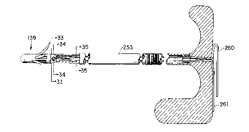
The drill tamper tool of the present invention is shown in Fig.'s 5-7, and
comprises an elongate rigid member 20 having a distal end which comprises a
conical boring tip 21. End 22 of conical boring tip 21 preferably has a cross
section similar in size and shape to the crater created in the pubic bone by thelaser. In this fashion, alignment of conical boring tip 21 within the craterwill be
relatively easy. It is also preferred that end 22 of conical boring tip 21 be blunt
SO that it will not pen~ dl~ soft tissue such as the bladder during normal use (i.e.,
not sufficiently sharp to penetrate soft tissue or organs during normal use).
Certainly, however, conical boring tip 21 should be sufficiently thin and blade-like
CA 02224640 1997-12-12
W O 97/00047 PCT~US96/10582
to permit boring tip 21 to create the bore in the soft cancellous bone by use ofhand force through the laparoscope. In this regard, conical boring tip 21 is
preferably shaped similar to a flat bladed screwdriver. Thus, boring tip 21 has
tapered side surfaces 23 and 24 which terminate in portion 25 which is of a
- 5 circular cross-section. The diameter of circular-crossection portion 25 is identical
to the diameter of the bore which will be created in the pubic bone. By rotatingthe drill tamper tool while simultaneously pressing boring tip 21 into the crater in
the pubic bone, the desired bore will be readily created therein. The diameter of
portion 25 is also approximately the same as the body of the anchor to be
inserted into the bore.
In order to ensure sufficient support for the bone anchors of the present
invention, it is also important that the anchor be seated deep within the pubic
bone. In order to ensure proper depth of the bore, therefore, collar 26 is
1 5 provided on the drill tamper tool. Collar 26 is of a larger diameter than conical
boring tip 21, and therefore will act as a stop preventing further penetration of the
drill tamper tool into the bone. Although collar 26 is shown as tapering in
diameter between conical boring tip 21 and intermediate portion 27, it is also
possible that collar 26 simply col"p,ise a non-tapered end of intermediate portion
27. Intermediate portion 27 has a diameter significantly greater than that of
conical boring tip 21, and is positioned on the opposite end of collar 26.
Intermediate portion 27 not only allows the provision of collar 26, but also adds
rigidity to the tamper tool. Intermediate portion 27, however, should be
significantly smaller in diameter than the operative channel of the laparoscope
so that sufficient vision of the operative region is provided. Preferably, the length
of conical boring tip 21 is between about 1, and 3 cm, and most preferably about1.4 cm. Intermediate portion 27 is preferably between about 2, and 6 cm, and
most preferably about 5 cm.
In order to provide stability during the boring procedure, cylindrical guide
portion 28 is also included on the drill tamper tool. Guide portion 28 has first end
29 and second end 30. First end 29 is attached to intermediate portion 27 at the
CA 02224640 1997-12-12
W O 97/00047 PCT~US96/10582
- 34 -
opposite end of collar 26. Cylindrical guide portion 28 preferably has a diameter
slightly less than the operative channel of the laparoscope. In this fashion, guide
portion 28 provides the necessary stability within the laparoscope to ensure
proper placement of the bores. Second end 30 of guide portion 28 is preferably
attached to handle 31. While handle 31 is shown as having a flat end portion 32
and curved hand grip surfaces 33, handle 31 can be of a variety of forms and still
be sufficient for purposes of the present invention. Handle 31 facilitates the
proper manipulation of conical boring tip 21 through the laparoscope, and
provides a sufficiently firm surface 32 upon which force may be applied to
complete the boring operation. Guide portion 28 preferably has a length between
about 50 and about 55 cm, and most preferably about 52 cm. The overall length
of the drill tamper tool of the present invention therefor permits sufficient access
to the pubic bone, while also providing an ergonomically-effective boring
operation through the laparoscope and ensuring that the tool does not interfere
with the anesthesiologist.
C. Insertion Of Bone Anchors In Pubic Bone
One preferred anchor for use in the present invention is shown in Figs. 3
and 4, and is identical to that disclosed in U.S. Patent No. 5,207,679, which isherein incorporated by reference. Anchor 9, which is preferably made of titaniumalloy or other suitable material, has a cylindrical body 40 and a conical end 44attached thereto. At least two flexible barbs 41 curve outwardly away from body
40. A groove 42 is provided on either side of body 40 at the end opposite to
conical end 44. In addition, cylindrical end 45 extends away from body 40
adjacent groove 42. The longitudinal axis of cylindrical end 45 is aligned with the
longitudinal axis of body 40. The diameter of cylindrical end 45 is preferably
equivalent to the diameter of body 40 within grooves 42 positioned on opposite
sides of body 40. As will be understood below, this structure facilitates the
attachment of anchor 9 to an insertion tool.
CA 02224640 1997-12-12
W O 97/00047 PCTAJS96/10582
- 35 -
As best shown in Fig. 4, body 40 and cylindrical end 45 have an aperture
43 provided therethrough. Aperture 43iS sized so as to accommodate a suture
appropriate for the fixation procedure of the present invention. It is preferred that
a size 0 GORE-TEX suture be employed, and thus anchor 9 and its
accompanying aperture 43 should be sized accordingly. The use of a GORE-
TEX suture is preferred for reasons of strength and non-elasticity. Certainly
other types of sutures could be employed if necessary. A portion of suture 10 isshown in Fig. 3 having been inserted through aperture 43.
The insertion of anchor 9 is relatively straightforward, and merely requires
that the anchor be pressed completely into the bore which has previously been
created in the pubic bone. Preferably, anchor 9 is inserted into the bore in thepubic bone until conical end 44 reaches the distal end of the bore. The bore
should be at least as long as the length of anchor 9, however, it is preferably
considerably longer to ensure sufficient support for the anchor. As anchor 9 is
pressed into the bore, flexible barbs 41 will be compressed against body 40 as
they are inserted past the hard periosteum and cortical bone surrounding the
bore. Once within the bore, however, flexible barbs 41 will tend to spring back
into the soft cancellous bone, thereby securing the anchor in place. A slight tug
on the tails of the suture 10 will also cause barbs 41 to further deploy.
In order to insert anchor 9 into the bore previously created in the pubic
bone, the anchor insertion tool shown in Figs. 8-10 may be employed. Thus,
after the drill tamper tool has been employed to create the necessary bores, theanchor insertion tool of the present invention having an anchor and threaded
suture loaded thereupon, is inserted into the laparoscope for proper seating of
anchor 9.
The anchor insertion tool of the present invention comprises a rigid
elongate member 50 having a handle 51 at one end, and an anchor-receiving tip
52 at the opposite end of elongate member 50. As was the case with the drill
tamper tool, handle 51 can be of any variety, and that shown is only the
CA 02224640 1997-12-12
W O 97/00047 PCTAJS96/10582
- 36 -
presently-preferred embodiment of this handle. Anchor-receiving tip 52 is similar
in construction to that shown in Figs. 4-6 of U.S. Patent No. 5,207,679. Anchor-receiving tip 52 is constructed so as to matingly receive anchor 9 in order to
facilitate insertion of anchor 9 into the bore. As will be apparent, the longitudinal
axis of anchor-receiving tip 52 should be aligned with the longitudinal axis of
elongate member 50. Anchor-receiving tip 52 is cylindrical in nature, having a
diameter approximately equivalent to body 40 of anchor 9. In this manner, at
least a portion of anchor-receiving tip 52 may pass through the bore in the pubic
bone during the anchor insertion process to properly seat the anchor completely
1 0 within the bore.
Anchor-receiving tip 52 has a pair of guide tabs 53 extending from the end
of anchor-receiving tip 52 on either side thereof. Guide tabs 53 are sized and
shaped so as to be ~ Lil Iyly received within grooves 42 positioned on either side
1 5 of anchor 9. Anchor-receiving tip 52 also has a cylindrical slot 54 aligned with
the longitudinal axis of tip 52. Cylindrical slot 54 should correspond in size and
shape to cylindrical end 45 of anchor 9 in order to matingly receive the same.
It is also preferable that the distance between guide tabs 53 be slightly smaller
than the distance between the corresponding grooves 42 on anchor 9. In this
fashion, guide tabs 53 as well as cylindrical slot 54 will apply compressive force
against anchor 9 thereby more securely holding anchor 9 in place when loaded
within anchor-receiving tip 52.
Since suture 10 will extend outwardly on either side of anchor 9, it is
preferable to provide a means for ensuring that suture 10 is not abraded by the
bone surrounding the bore during the insertion process. Thus, a pair of tapered
grooves 55 are provided on either side of the anchor insertion tool, and extend
from the end of anchor-receiving tip 52 along at least a portion of the length of
elongate member 50. As best shown in Fig. 10 wherein anchor 9 has been
loaded upon the insertion tool, grooves 55 ensure that the sutures will be
protected by anchor-receiving tip 52 and a portion of elongate member 50 during
the insertion process. Since any nicks in the suture may compromise the
CA 02224640 1997-12-12
W O 97/00047 PCTAUS96/10582
- 37 -
strength and permanency of the fixation, it is important to ensure that the suture
is not damaged in any fashion.
Anchor-receiving tip 52 should also be of the proper length to ensure deep
placement of anchor 9 completely within the bore. Thus, the length of the
combination of anchor-receiving tip 52 and bone anchor 9 when loaded in the
manner shown in Fig. 10 should be equivalent to the size of the bore created in
the pubic bone. Since the diameter of elongate member 50 is significantly
greater than that of anchor-receiving tip 52, distal end 56 of elongate member 50
1 0 will firmly abut the pubic bone once the anchor has been completely inserted into
the bore. In this manner, the surgeon can be certain that the anchor has been
seated to its complete and proper depth.
As was the case with the drill tamper tool of the present invention,
1 5 elongate member 50 further comprises an intermediate section 57 and a guide
portion 58. Intermediate portion 57 should have a diameter sufficiently less than
that of the laparoscope in order to provide adequate vision for the surgeon, andintermediate portion 57 also has distal end 56 described above. Preferably,
intermediate portion 57 has a length between about 2 and about 6 cm, most
preferably about 5 cm. It should be kept in mind that, as shown in Fig. 10, suture
10 will extend along the length of intermediate portion 57 on either side thereof.
This consideration must be kept in mind when sizing the diameter of intermediateportion 57 to ensure not only that it can be easily inserted through the
laparoscope, but also to ensure adequate vision.
Guide portion 58 will necessarily be slightly smaller than the diameter of
the guide portion for the drill tamper tool previously described, since, as shown
in Fig. 10, suture 10 will extend along either side of guide portion 58. With this
in mind, the total of the diameter of guide portion 58 and twice the diameter ofsuture 10 should be only slightly less than the inside diameter of the operativechannel of the laparoscope. In this fashion, guide portion 28 provides a rigid
support for the surgeon during the anchor insertion process. Preferably, guide
CA 02224640 1997-12-12
W 097/00047 PCTAUS96/10582
- 38 -
portion 58 has a length between about 50 and 55 cm., most preferably about 52
cm. This provides an overall inserter length comparable to that of the drill tamper
tool, thereby providing the same advantages with regard to the tamper tool
length.
In order to effectively employ the procedure of the present invention
laparoscopically, it is necessary that the two tails of suture 10 be controlled as
much as possible. If the sutures are permitted to hang away from anchor 9 or
the anchor insertion tool, the tails will generally become balled within the space
of retzius, thereby making the suture tying procedure difficult, if not impossible.
Although the sutures may be held against the anchor insertion tool by hand,
Applicant has developed a more effective manner of accomplishing this. Thus,
first and second shouldered depressions 59 and 60 are preferably provided
about the circu~r~ llce of guide portion 58. A small rubber band 61 (as shown
in Fig. 10), or other suitable elastic band, may be held within each of the two
shouldered depl~:ssions, with the two tails of suture 10 held beneath rubber band
61 on either side of guide portion 58 as shown. First shouldered depression 59
is preferably positioned approximately 10 cm from the ends of guide tabs 53.
Second shouldered depression 60 is preferably positioned about 40 cm from
guide tabs 53. Both shouldered depressions act, in conjunction with an elastic
band contained therein, to hold suture 10 in place on the sides of the anchor
insertion tool.
In addition to holding the suture in place on the anchor insertion tool, the
shouldered depression/elastic band combination further improves the
laparoscopic procedure of the invention by not only assisting in seating the
anchor, but also in the suture retrieval process. As shown in Fig. 20, the anchor
insertion tool of the present invention, with a pre-loaded anchor and suture
assembly attached thereto, is inserted through the laparoscope for placement of
the anchor as previously described. Once the anchor has been seated within the
bore, the anchor insertion tool is then pulled outwardly utilizing handle 51
contained thereon. The combination of shouldered depressions 59 and 60 and
CA 02224640 1997-12-12
W O 97/00047 PCTAUS96/lOS82
- 39 -
rubberband 61 act to provide tension in suture 10 which in turn pulls outwardly
on anchor 9 which is now contained in the bore. This outward force on anchor
9 will cause flexible barbs 41 to extend outwardly into the cancellous bone
surrounding the bore, thereby further securing anchor 9 in position. In other
words, this outward force on suture 10 by the drag created by the shoulder
depression/elastic band combination will act to deploy the previously compressedbarbs 41 on anchor 9, thereby rigidly securing anchor 9 within the bore.
In order to facilitate the surgical procedure of the present invention, the
drill tamper tool and the anchor-insertion tool described above rnay be providedin the form of a reusable surgical kit.
D. Fixation of Periurethral Tissue At the UVJ Via Suturinq
Once the anchor and suture assembly have been secured within the bore
created in the pubic bone, it is next necessary to utilize the two tails of the suture
to elevate the periurethral tissue on the corresponding side of the urethra at the
UVJ. Applicant has found that the most effective means for accomplishing this
is to pull each tail of the suture through the periurethral tissue into the vagina.
In this fashion, the two tails may then be easily tied to one another within thevagina to provide the necessary support, and eliminating any need for
laparuscopic suture tying. Obviously, however, a means for retrieving the suturetails must be provided.
The method of suture retrieval doing is best shown in Fig. 20, which is a
perspective view of the procedure with portions of the patient's anatomy cut-
away for purposes of clarity. As shown in Fig. 20, as the anchor insertion tool of
the present invention is partially withdrawn from the patient through the
laparoscope, first shouldered depression 59 in combination with rubberband 61
- 30 will cause both suture tails to be tensioned between the anchor and the anchor
insertion tool as shown. Were this not the case, the suture tails would merely fall
into the space of Retzius similar to a ball of yarn, and thereby be difficult (if not
CA 02224640 1997-12-12
W O 97/00047 PCTAUS96/10582
- 40 -
impossible) to retrieve. While the suture tails could be tensioned between the
anchor and the anchor insertion tool by pulling outwardly on the sutures, this
would unnecessarily require an additional pair of hands. Thus, the shouldered
depression/rubber band combination is also effective in this regard.
Once the anchor insertion tool has been partially removed in order to
tension the suture tails in the manner shown in Fig. 20, a suture retrieving tool
may be inserted into the vagina and then pressed upwardly on one side of the
urethra into the space of Retzius in order to retrieve one of the suture tails in the
manner shown. Thus, the suture retrieving tool must have a sharp point capable
of passing completely through the full thickness of the periurethral fascia and
vaginal mucosa adjacent the urethra. The suture retrieval tool must also have
a means for grasping the suture tail and pulling the tail back through the full
thickness of the periurethral and vaginal mucosa by means of the same entry
1 5 hole created by the sharp point. Each tail is pulled into the vagina in this fashion
at the proper location. Pl~:r~r~bly, one tail is pulled into the vagina approximately
1 cm from the urethra at the UVJ, and the other tail is pulled into the vagina
approximately 2 cm lateral from the urethra. In other words, each tail penetrates
the periurethral tissue along an imaginary line extending substantially
perpendicularly away from the urethra. Each tail may then be pulled out of the
vagina for purposes of tying.
The suture tails are tied to one another using a series of standard
surgeon's knots, and each knot is slid by hand to the point in the vagina at which
the suture tails were previously retrieved. The tails are tied to one another in a
sufficiently tight fashion so that suture 10 creates an upward force on the
periurethral tissue adjacent the urethra in order to elevate the urethra at the UVJ
and restore the urethra to its proper angle. The position of the urethra can be
readily observed by the surgeon as this procedure is performed, thereby
ensuring that the urethra is restored to the desired angle. The entire procedure(anchor insertion, suture retrieval, etc.) is then repeated for the anchor placed on
the opposite side of the pubic symphysis, and the suture tails of that anchor are
CA 02224640 1997-12-12
W O 97/00047 PCT~US96/10582
- 41 -
pulled into the vagina through the periurethral tissue on the opposite side of the
urethra as the first suture tails. Tying is then performed in the same fashion,
thereby elevating the other side of the urethra to thereby completely restore the
urethral angle at the UVJ.
The result of this process is best shown in Fig. 2, wherein it is shown that
suture 10, and the corresponding suture on the opposite side of the urethra haverestored the urethra to its proper angle. It should be noted that at no time do the
sutures pull upward directly beneath the urethra, since doing so would create the
risk that the suture would cause urethral blockage. After tying, the remaining
tails of suture 10 are cut at the knot. The portion of suture 10 remaining in the
vagina will epithelize within three to four days, and the patient will no longersense that the sutures are in place. The result is a permanent fixation of the
periurethral tissue on both sides of the urethra, thereby restoring the urethra to
its correct angle and eliminating the SUI. After completion of the tying process,
the surgical area within the patient is flushed with a dilute lidocaine solution, the
laparoscope and trocars removed, a stronger lidocaine solution is applied to theincision sites, and the incisions are closed in the usual fashion. The Foley
catheter may then be removed, and the patient permitted to recover in the usual
fashion. Normal everyday activities may be resumed within 2-3 days.
E. Suture Retriever
In order to pull the tails of suture 10 through the entire thickness of the
periurethral fascia and vagina mucosa, various tools can be employed. For
example, a U.S. Surgical Auto-Stitch tool may be effectively employed for this
purpose. It is critical, however, that the tool employed being capable of readily
be inserted through the periurethral tissue from the vagina into the space of
Retzius, while also being capable of grasping the suture tails. It is also critical
- 30 that the surfaces contacting suture 10 be perfectly smooth in order to eliminate
the risk of nicks or cuts in suture 10 which would obviously compromise the
effectiveness of the procedure. Applicant has developed a novel suture retriever
CA 02224640 1997-12-12
W O 97/00047 PCTAUS96/10582
- 42 -
for accomplishing this purpose which provides a convenient and simple means
of retrieving the suture tails.
The suture retrieval tool of the present invention is depicted in Fig.11, and
comprises metal retrieving end 65, midshaft 66, and handle 67. Midshaft 66 and
handle 67 may be singularly molded from polycarbonate or a similar FDA-
approved material in the typical fashion. As shown in Fig.12, handle 67 is also
preferably knurled in order to facilitate grasping and manipulation of the retriever.
Metal retrieving end 65 is preferably made of stainless steel and can be securely
1 0 molded into distal end 68 of midshaft 66.
Metal retrieving end 65 comprises a rigid, rod-like shaft 69, and a sharp
tip 70 capable of penetrating the periurethral tissue. The diameter of distal end
68 is preferably significantly greater than that of shaft 69, and will act as a stop
1 5 in order to limit the penetration of the suture retrieval tool into the space of
Retzius. Thus, the length of shaft 69 and tip 70 may be selected so as to ensurethat when the suture retrieval tool is inserted into the space of Retzius through
the vagina that sharp tip 70 will generally be incapable of striking any
surrounding soft tissue.
Retrieving end 65 further comprises a return leg 71 which extends away
from sharp tip 70 in the same direction of shaft 69. Shaft 69, retum leg 71 and
the underside of sharp tip 70 thus create an inverted U-shaped region capable
of ensnaring suture 10 for retrieval purposes. The distance between return leg
71 and shaft 69 in the region of the inverted U-shape is preferably approximately
the same diameter as suture 10. In this fashion, and as shown in Fig. 20, after
sharp tip 70 has penetrated the periurethral tissue adjacent the vagina in orderto enter the space of Retzius, the inverted U-shape may be pulled downwardly
over a suture tail, thereby snaring the suture. Metal retrieving end 65 may thenbe pulled back through the periurethral tissue, and the tail of suture 10 will
remain snared between return leg 71 and shaft 69 directly beneath sharp tip 70.
CA 02224640 1997-12-12
W O 97/00047 PCTrUS96/10582
- 43 -
Although the suture tail will slide within the U-shaped region, it will nevertheless
be pulled into the vagina.
One critical feature of the suture retriever of Fig. 12 is that the inner
- 5 surfaces of metal retrieving end 65 which contact suture 10 must be rounded and
smooth in order to permit suture 10 to freely slide within the inverted U-shapedportion as the retriever is withdrawn. This prevents nicking or fraying of the
suture while still permitting the suture tail to be withdrawn. Thus, the only sharp
portion of metal retrieving end 65 is sharp tip 70.
1 0
While it is possible that handle 67, midshaft 66, and metal retrieving end
65 may all be positioned along the same longitudinal axis thereby forming a rigid,
elo"g~le structure, Applicant has found that the process of the present invention
can be simplifed if the elements of the retriever have the angular relationship
indicated in Fig. 11. This is particularly true when the template of the presentinvention (to be described further herein) is employed. Thus, metal retrieving
end 65 preferably is positioned at an angle of between about 50 degrees and
about 80 degrees, most preferably about 60 degrees, to midshaft 66 (this angle
is indicated as A in Fig.11). Likewise, handle 67 is preferably positioned at anangle of between about 50 degrees and about 80 degrees, most preferably about
60 degrees, to handle 67 (this angle is indicated as B in Fig. 11). Thus, the
longitudinal axis of shaft 69 of metal retrieving end 65 will be parallel to thelongitudinal axis of handle 67. In this fashion, when the retriever of the present
invention is employed midshaft 66 will generally be positioned parallel to the
longitudinal axis of the vagina. Metal retrieving end 65 will then extend upwardly
through the periurethral tissue into the space of Retzius as desired. As best
shown in Fig. 20, handle 67 will thereby extend downwardly outside of the
vagina, and thus sharp tip 70 may be readily forced through the periurethral
tissue as desired merely by pushing upwardly on handle 67.
As shown in Fig.11, the transitions between handle 67 and midshaft 66
as well as between midshaft 66, and retrieving end 65 should be gently curved
CA 02224640 1997-12-12
W O 97/00047 PCTAUS96/10582
- 44 -
in order to ease placement. It should be noted that angles A and B can vary
significantly, however it is preferred that they be as close to one another as
possible so that the parallel relationship of handle 67 and retrieving end 65is
maintained. In addition, the present configuration ensures that the retrieving end
65 will enter the space of Retzius at an angle of approximately 90 degrees to the
tensioned suture tails (i.e., the suture tails tensioned between the anchor and the
anchor insertion tool). This perpendicular relationship allows for effective snaring
of the suture, as well as improved vision of the retrieval through the laparoscope.
Preferably, midshaft 66is between about 2 and about 6 cm. in length,
most preferably about 4 cm. The cross-sectional area of midshaft 66 should
merely be a size chosen to provide the necessary rigidity while also not blocking
vision within the vagina during the procedure. Likewise, handle 67is preferably
about 4 cm long, however the length is not as critical since handle 67 remains
1 5 compl~tely outside of the body. The diameter of handle 67 should be chosen so
as to provide a comfortable and secure means of grasping the suture retrieval
tool. Preferably, handle 67is about 3 cm in diameter.
F. Surgical Template
While the placement of the sutures at the UVJ may be accomplished by
merely feeling for the correct location within the vagina by hand, particularly in
relation to the ball of the Foley catheter, Applicant has developed a novel
template which greatly simplifies proper placement of the sutures. This device
not only prevents bladder injury, urethral injury, or vascular accidents, the
template also ensures a proper distance between the two suture tails to ensure
that there is adequate periurethral tissue between the tails to provide the
necessary support. Obviously if the suture tails are placed too close to one
another, there is a risk that the suture will tear through the periurethral tissue and
eliminate the fixation. Thus, a template is provided wherein the template has atleast two apertures which may be properly positioned on either side of the
urethra. The suture retriever may then be inserted through these apertures and
CA 02224640 l997-l2-l2
W 097/00047 PCTrUS96/10582
- 45 -
thereafter through the periurethral tissue in order to snare the suture tails in the
manner described previously.
The presently preferred template is depicted in Figs. 13-17. The template
~ 5 comprises a trough 80 of arcuate cross-section, wherein trough 80iS sized so as
to cradle the patient's urethra when properly positioned. First and second wing
members 81 and 82 extend away from opposite sides of trough 80, preferably
perpendicularly to the longitudinally axis of trough 80 in the manner shown. Most
preferably wing members 81 and 82 extend perpendicularly away from opposite
sides of trough 80 at the upper most edges 83 and 84 of trough 80. First and
second suture guide apertures 85 and 86 are positioned in each of the wing
members 81 and 82 as shown. The guide apertures are positioned so that when
the urethra of the patient is properly positioned within trough 80, guide apertures
85 and 86 on each wing will indicate the proper location for the sutures. First
suture guide aperture 85 iS positioned so that the first tail of suture 10 will
penetrate the periurethral tissue approximately 1 cm from the urethra adjacent
the UVJ. Second guide aperture 86is preferably positioned about 1 cm further
away from the urethra along a line perpendicular to the longitudinal axis of trough
80. In other words, the distance between first suture guide aperture 85 and
second suture guide aperture 86iS preferably about 1 cm. In this manner, the
surgeon can be confident that sufficient periurethral tissue will be present
between the two suture tails.
It is certainly possible that the template of the present invention may
merely be held in place by hand, and in fact the downwardly sloping nature of the
underside wing members 81 and 82 are suitable for placement of the surgeon's
fingers thereunder. Additional alignment means are preferably provided,
however at a minimum, end wall 87is provided at the end of trough 80 furthest
away from the suture guide apertures. Thus, as long as the template is properly
sized for the length of the patient's urethra, the surgeon may hold the templatein place with end wall 87 abutting the outermost end 89 of urethra 4 in order toensure proper alignment. For example, if the length of the patient's urethra is
CA 02224640 l997-l2-l2
W 097/00047 PCTAUS96/10582
- 46 -
determined to be three cm, the distance between end wall 87 and first and
second guide apertures 85 and 86 should be between about 2.5 and about 3.2
cm, most preferably about 2.8 cm. The guide apertures should also be between
about 0.25 and about 0.5 cm from distal end 91 of the wing members (Fig. 13).
As long as the template is positioned with end wall 87 abutting the outermost
portion of the urethra, the surgeon will be assured that the sutures will be
properly placed without risk of puncturing the bladder, or urethra.
While the surgeon may employ two fingers beneath wing members 81 and
82 to hold the template in the proper position, Applicant has found that the
provision of arcuate alignment member 88 secured to end wall 87 may be
effectively employed for securing the template in place without the need for thesurgeon to hold the template in any manner. Alignment member 88iS preferably
positioned parallel to trough 80, with the center line of alignment member 88
aligned with the center line of trough 80. Alignment member 88 may either
extend away from trough 80 as shown in Fig. 13, or alternatively may extend
away from end wall 87 directly along the interior of trough 80 as shown in Fig. 18.
When the embodiment of Figs. 13-16 is employed, alignment member 88
is preferably sized so that when the template is manufactured from a resilient
material, alignment member 88 may be rigidly snapped about Foley catheter 14
as shown in Fig. 20. Thus, the shaft of Foley catheter will be securely held within
alignment member 88, and the surgeon need only pull outwardly on the Foley
catheter while sliding the template into the vagina towards the urethra until end
wall 87 abuts end 89 of urethra 2. The ball of the Foley catheter will thus be
positioned at the UVJ, and the template will likewise be positioned at the
appropriate location assuming that a template of the proper size has been
selected based upon the length of the urethra.
As an alternative to providing various sizes for the template of the present
invention, a single, larger-sized template may be employed provided that
alignment member 88 iS snapped about the shaft of Foley catheter 14 in the
CA 02224640 l997-l2-l2
W O 97/00047 PCTAUS96/lOS82
- 47 -
proper location. Thus, instead of abutting end wall 87 against end 89 of urethra2, end wall 87 is instead aligned with the appropriate graduation along the shaft
of Foley catheter 14 based upon the previously-measured length of the urethra.
Likewise, end 90 of alig~ Ime~ ll member 88 could alternatively be aligned with the
- 5 appropriate graduation along the shaft of Foley catheter 14 in order to provide
the proper placement of the template based upon the length of the patient's
urethra.
Fig. 18 depicts yet another altemative embodiment for the template of the
present invention in which alignment member 88 extends along the interior of
trough 80. In employing this embodiment, alignment member 88 is inserted into
the urethra along with Foley catheter 13 until end wall 87 of the template abutsend 89 of urethra 2. As will be understood, therefor, an appropriately-sized
template will ensure proper placement of the sutures.
1 5
Finally, the template of the present invention can be manufactured of any
suitable material such as polycarbonate or other FDA-approved plastic. The
template may be readily molded using known technology, and is preferably
manufactured as a disposable, single-use item. The drill tamper tool and anchor
insertion tools, on the other hand, should be made from medical-grade stainless
steel. The handles, however, may be polycarbonate or other FDA-approved
plastic.
G. Alternative. and Presently Preferred Anchor and Anchor-lnsertion Tool
As mentioned previously, and as best shown in Fig. 19, pubic bone 12
falls away from the laparoscope at an angle of about 45~ to 60~. Thus, it is
impossible to create a bore in the pubic bone which extends perpendicularly fromthe surface of the bone. Thus, the bore produced in pubic bone 12 extends
- 30 inwardly at an angle of between about 20~ and about 60~. In other words, as
shown in Fig. 23 which is a cross-sectional view of pubic bone 12, when bore 13
is viewed in cross-section as shown, the angle between surface 100 of the pubic
=
CA 02224640 1997-12-12
W O 97/00047 PCTAUS96/10582
- 48 -
bone and bore 13 at X wiil be between about 20~ and about 60~, preferably about
45~. During the laparoscopic surgical procedure of the present invention, the
surgeon may readily modify angle X in order to provide an angle which
corresponds to that of the anchor to be described (preferably about 45~). This
may be accomplished by inflating or deflating the abdominal cavity as needed in
order to change the angle of the laparoscopic channel in relation to the pubic
bone. Since interior surface 102 of cortical bone 101 is parallel to outer surface
100 of pubic bone 12, this same angular relationship will exist within the interior
of the bore. When an anchor such as that disclosed in Fig's 3 and 4 is
employed, one will immediately recognize that this angular relationship of surface
102 and bore 13 will only permit the lower barb of the anchor to contact surface102 Since cancellous (or tribecular) bone 103 offers very little support for theanchor, only the contact between the lower most barb of the anchor of Fig's 3
and 4 and surface 102 provides the principle support which prevents the anchor
from being pulled out of the bore.
When anchors of the type shown in figures 3 and 4 are tested for pull-out
strength, they are typically pulled only in the direction of arrow B. After the
urethropexy procedure of the present invention is accomplished, however, any
force on the anchor is most often in the direction of arrow A. As will be readily
understood, due to that fact that only the lower barb will be in contact with
surface 102, it is possible for the anchor to be pulled out of the bore due to forces
acting in the direction of arrow A. This results from the fact that the end of the
anchor to which the suture is secured is able to pivot downwardly in the direction
of ~rrow A since cancellous bone 103 offers little support.
In order to overcome the problems of the prior art anchor designs noted
above, the present applicant has developed a new anchor design shown in Fig's
23-30. The shortcomings of the prior art are overcome by modifying the
orientation of the wing members or barbs to ensure that the external ends of thewing members (i.e., the portions which contact inner surface 102 of the corticalbone) have an angular relationship corresponding to the angular relationship
CA 02224640 1997-12-12
W O 97/00047 PCT~US96/10582
- 49 -
between bore 13 and surface 102 (to be further described in detail below). Thus,as best shown in Fig's 24 and 25, anchor 139 comprises cylindrical body 140, at
least three wing members (barbs 141,142 and 143), and aperture 150 positioned
in tab member 151. Body 140 is similar to that of the prior art designs, howeverdistal end 160 may be rounded since it is relatively easy to urge distal end 160into cancellous bone as needed. The conical shape of the prior art design,
however, will certainly work. Proximal end 161 is preferably perpendicular to
longitudinal axis 162 of body 140 to thereby provide a flat proximal end surfacewhich may cooperate with the new anchor-insertion tool to be described herein.
Aperture 150 serves the same purpose described previously (i.e., a means for
attaching a suture to the anchor), and once again the surfaces and edges of
aperture 150 should be as smooth as possible to prevent fraying of the suture.
In fact, aperture 150 may even be highly polished or even coated to reduce
friction.
Tab member 151, as shown in Fig's 24-26, preferably has a height 163
corresponding approximately to the diameter of body 140 and a width 164 less
than the diameter of body 140. The length 165 of tab member 151 should be
sufficient for aperture 150 to permit a size 0 suture to be passed therethrough.It is also preferred that the length of tab member 151 extend perpendicularly
away from proximal end 161 to facilitate cooperation and mating with the anchor-insertion tool to be described.
As mentioned above, the most significant feature of the anchor shown in
Fig's 23-30 is the arrangement of the wing member or barbs. As is the case with
the prior art anchor design, the barbs may be elastically flexed (i.e., have
memory) from the normal deployed position (as shown in the figures), to a
compressed position. In the deployed position, the barbs preferably curvilinearly
extend radially and axially away from body 140, as shown. In the compressed
position, the barbs are substantially ~ ;yhLe,1ed to pemmit insertion of the anchor
into a bore created in bone in the manner of the prior art (see, for example, U.S.
Patent No's 5,207,679 and 5,217,486 both of which are herein incorporated by
CA 02224640 l997-l2-l2
W O 97/00047 PCTAJS96/10582
- 50 -
way of reference). To facilitate cor,,~ur~ssion and insertion, a plurality of channels
170 are provided along the length of body 140. One of channels 170 is
positioned beneath each of the wing members, and is aligned therewith. Each
of the channels is further sized so as to accept at least a portion of, and
preferably the entirety of the corresponding wing member aligned therewith. In
this manner, when a wing member is compressed during the anchor insertion
process, the compressed and straightened wing member will be positioned
entirely within channel 170 positioned adjacent the wing member.
With the wing members in this compressed state, the diameter of the
anchor will be equivalent to that of body 140, thereby permitting the anchor to be
inserted into a bore having a diameter only slightly greater than body 140. As the
wing members extend into the bore beyond interior surface 102 of the cortical
bone, the wing members will return to their deployed position, since the
cancellous bone is not sufficiently hard to resist the spring-back of the wing
members due to their inherent el~ticity (or memory). As shown in Fig. 23, since
the diameterof bore 13 is only slightly greaterthan body 140, the external ends
of the wing members will abut against interior surface 102 thereby preventing
removal of the anchor from the bore. For example, external end 144 of barb 141
is shown in Fig. 23 as resting against interior surface 102.
The cross-sectional profile of channels 170 is shown in Fig. 30, which is
a cross-sectional view of the anchor of Fig. 26. As shown in this view, channels170 are preferably sufficiently deep to permit an entire wing member to be
compressed into the channel. In addition, as shown in Fig.24, the length of eachchannel 170 is only sufficiently long to contain the entire corresponding wing
memberwhen the latter is compressed, Thus, channels 170 may be of different
lengths depending upon the various lengths of the corresponding wing members.
End 171 of channel 170 (see Fig. 24) may also be angled in relation to the
longitudinal axis 162 as shown in orderto correspond with the external ends of
the wing members, should these external ends be angled in the manner to be
described.
CA 02224640 1997-12-12
W O 97/00047 PCTAJS96/10582
- 51 -
In order to ensure that at least a portion of the external ends of each wing
member contacts interior surface 102 of the cortical bone adjacent bore 13 when
bore 13 is positioned non-perpendicularly to outer surface 100 of the pubic bone,
at least a portion of the external end of each wing member is aligned along first
imaginary plane 175 extending through body 140 (see Fig. 25a). In the prior art
anchor designs this imaginary plane is perpendicular to longitudinal axis 162,
thereby evenly aligning all of the external ends of the wing members. In the
anchor of the present invention, however, first imaginary plane 175 extends
through said body at an angle to transverse cross-section 176 through body 140,
wherein cross-section 176 is perpendicular to longitudinal axis 162. As will be
understood, angle F of Fig. 25a is preferably substantially equivalent to angle X
of Fig.23, thereby ensuring that at least a portion of the extemal end of each barb
will contact interior surface 102 of the cortical bone adjacent bore 13. Thus,
angle F is preferably between about 20~ and about 60~, most preferably about
45~. The external ends of the wing members are more preferably tapered as
shown so that the entirety of each external end will lie along first imaginary plane
175. Thus, external ends 144, 145 and 146 of corresponding barbs 141, 142,
and 148 are aligned along plane 175, thereby adding further support when the
anchor is secured within bore 13.
As will be more fully understood later, it is helpful at this point to also
define the top of the anchor by a top-line 166 which extends along the surface
of body 140 parallel to longitudinal axis 162. First imaginary plane 175 intersects
top-line 166 at the point of intersection between the surface of body 140 and
imaginary plane 175 which is nearest to proximal end 161. In other words,
therefore, the barb extending from body 140 at point nearest to top-line 166 will
also have an external end which is closest to proximal end 161. The further fromtop-line 166 that a barb extends from body 140, the further its external end will
be from proximal end 161. The upper portion of body 40 is defined as that half
- 30 of body 40 nearest to top-line 166, and the lower portion is therefore the
remainder.
CA 02224640 1997-12-12
WO 97/00047 PCT~US96/10582
- 52 -
To provide the alignment described above, the wing members may extend
from the anchor body in a number of fashions. Most preferably, however, each
barb should be of a similar length in order to provide similar flexing
characteristics for each barb thereby ensuring that the anchor will be urged
straight into the bore. Thus, each wing member preferably extends from body
140 along a second imaginary plane extending through body 140 at an angle to
cross-section 176. As will be understood, the angle between this second
imaginary plane and cross-section 176 should be approximately equivalent to
angle F of Fig. 25a. Alternatively, the wing members may extend from the
1 0 anchor body at points equidistant from proximal end 161 (i.e., along a plane
extending perpendicularto longitudinal axis 162). In orderto provide the proper
alignment for the external ends of the barbs, however, the length of the barbs in
this embodiment would necess~rily not all be equivalent to one another.
In order to provide proper support during use, various configurations and
numbers of barbs may be employed. Three preferred arrangements are shown
in Fig's 27-29. In the embodiment of Fig. 27, which corresponds to that of Fig's23-26, five barbs are employed. Thus, barbs 141,142, and 143 extend upwardly
away from the upper portion of body 40, while barbs 148 and 149 extend
downwardly away from the lower portion. When the anchor is viewed from the
proximal end towards the distal end (Fig. 27-29), it is seen that barb 141 is
substantially aligned with the top-line of body 140. To simplify the description,
this will be defined as the 12 o'clock position in temms of a standard clock face.
Thus, barb 141 is at about 12 o'clock, barb 142 is between about 10 and 11
o'clock, barb 143 is between about 1 and 2 o'clock, barb 149 is between about
4 and 5 o'clock, and barb 148 is between about 7 and 8 o'clock. It should be
pointed out that whenever an odd number of barbs are employed, it is preferable
that a greater number of barbs extend from the upper portion of the body rather
than the lower portion, since further support is needed in this area.
In the embodiment of Fig. 28, three barbs (152, 153 and 154) are
employed, two of which extend from the upper portion of body 140. Preferably,
CA 02224640 l997-l2-l2
W O 97/00047 PCTAJS96/10582
- 53 -
barb 153 is between about 10 and 11 o'clock, barb 152 is between about 1 and
2 o'clock, and barb 154 is at about 6 o'clock. In the embodiment of Fig. 29, four
barbs (155, 156, 157 and 158) are utilized, two extending upwardly away from
the upper portion of body 140, and two extending downwardly away from the
lower portion of body 140. Preferably, barb 155 is between about 10 and 11
o'clock, barb 156 is between about 1 and 2 o'clock, barb 157 is between about
4 and 5 o'clock, and barb 158 is between about 7 and 8 o'clock. It should be
noted that any of a number of alternative configurations are possible, and the
three shown are merely exemplary of the simplest and preferred embodiments.
1 0
The anchor of the present invention may be made of any of a number of
materials, however titanium or titanium alloys are preferred. The anchor is alsopreferably singularly molded, however the wing members may be formed
separately and attached to body 140 during the assembly process. For example,
the wing members or barbs may be attached to body 140 in the manner
described in U.S. Patent No's 5,207,679 and 5,217,486 both of which are herein
incorporated by way of reference. It is preferred, however, that the barbs
comprise wire-like members, and may even be formed from titanium or nickel-
titanium wires in a known fashion.
As also discussed previously, aperture 150 of anchor 139 is designed so
that suture 180 may pass therethrough. In this fashion, two suture tails will
extend from aperture 180, and may be used in the manner previously described
to elevate the periurethral tissue at the UVJ, for example. It is important during
such a procedure, however, that the suture be protected from nicks and other
abrasions which would compromise suture strength. While the proximal end of
the anchor shown in Fig's 23-30 may be modified to that shown in Fig's 34 so
that the anchor-insertion tool of Fig's 8-10 can be employed therewith, Applicant
has developed yet another anchor-insertion tool which can be employed with the
anchor design shown in Fig's 23-30. This new anchor-insertion tool not only
protects the suture, it also facilitates proper alignment of the anchor within the
CA 02224640 l997-l2-l2
W 097/00047 PCT~US96/10582
- 54 -
bore and also provides a means for tensioning the suture tails in the manner
shown in Fig. 20.
An anchor-insertion tool according to the present invention is shown in
Fig's 31-35, and comprises a rigid elongate member 250 having:
-anchor-receiving tip 252
-hollow cylindrical intemmediate portion 253 having distal end 260 adjacent
anchor-receiving tip 252, intermediate portion 253 preferably having a
diameter greater than the diameter of tip 252
-handle 251 attached to intermediate portion 253 at the end opposite
anchor-receiving tip 252
-passageway 254 providing communication between the interior of
intermediate portion 253 and the exterior of the tool
Since anchor-receiving tip 252 is also preferably hollow and its interior is in
communication with the interior of intermediate portion 253, an anchor having a
suture extending therefrom may be secured on tip 252, and the suture
(preferably a pair of suture tails) will extend through tip 252, through at least
intermediate portion 253, and through passageway 254. As shown in Fig. 31,
passageway 254 is preferably provided in handle 251 and terminates on rear
face 255 of handle 251. Since passageway 254iS in communication with the
interior of intermediate portion 253, the suture tails may extend along the entire
interior length of the tool, and exit at rear face 255 of handle 251. Thus, as
shown in Fig. 32, suture tails 260 and 261 extend from anchor 139 held on tip
252, through the entire interior of the tool, and exit at rear face 255. Although the
suture tails may exit from the interior of the tool at various other locations, rear
face 255 provides a location convenient for the surgeon during laparoscopic
urethropexy and similar procedures. In addition, the sutures are completely
enclosed within the tool, and thus nicking or fraying cannot occur during the
insertion process. It should be noted that the length of suture tails 260 and 261
has been shortened for purposes of clarity.
CA 02224640 1997-12-12
W O 97/00047 PCT~US96/10582
Anchor-receiving tip 252 should be structured so as to matingly engage
the particular anchor being employed. In addition, as described previousiy, the
diameter of tip 252 should be equivalent to that of the anchor body to facilitate
anchor insertion. In the embodiment of Fig. 31-32, tip 252 has a slot 256
extending across the diameter of the tip and at least partially along its length, slot
256 capable or matingly receiving tab member 151 of anchor 139. Since it is
critical that anchor 139 be properly aligned within the bore in the pubic bone, slot
256 prevents rotation of the anchor during the anchor insertion process. Slot 256
is preferably sized equivalent to or slightly larger than tab member 151 of anchor
1 0 139, so that tab member 151 may be placed within slot 256. Tab member 151
may be wedged within slot 256 to hold the anchor oh tip 252, or alternatively, end
258 of tip 252 may be secured to proximal end 161 of anchor 139 by means of
a releasable adhesive. Any FDA-approved epoxy or other adhesive may be
employed, as long as the bond between end 258 and proximal end 161 may be
1 5 readily broken after the anchor has been seated within the bore merely by pulling
the insertion tool away from the bore. In addition, since the suture tails are
preferably tensioned between the anchor and handle 251 in a manner to be
described, suture tails 260 and 261 will also act to keep tab member 151 held inplace in slot 256.
As best shown in the cross-sectional views of Fig's 33-34, the wall
thickness of tip 252 should be such that suture 180 which extends through
aperture 150 of the anchor tab member will not be compressed by the interior
walls of tip 252 when the anchor is in place on tip 252. In other words, the
interior diameter of anchor-receiving tip 252 is greater than the sum of the width
of tab member 151 plus twice the diameter of suture 180. In this manner, suture
180 will be further spared from damage during the anchor insertion process.
As will be apparent, in order to properly seat anchor 139, anchor-receiving
- 30 tip 252 should have a length sufficient to ensure that all of the barbs of the
anchor are inserted past the cortical bone for proper barb deployment. Since it
is preferred that proximal end 161 of anchor 139 be approximately aligned with
CA 02224640 l997-l2-l2
W 097/00047 PCTnUS96/10582
- 56 -
interior surface 102 of the cortical bone surrounding the bore when the anchor
is properly seated within a bore (i.e., external ends of all barbs resting against
interior surface of cortical bone), it will be apparent that anchor-receiving tip 252
should have a length approximately equivalent to, or slightly greater than the
thickness of the cortical bone (between about 5 mm and about 8 mm). End 260
of intermediate portion 253 not only acts to drive the anchor into the bore, butalso acts as a stop since its diameter is preferably greater than the anchor body
and the bore into which it is being inserted. Thus, the proper length for tip 252
will ensure not only that the anchor is driven sufficiently deep within the bore to
fully deploy all of the barbs, but also to ensure that the anchor is not driven too
far into the bore. In the latter situation, anchor stability may be compromised,and the suture may be damaged by the edges of the bore once the surgical
procedure has been completed.
In order to facilitate manipulation of the anchor-insertion tool within the
laparoscope, a plurality of ridges 259 are preferably provided about the
circumference of intermediate portion 253. Ridges 259 are preferably positioned
adjacent handle 151, most preferably between about 5 cm and about 10 cm from
the handle. Since the diameter of intermediate portion 253is preferably slightlysmaller than the diameter of the lapar~Jscope to be employed (preferably 7.8 mm
and 8.0 mm respectively), ridges 259 will assist in controlling the insertion tool
during the procedures of the present invention. Preferably, ridges 259 are sizedso as to cooperate with the standard rubber grommet of a laparoscope.
Handle 252 may be secured to intermediate portion 253 by any of a
number of means, however it is preferable that handle 252 be threadably
secured to intermediate portion 253. Thus, corresponding male and female
threads are provided on the handle and the intermediate portion. Once again
handle 251 preferably has curved hand grip surFaces . In this embodiment,
however, handle 251 preferably has dissimilar top and bottom portions 271 and
272, respectively. If anchor 139is secured to tip 252 such that the top-line of the
anchor is aligned with top portion 271 of handle 251, the surgeon can readily
CA 02224640 1997-12-12
W 097/00047 PCTAUS96/10582
ensure that the anchor will be properly inserted into the bore. As long as top
portion 271 extends directly upwardly during anchor insertion, proper placement
will be guaranteed. In order to f~ t~te alignment, bottom portion 272 of handle
251 is preferably significantly heavier than top portion 271. This weight
differential will further guarantee alignment, since gravity will cause the insertion
tool to rotate within the lapa~oscopic channel to the correct position. It should be
appreciated, however, that this alignment means (i.e., and oriented handle)
requires that the engagement of handle 251 with intermediate portion 253 be
precise. Of course other ..li~"",e,ll means may be provided, such as guide lines,
leveling bubbles, and the like.
As mentioned previously, providing tension in the suture tails extending
from the anchor will not only help in maintaining the anchor on anchor-receivingtip 252, but will also assist during the suture retrieval step shown in Fig. 20 (i.e.,
when the insertion tool has been partially removed from the laparoscopic
channel). Proper tensioniilg can be achieved by means of at least one notch intowhich the suture tail(s) may be wedged. Preferably, the notch(es) are positionedadjacent said passageway so that the suture tails may extend through the
passageway directly into the notch(es). Thus notches 280 and 281 are
preferably provided on rear face 255 of handle 251. In fact, passageway 254
preferably exits handle 251 in an elongated diamond-shaped exit 279, and thus
notches 280 and 281 may comprise the opposing narrow corners of exit 279 as
shown. When suture tails 260 and 261 leave passageway 254 through exit 279,
each tail may be wedged within notches 280 and 281, respectively. In this
manner, the tails will be tensioned between notches 280 and 281 and aperture
150 of the anchor. After the anchor has been inserted into the bore, and as the
insertion tool is pulled partially out of the laparoscopically channel, suture tails
260 and 261 will slide within their respective notches, thus maintaining the
desired tension in the suture tails as needed for the suture retrieval step. To
~ 30 further enhance the tensioning of the sutures, passageway 254 may taper from
intermediate portion 253 towards exit 279, thereby providing an additional
wedging of the suture tails within the final portions of passageway 254.
CA 02224640 1997-12-12
W O 97/00047 PCTAJS96/10582
- 58 -
It should be noted that intermediate portion 253 pre~erably has a length
of between about 52 cm and about 55 cm to facilitate its use through a
laparoscope.
To facilitate a quick and proper anchor insertion process, anchor 139 and
the anchor-insertion tool of the present invention may be provided to the surgeon
in the loaded form of Fig. 32 (with suture attached and wedged for tensioning asshown). The anchor may even be rele~s~hly adhered to the anchor-receiving
tip as shown. In this manner, the surgeon can merely open a sterile surgical kitcontaining preferably two pre-loaded insertion tools (i.e., anchor 139 engaged
with anchor-receiving tip and suture tensioned between anchor and notches on
handle 251). The pre-loaded insertion tools are surricYe,llly inexpensive to permit
one-time use, or the manufacturer could provide a credit for the return of used
anchor-insertion tools which can be sterilized and re-loaded. The surgical kit for
pel rul " ,i"g laparuscopic urethropexy can also include a drill tamper tool, a suture
retrieval tool, and/or one or more variously-sized templates (all of which have
been described previously). In this fashion, the surgeon will have all of the tools
necessary for a quick and simple urethropexy within a small sterile kit.