Note : Les descriptions sont présentées dans la langue officielle dans laquelle elles ont été soumises.
CA 02232163 1998-03-16
.
FULLY A~TONATED METHOD AND ~NT COMPOSITION ~H~ OR
FOR RAPID ID~ ~lcATIoN AND ~U~ T~RIZATION OF
RETIC~LOCYTE8 ~KY-.nKOCYTES AND PLATELETS IN WHOLE BL'OOD
FIELD OF THE lNV~ lON
The present invention relates to fully automated
hematology analysis methods and reagent compositions used
therein for identifying and characterizing cells in
samples of whole blood, and more particularly for (i)
identifying reticulocytes and erythrocytes; (ii)
identifying and distinguishing platelets; and (iii)
simultaneously measuring the volume, hemoglobin
concentration and hemoglobin content of large numbers of
individual reticulocytes and erythrocytes, in a whole
blood sample by light scatter and absorption flow
cytometry techniques.
BACRGROUND OF THE lNV~ ON
In all higher animals, blood consists of an
aqueous fluid part (the plasma) in which are suspended
corpuscles of various kinds: the red blood cells
(erythrocytes), the white blood cells (leukocytes) and the
blood platelets. Plasma has a composition comprising
roughly 90% water, 9% protein, O.9% salts and traces of
other materials such as sugar, urea, uric acid, and the
like.
The cells or corpuscles of the peripheral blood
(i.e. the blood outside of the bone marrow) are divided
into two main groups: erythrocytes whose primary object is
to transport oxygen, and leukocytes whose primary function
relates to the immune system and the destruction of
materials foreign to the body. In addition to these two
main groups, the blood also contains the so-called blood
platelets which are important in hemostasis.
The final stages of erythrocyte maturation occur
after their release from the bone marrow while these cells
CA 02232163 1998-03-16
are circulating in the peripheral blood. These young red
cells, or "reticulocytes", have lost their nuclei, and
thus, their ability to divide or to synthesize ribonucleic
acid (RNA). Although these functions have ceased,
reticulocytes are still metabolically active and are
S capable of synthesizing protein, taking up iron for the
synthesis of heme, and carrying out the necessary
metabolic reactions required to maintain an energy-rich
state. These cells are usually most easily distinguished
from mature erythrocytes by exposing them to solutions of
cationic dyes which react with the anionic RNA in the
reticulocytes and precipitate into a fine or coarse
stained "reticulum" within the reticulocytes, which gives
the reticulocytes their name.
Although reticulocytes normally comprise about
0.5 to 2 percent of the total red blood cell population,
this percentage can change dramatically under abnormal
conditions. For example, reticulocyte counts have been
used for many years as a diagnostic aid in studying blood
dyscrasias, as an index of red blood cell regeneration
following hemorrhage, as well as for monitoring early
toxicity in chemotherapy of certain malignant diseases.
Nucleic acids ~RNA and DNA) are polyanions which
can be stained with practically any cationic dye. The RNA
in reticulocytes can be stained with only a few cationic
dyes, including, for example, Brilliant Cresyl Blue (BCG),
New Methylene Blue (NMB), Auramine O (AuO), Acridine
Orange (AO), Thiazole Orange (TO), Oxazine 750, and
Pyronine Y (PY). Among these dyes, only a sub-set can be
made to penetrate the cells (and therefore stain) rapidly.
The rate and degree of staining of reticulocytes depend
upon the extracellular concentration of the dye, the rate
of penetration of the dye through the reticulocyte
membrane, and the strength of the specific binding
constant between the cationic dye and the reticulocyte
RNA. The latter two properties are different, and are not
CA 02232163 1998-03-16
easily predictable for each dye, so that trial and error
~ are necessary to discover useful reticulocyte stains.
Several semi-automated methods are available
which can be used for counting the percentage of
reticulocytes in an anticoagulated sample of whole blood
using fluorescent based methods and systems. In such
methods, a diluent containing an organic cationic dye,
such as AO, AuO or TO, is used to stain the RNA within the
reticulocytes. The dye penetrates the cell membrane and
binds to the RNA and usually precipitates a "reticulum"
within each reticulocyte. The amount of the signal from
stained RNA is roughly proportional to the RNA content.
After proper staining, a fluorescence flow cytometer
(rather than an absorption flow cytometer), equipped with
the proper excitation light source (typically an argon ion
laser emitting at 488 nm), and emission detection system,
can be used to determine the percentage of reticulocytes
in the effluent.
Although fluorescent cytometric methods and
fluorescent dyes have been used in the art to
differentiate reticulocytes in whole blood samples, there
is a dearth of very rapid, accurate, and reliable
cytometric methods using fully automated
scatter/absorption flow cytometry techniques for
determining reticulocyte, red blood cell and platelet
counts, and for assessing further parameters of
reticulocytes and red blood cells. However, the present
invention, which comprises a method and reagent designed
for rapid whole blood sample analysis via light scatter
and absorption fully automated flow cytometry, provides
the art with a needed and highly advantageous blood
analysis method and reagent for use with fully automated
hematology systems. The invention speeds up the process
of blood cell analysis to allow more samples to be tested
and analyzed and to provide a much faster turnaround time
for processing and, ultimately, informing clinicians,
CA 02232163 1998-03-16
physicians, and patients of the results.
A difficulty in monitoring reticulocyte counts
with a flow cytometer is the problem of differentiating
between reticulocyte detection signals, mature red blood
cell signals, and system noise. The stained strands of
RNA are numerous in young reticulocytes, and generate
signals of relatively large magnitude when detected by a
flow cytometer. However, more mature cells contain less
stained RNA and generate smaller signals which may be
masked by the noise of the flow cytometer measuring
system. A need exists for fast and accurate methods and
reagents used therefor for rapidly identifying
reticulocytes and simultaneously measuring the volume,
hemoglobin concentration and hemoglobin content of
reticulocytes and erythrocytes in a whole blood sample by
lS light scatter and absorption flow cytometry techniques in
fully automated systems, which rapidly process samples.
Illustrative methods for differentiating
reticulocytes in whole blood samples using fluorescent
dyes and fluorescent-based flow cytometric methods, which
are unlike the present invention and which frequently
require many seconds to several minutes of sample
incubation prior to analysis, are disclosed in the patent
literature. For example, U.S. Patent No. 3,684,377 to
Adams and Kamentsky discloses a dye composition for
differential blood analysis including an aqueous solution
of A0, and having a pH factor and osmolality within normal
physiological ranges for human blood. The dye composition
can be used for counting reticulocytes by measuring the
presence or absence of a fluorescence signal with an
erythrocyte scatter signal.
U.S. Patent No. 3,883,247 to Adams discloses a
similar method to that of Adams and Kamentsky using a dye
composition including A0 having a concentration of between
10-6 and 105 grams per ml.
U.S. Patent No. 4,336,029 to Natale discloses a
CA 02232163 1998-03-16
reagent composition comprising an aqueous solution of the
dye A0, citrate ion and paraformaldehyde at a pH of about
7.4 and an isotonic osmolality. The concentrations of the
various ingredients were selected to maximize dye uptake
of the reticulocytes and platelets, and resulted in dye
S uptake being achieved within 2-5 minutes of mixing the
blood sample and reagent composition. An automated method
for detection of platelets and reticulocytes utilizing the
Natale reagent is disclosed in U.S. Patent No. 4,325,706
to Gershman et al.
In the reagent disclosed in U.S. Patent No.
4,707,451 to Sage, Jr., reticulocytes are stained with
thioflavin T or chrysaniline. A whole blood sample was
found to be effectively stained by mixing a 25 ~1 aliquot
of the dye in an isotonic saline solution (0.2 mg/ml) with
10 ~1 of anticoagulated whole blood with the mixture
incubated for about 7 minutes.
U.S. Patent No. 4,883,867 to Lee et al.
discloses a dye composition for staining RNA or DNA. The
st~;n;ng composition includes T0 as the preferred dye
compound. The reticulocytes are stained in a minimum time
of 30 minutes.
A reagent for reticulocyte counting with flow
cytometric techniques is described in U.S. Patent No.
4,971,917 to Kuroda and contains a carbonate salt to
reduce the non-specific staining of the mature
erythrocytes by the dye, e.g. AuO, to prevent the mature
erythrocytes from being erroneously counted as
reticulocytes when analyzed by fluorescence flow
cytometry.
U.S. Patent No. 4,981,803 describes a reagent
for reticulocyte counting which comprises two solutions,
namely a stock solution for staining in which a dye AuO is
dissolved in a non-aqueous solvent and a buffer solution
which satisfies the optimum staining conditions.
Another reticulocyte staining reagent for
CA 02232163 1998-03-16
O -6-
fluorescence flow cytometric techniques including AuO is
disclosed in U.S. Patent No. 4,985,176 to Kuroda et al.
This patent discloses an incubation time of the reagent
and sample for fluorescent analysis from between the wide
range of 30 seconds and 20 minutes.
Quaternized AO derivatives for quantifying
reticulocytes are described in U.S. Patent No. 5,075,556
to S. Fan and G. Fischer. The Fan et al. reagent contains
10-6 gram per ml of an AO derivative in a buffer solution
including paraformaldehyde and potassium oxalate and
stains reticulocytes to enable the quantitative
fluorescence flow cytometric analysis of reticulocytes in
a blood sample.
None of the above-mentioned reagents contain a
sphering agent to prevent orientational noise problems as
discussed below, and none permit the simultaneous
determination of other diagnostically significant
parameters such as volume and hemoglobin concentration of
the reticulocytes and erythrocytes on a cell-by-cell
basis. Moreover, several of the above-described methods
require sample preparation, and reaction or incubation
time with reagent solution, which are not suitable for a
very rapid method for use in fully automated systems that
involve quicker and faster rapid sample processing times.
Shapiro and Stephens disclose the use of Oxazine
750 for the determination of DNA content by flow cytometry
in "Flow Cytometry of DNA Content Using Oxazine 750 or
Related Laser Dyes With 633 nm Excitation", Cytometry,
Vol. 7, pp. 107-110 (1986). The cells are stained by 10
~M to 30 ~M of Oxazine 750, and are fixed by the addition
of ethanol for the DNA determination. Shapiro and
Stephens claim that Oxazine 750 does not appear to stain
RNA within the cells. Moreover, such protocols with
Oxazine 750 do not permit reticulocyte counting or
simultaneous determination of other diagnostically
significant red blood cell parameters such as volume and
CA 02232163 1998-03-16
' '
hemoglobin concentration on a cell-by-cell basis.
U.S. Patent Nos. 4,575,490 and 4,412,004 to Kim
and Ornstein teach a method and reagent for the
elimination of orientational noise in the measurement of
the volume of red blood cells in a flow cytometer. The
disclosed method involves isovolumetric sphering of
unstained red blood cells to eliminate any orientational
differences between the cells to permit more precise and
accurate measurement of cell volume. Each red blood cell
is converted from a biconcave shape to a perfect sphere by
a surfactant sphering agent. A "buffering" protein and/or
an aldehyde fixing agent are disclosed to be required for
use with the sphering agent to prevent lysis of the
erythrocytes. The anionic surfactant sphering agents in
the reagents described by Kim and Ornstein cannot be used
with reticulocyte stains because they react rapidly with
and precipitate the cationic dyes used to stain and
precipitate the reticulum.
U.S. Patent Nos. 5,360,739 and 5,411,891 to S.S.
Fan et al. disclose methods and reagent compositions for
reticulocyte determination using cationic dye and
fluorescence/scatter flow cytometry analysis.
U.S. Patent No. 4,735,504 to Tycko discloses the
red blood cell channel of the TECHNICON H-l~ system, a
flow cytometer which provides a fully automated method and
means for determining the individual and mean erythrocyte
volumes (MCV), and individual and mean corpuscular
hemoglobin concentrations (MCHC) of the erythrocytes in an
anticoagulated whole blood sample. In this method, the
red blood cells in a two microliter aliquot of a whole
blood sample are first diluted, and then isovolumetrically
sphered using methods known in the art. After a twenty
second incubation period, these cells are passed,
essentially one at a time, through the illuminated
measurement zone within the red cell channel of the
analyzer. The method of Tycko does not distinguish
CA 02232163 1998-03-16
.
between reticulocytes and non-reticulocytes and does not
determine separately the diagnostically significant
parameters of the reticulocytes and erythrocytes, such as
volume and hemoglobin concentration on a cell-by-cell
basis.
S The present invention affords significant
advantages to the art by providing an improved and more
rapid method of blood cell determination which requires no
manual sample preparation time and which allows an
incubation time of less than about twenty seconds in
reaction solution on fully automated hematology analyzer
systems prior to blood sample analysis. In addition, the
invention affords reproducible, reliable and accurate
results using scatter/absorption flow cytometry
techniques. Such results are attained demonstrably faster
than can be obtained with existing scatter/absorption flow
cytometry methods.
81JMMARY OF THE lNV~ lON
Accordingly, it is a principal object of the
present invention to provide an improved and rapid (i.e.,
about 20 seconds or less reaction time) method and reagent
composition for differentiating reticulocytes, red blood
cells, and platelets from other cells in a whole blood
sample by absorption flow cytometry techniques using fully
automated sample analysis and high-speed automated
hematology analyzers.
Another object of the invention is to provide
rapid and sensitive method and reagent composition used
therein for enumerating reticulocytes, red blood cells and
platelets in a whole blood sample employing fully
automated absorption flow cytometry analyzers in which no
manual sample preparation, and thus, no additional sample
preparation time, are required prior to the automated
blood sample analysis.
A further object of the invention is to provide
a fully automated and rapid method and reagent composition
CA 02232163 1998-03-16
therefor for the simultaneous sphering of red blood cells
and reticulocytes and staining of reticulocytes and for
obtaining reticulocyte counts and indices and platelet
counts within only a few seconds, compared with other
automated methods of blood cell analysis employing
scatter/absorption flow cytometry.
A further object of the invention is to provide
a fully automated and rapid method and reagent composition
therefor to determine reticulocyte and red blood cell
indices, such as mean cellular volume, hemoglobin
concentration and hemoglobin content in a whole blood
sample by absorption and scattered light flow cytometry.
The invention further provides the distributions of the
above cellular parameters.
Still yet another object of the invention is to
provide a method and reagent composition as above for
simultaneously discriminating between and counting each of
the red blood cells, reticulocytes, and platelets within a
blood sample, and for determining the volume, hemoglobin
content, hemoglobin concentration, mean erythrocyte
volume, and mean corpuscular hemoglobin concentration of
each red blood cell type determined from measurements on a
cell-by-cell basis.
DESCRIPTION OF THE DR~WINGS
The above and other objects and significant
advantages of the invention are believed to be made clear
by the following detailed description thereof taken in
conjunction with the accompanying drawings wherein:
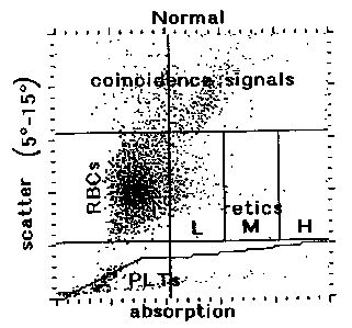
FIGS. lA-lD show the results of scatter/
absorption flow cytometry analyses as performed in
accordance with the present invention. Figs. lA and lB
depict cytograms presenting the scatter versus absorption
patterns associated with whole blood sample analysis. The
cytogram regions corresponding with various cell types and
report screen areas are specifically identified in Fig.
lA. The regions include: red blood cells (RBCs),
CA 02232163 1998-03-16
--10--
reticulocytes (retics), platelets (PLTs) and coincidence
signals. Coincidence signals result from the presence of
two or more particles in the counting region at one time.
Figs. lC and lD depict histograms showing the distribution
of platelet volumes within the samples. Fig. lA
S represents a normal blood sample; Fig. lB represents an
abnormal blood sample.
FIG. 2 presents a scatter/absorption cytogram
displaying typical results for a normal blood sample
analyzed in accordance with the invention using NaN3 in a
reagent formulation as shown in Table l herein. The
cytogram descriptors are as set forth in Figs. lA and lB
and as described hereinbelow.
FIG. 3 shows the highly correlated results of
comparative studies employing the rapid automated method
and composition of the invention and a manual method of
whole blood cell analysis to perform reticulocyte
determinations using scatter/absorption flow cytometry.
c The least squares equation and the correlation coefficient
(r) are presented on the graph.
FIGS. 4A and 4B show the results of red blood
cell (4A) and platelet (4B) determinations using the rapid
automated method as described herein. Fig. 4A shows a
comparison of red blood cell counts using the method in
accordance with the invention and a standard automated
method, for example, the BAYER H-3~ Hematology Analyzer.
Fig. 4B shows a comparison of platelet counts obtained
usin~ the rapid automated method of the invention versus a
standard automated method. For Figs. 4A and 4B, ten
normal samples were run in duplicate in each method. The
least-squares equation and the correlation coefficient (r)
are presented on the graphs.
DETAILED DESCRIPTION OF THE lNv~ lON
In the area of hematology analysis, there is an
ever-present need for more rapid assays and analysis
methods for determining absolute and percent red blood
CA 02232163 1998-03-16
cell and reticulocyte counts, and for determining platelet
counts in a blood sample drawn for testing.
Semi-automated hematology analyzers as used in
the art routinely provide sample data after a blood sample
containing blood cells is mixed with an appropriate
reagent solution to form a reagent mixture (i.e., a
reaction suspension), either within or outside of the
analyzer system. After a period of incubation of the
reagent mixture, the mixture is analyzed in a hematology
analysis device. Thus, sample preparation and incubation
are limiting factors contributing to the length of time
needed to analyze a blood sample in semi-automated
hematology devices.
For fully automated, high-speed analyzers, such
as the TECHNICON H- System series of analyzers, for
example, more rapid, automated hematology and whole blood
sample analysis methods are needed for accurate and
reliable integration and processing in the faster systems.
A novel method which requires no off-line, manual sample
preparation and is thus essentially a one-step process of
sample analysis which occurs within the automated analyzer
is afforded by the present invention. The method and
reagent used therefor eliminate manual sample preparation
and processing times and thus significantly and
advantageously decrease the overall blood cell
identification and determination times, i.e., by at least
an order of magnitude, when compared with those of other
scatter/absorption methods in the art.
In an embodiment of the invention, a rapid
method is provided for determining reticulocyte, red blood
cell and platelet counts and for determining reticulocyte
and red blood cell indices and parameters using fully
automated analyzers based on measurements of light scatter
and absorption. The use of light scatter and absorption
methodology for the very rapid automated sample analysis
as described herein provides an improved process to the
CA 02232163 1998-03-16
art which routinely employs fluorescence-based
methodology.
The method of the invention comprises the use of
a reticulocyte staining reagent which comprises a cationic
dye at a concentration effective to stain reticulocytes in
a whole blood sample for enumeration and characterization
of the cells within about 5 to 60 seconds, preferably,
within about 20 to 50 seconds, and more preferably, within
about 20 to 30 seconds or less, of the initial reaction
with an aliquot of the whole blood sample. In accordance
with the invention, a blood sample can be analyzed in an
automated hematology analyzer in about 30 seconds, which
includes approximately 20 seconds of reaction time of
sample in the reagent composition and approximately 10
seconds of counting time/ with less than approximately 0.1
lS second of mathematical analysis time.
The simplicity and rapidity of the method are
advantageous for the skilled practitioner. Because no
sample preparation is involved, even the least skilled
personnel may operate the fully automated analyzers,
employ the method of the invention, and obtain accurate
and reliable blood cell counts and indices. In addition,
many more samples can be analyzed in the same period of
time that was previously needed to perform fewer blood
sample analyses using semi-automated systems; for example,
in accordance with the present invention, blood sample
analysis occurs within less than about 20 to 30 seconds in
a fully automated system versus at least about 150 seconds
for a semi-automated method.
The aqueous reagent composition involved in the
method comprises a buffer to maintain the pH of the
reaction mixture at a value that will prevent non-specific
staining of mature red blood cells from interfering with
the reticulocyte and erythrocyte enumeration and
characterization analyses. Blood samples may be anti-
coagulated as conventionally known in the art. The
CA 02232163 1998-03-16
~, -
reagent further comprises a sphering agent, which is
preferably a zwitterionic surfactant as described herein,
to enable the determination of reticulocyte indices; the
reagent is also optimized with respect to osmolality of
the buffered reaction solution.
S Those skilled in the art will appreciate that
not all cationic compounds are capable of penetrating
intact red cell (and reticulocyte) membranes, and the
nature of the anions which necessarily accompany the
cations can affect whether or not the cationic compound
penetrates rapidly, slowly, or not at all. Hydrophobic
molecules generally penetrate red cell membranes faster
than hydrophilic molecules, and small molecules generally
penetrate membranes faster than large molecules. Only a
sub-set of salts or buffers mixed with those cationic dyes
which can stain reticulocytes permit rapid staining; that
is, the "right" dye with the "wrong" buffer can take an
excessive amount of time to stain reticulocytes. Again,
trial and error are necessary to discover useful
formulations of reticulocyte staining mixtures. Thus,
despite various "rules" which can be used as guides, it is
difficult to predict, a priori, whether and under which
conditions, any particular cationic dye may rapidly
penetrate and stain reticulocytes.
The fully automated apparatus for performing
blood sample analyses using the method and reagent of the
invention generally comprises 1) blood and reagent
metering devices to provide the proper volume of each of
the reaction components; 2) a means for adequately mixing
the components together; 3) a reaction chamber; and 4) a
means for transferring the mixture to the measurement
device. The measurement device comprises a means for
providing a metered flow of the cell-containing reaction
mixture through a flow cell for counting and enumeration.
In general, light from a Helium-Neon laser or laser diode
is incident upon the flow cell and this light is
CA 02232163 1998-03-16
interrupted by the passage of blood cells through the flow
cell. The blood cells scatter light as they intercept it,
and, in the case of stained reticulocytes, absorb light as
well.
The measurement device includes three optical
detectors. Two of the detectors detect light scattered at
1~-3~ and 4~-20~, respectively, preferably at 2~-3~ and 5~-
15~, respectively, from the axis of incidence. The third
detector determines the fraction of light absorbed. The
signals from the three detectors are analyzed by a
computer, which uses Mie Scattering Theory-derived tables
to convert the signals into cell volume, hemoglobin
concentration and hemoglobin content data for each cell
which passes through the flow cell. The computer also
displays a cytogram of the 5~-15~ scatter versus
absorption for the cell suspension and uses mathematical
algorithms to distinguish and differentiate among red
blood cells, reticulocytes, platelets and coincidence
signals.
For example, report screens obtained from the
above-described analysis are shown in Figs. lA and lB.
One of the screens reports the data obtained for a blood
sample having a normal percentage of reticulocytes (i.e.,
1.4); the second screen reports the data obtained for an
abnormal blood sample in which the percentage of
reticulocytes is 11Ø The reports include the
reticulocyte indices. For Figs. lA and lB, the reported
results, which correlate with the cytograms, are as
follows:
For Fig. lA:
SAMPLF RETICS
CHCM 30.04 g/dl CHCM 27.43 g/dL
MCV 93.86 fl MCV115.60 fl
MCH 27.60 pg MCH30.98 pg
HDW 2.41 g/dl HDW2.38 g/dl
RDW 13.03 % RDW17.61
CA 02232163 1998-03-16
--15--
RBC 5.19 x 10⁶/~L
RETIC 71.97 x 10⁹/L
POS 251
NEG 17860
%POS 1.39%
L 96.81%
M 2.79%
H 0.40%
For Fig. lB:
SAMPLE RETICS
CHCM30.01 g/dl CHCM26.81 g/dL
MCV 96.94 fl MCV113.15 fl
MCH 28.35 pg MCH29.56 pg
HDW 4.02 g/dl HDW 3.76 g/dl
RDW 20.21 % RDW18.53 %
RBC 2.89 x 10⁶/~L
RETIC318.13 x 10 /L
POS1769
NEG14280
%POS11.02%
L 60.15%
M 29.79%
H 10.06%
These reported results for Figs. lA and lB are as follows:
SAMPLE=the total number of cells analyzed; RETICS=cells
counted as reticulocytes; CHCM=Cellular Hemoglobin
Concentration Mean, which measures the same cellular
property as MCHC or Mean Cellular Hemoglobin
Concentration; MCV=Mean Cellular Volume (of red blood
cells); MCH=Mean Cellular Hemoglobin; HDW=Hemoglobin
Distribution Width (a measure of the variability of
cellular hemoglobin concentration with a sample);
RDW=Red(blood cell) Distribution Width; RBC=Red Blood Cell
Count; RETIC=Reticulocyte Count; POS=number of
reticulocytes counted; NEG=number of red blood cells
counted; ~POS=reticulocyte percentage in sample; L, M,
H=percentage of low, medium, and high-staining
CA 02232163 1998-03-16
-16-
reticulocytes; PLT=Platelet count; MPV=Mean Platelet
Volume; PCT=Platelet Crit (the percentage of sample volume
occupied by platelets); PDW=Platelet Distribution Width
(the distribution of platelet volumes within the sample).
An illustrative measurement device and system
suitable for the analyses of the invention is as described
in U.S. Patent No. 4,735,504 to Tycko, which is
incorporated by reference herein. It is to be understood
that the Tycko method and system may be modified as
necessary by those in the art for performance of the
improved, rapid method of the invention.
To utilize Tycko's method, a light source which
emits monochromatic light in a region where hemoglobin is
very transparent is required; typically a light source
like a red helium neon (HeNe) laser, or a laser with even
lS longer wavelength. This means that if that wavelength is
also to be used for the absorption measurement, the dye
must-be a blue dye with a strong absorption of red light.
The dye Oxazine 750 ser~es as a preferred dye and use in
the improved and rapid absorption/scatter flow cytometric
method of the invention for determining reticulocyte RNA
concentration, reticulocyte cell count, platelet count and
mature red cell and reticulocyte volume and hemoglobin
content on a cell-by-cell basis.
More particularly, the fundamental concept of
flow cytometry is essentially the passing of cells, one at
a time, through a specific sensing region. Typically, by
means of hydrodynamic focusing, single cells are passed
through the sensing zone, which consists of a focused
light source and a detection system for the measurement of
scattered and absorbed light.
The effect that a particle has on the light it
intercepts can be detected in a number of ways. In
general, the particle has a refractive index which is
different from that of the medium in which it is
suspended. The refractive index, n, consists of two
CA 02232163 1998-03-16
.,
parts: the real part, nr, and the so-called imaginary
part, nj. Non-zero values of nj are associated with light
absorption. A particle will therefore scatter and absorb
light with which it is illuminated through a range of
angles, and with varying intensities, that depend upon
that refractive index difference, the particle's size, its
shape and any internal variations in refractive index and
structure, as well as upon the wavelength of the
illuminating light. (For homogeneous spheres, Mie
Scattering Theory provides a complete description of the
distribution and intensities of scattered light).
As stated above, a particle may also absorb some
of the incident light. In the latter case, a portion of
the absorbed light may or may not be re-emitted as
fluorescence, typically at a longer wavelength than the
wavelength of the absorbed light. These and other effects
can be measured with light detectors arranged to measure
different angular intervals of scattered light,
unscattered light, fluorescent light and the fraction of
light absorbed by the particle.
When particles are as small as blood cells,
typically less than 15 micrometers in diameter, the number
of photons in the illuminating beam affected by their
passage at high speed (typically hundreds to thousands of
widely-spaced cells per second) can be very small,
especially when compared with the number of photons per
second falling on the illuminated part of the suspension
stream, and compared with the background illumination of
an absorption detector. Therefore, the limits of
sensitivity of detection of small particular differences
between particles depends critically on the photon flux
(which depends at least on the intrinsic "brightness" of
the light source) and how large the perturbations of the
photon flux are that are produced by other small and large
differences between particles.
The main sources of interfering noise in
CA 02232163 1998-03-16
-18-
absorption and scatter flow cytometry signals can be guite
different for each kind of signal. To a first order
approximation, the magnitudes of scatter and absorption
signals from stained or unstained cells are very strongly
influenced by shape or orientation of the cells from which
the signals arise. As an extreme example, the native
biconcave shape of human erythrocytes has a profound
effect on the absorption and scatter signals they
generate; such an effect is larger than the small
absorption signals of typical classically stained
reticulocytes. A description of scatter/absorption flow
cytometric analyses for reticulocyte determination is
described in U.S. Patent Nos. 5,350,695 and 5,438,003 to
G. Colella et al., the contents of which are incorporated
by reference herein.
As noted above, only a small sub-set of cationic
dyes selectively stains reticulocytes, and only a smaller
sub-set of these dyes rapidly penetrates reticulocytes.
The concentration of cationic dye compound combined with
the optimal pH and ionic strength of the reagent
composition used in the method of the invention achieve
the rapid staining of reticulocyte RNA and sample analysis
in seconds. This allows reticulocyte, red cell and
platelet analyses by fully automated flow cytometry to be
performed in less than about 20 to 30 seconds after the
blood sample and the reagent composition employed in the
method are mixed together, thus contributing to the speed
of the present invention and its successful and accurate
performance in fully automated procedures. The method and
reagent of the invention allow blood sample analyses at
least an order of magnitude faster than current
scatter/absorption flow cytometry methods in the art. By
contrast, former reticulocyte RNA staining procedures
require at least several minutes for sample and reagent
mixture and incubation.
The present invention significantly improves
CA 02232163 1998-03-16
o --19--
upon former methods by achieving a significant reduction
in reaction time between a blood sample and the presently
described reagent composition with which it is mixed prior
to automated hematology analysis. The inventive discovery
of the combination of increased cationic dye
concentration, reduction of the pH of the reagent
composition and an optimal osmolality range used in the
method was found to reduce the reaction time of the blood
sample with the reagent composition, which collectively
comprise the reagent mixture for analysis. This novel
combination of parameters favored the staining of RNA in
reticulocytes over the non-specific staining of hemoglobin
in mature red blood cells as described hereinbelow.
Interestingly, increasing the dye concentration
in the composition did not, by itself, reduce the reaction
time of the blood sample mixed with the reaction medium.
This was because within the first 30 seconds, mature red
blood cells as well as reticulocytes significantly bound
to the dye (e.g., Oxazine 750) at the higher
concentrations required for use in the present invention.
With a longer time of incubation of blood sample in the
reaction medium (e.g., over 30 seconds), the binding of
dye by the mature red cells declined. Thus, the longer
reaction times of other methods were required not only to
increase the binding of the cationic dye to RNA, but also
to decrease the binding of dye to the cellular hemoglobin
of mature red blood cells, thus causing non-specific
staining of red blood cells. Such non-specific staining
reduces the distinction between red cells and
reticulocytes with respect to absorption signals.
In spite of the foregoing, the present method
and composition used therein were able to achieve a fast
reaction time without the adverse effect of non-specific
staining by cells other than reticulocytes. Affinity of
the dye for red blood cells was reduced while adequate
affinity for reticulocyte RNA was maintained by the
CA 02232163 1998-03-16
-20-
invention within a 20 second or less reaction time period
by the above-described reduction of pH (e.g., reducing the
pH of the reaction medium from approximately 8.1 to
approximately 7.4) combined with an increased cationic dye
concentration. In accordance with the invention, the
increased hydrogen ion concentration of the reagent
composition may reduce the affinity of hemoglobin for the
positively-charged dye compound.
Moreover, it was determined that a reduction of
the ionic strength of the reaction medium favored the
staining of RNA over red cell hemoglobin, thereby reducing
the reaction time. The reduced ionic strength may favor
the binding of RNA to the positively-charged dye rather
than to other ionic constituents in the reaction mixture.
In another aspect of the invention, the binding
of cationic dye, e.g., Oxazine 750, to mature red blood
cells was reduced, without adverse effects on RNA binding,
by converting hemoglobin to its oxidized (Met) form in the
presence of nucleophiles such as azides (N3-), e.g.,
sodium azide, or cyanate (OCN-) ion, e.g., sodium cyanate.
See Table 1 and Fig. 2. As a result, as little as about
one-fifth of the amount of dye used in the absence of such
nucleophiles can be used in this embodiment of the present
invention. Without wishing to be bound by any particular
theory, it is possible that the conformational structural
change to hemoglobin associated with the oxidized form may
reduce the affinity of hemoglobin for the positively
charged dye component of the reagent composition. As a
general guide, nucleophiles are used at a concentration of
about approximately 20 mM, provided that there is a 1:1
ratio of N3- or OCN- ions to home sites. Because reagents
which include both nucleophiles and dyes such as Oxazine
750 are not typically stable during storage, the reagents
can be stored in two parts, which when added together
yield the required final concentrations of components. An
illustrative example of such a two-part reagent system is
CA 02232163 1998-03-16
-21-
shown in Table 1:
' TABLE 1
Reagent Part 1 Range Part 2 Range
Component(Per (Per (Per (Per
liter) liter) liter) liter)
TRIS 0.81 g 0.7-0.9 g 0.81 g 0.7-0.9 g
TRIS-HCl 6.83 g 6.0-8.0 g 6.83 g 6.0-8.0 g
NaCl 6.40 g 5.25- 5.82 g 4.67-
7.30 g 6.72 g
Proclin 0.25 ml 0.15-0.35 -- --
300 ml
NaN3 or -- -- 1.303 g 1.10-
NaOCN 1.50 g
TDAPS 16.5 ml 8.2- -- --
22.0 ml
Oxazine 2.2 ml 1.8- -- --
750~ 6.6 ml
Deionized950 ml -- 950 ml --
H20
: Amount of stock solution. 1 mg/ml TDAPS (tetradecyl-
N,N-dimethyl-3-ammonio-1-propanesulfonate) in distilled
H20 .
: Amount of stock solution. 14.82 mg oxazine 750 dye
per 4.81 ml dimethyl formamide.
The cytogram of Fig. 2 is representative of a normal
sample analyzed using the sodium azide formulation
presented in Table 1. The reaction time for the analysis
was 20 seconds. In the Fig. 2 cytogram, the parameters,
which are defined above for Figs. lA and lB, are as
follows:
SAMPLE RETICS
CHCM 27.40 g/dl CHCM 24.13 g/dL
MCV 96.22 fl MCV119.13 fl
MCH 25.74 pg MCH27.84 pg
HDW 2.71 g/dl HDW3.62 g/dl
RDW 14.31 % RDW17.50 %
CA 02232163 1998-03-16
-22-
RBC 4.15 x 10⁶/~L
- RETIC62.94 x 109/L
POS276
NEG17917
%POS 1.52%
As described above, investigations into
improving and accelerating the process of
absorption/scatter flow cytometry analysis, and developing
the method and reagent used therein to detect
reticulocytes, erythrocytes and platelets resulted in the
present, extremely rapid method which involved staining of
the reticulum of reticulocytes by a cationic dye without a
requirement for sample preparation for particular use with
fully automated hematology analyzers. In addition, in the
improved and rapid absorption method of the invention, the
lS isovolumetric sphering of red cells was used to eliminate
orientational noise. By using isovolumetric sphering and
scatter/absorption methods, reticulocyte and mature red
4 cell volume and hemoglobin could be simultaneously
measured and quantified on a cell-by-cell basis using a
reagent which also selectively stained reticulocytes.
Those skilled in the art will appreciate that if the
sphering is complete, and not isovolumetric, but has some
known factor X of isotonicity, then by using Tycko's
method with a correction by l/X for volume and a
correction by X for protein, e.g., hemoglobin
concentration, original values can be calculated.
In the reagent solution employed for use in the
current method, a zwitterionic surfactant was used as a
red cell and reticulocyte sphering agent, since it is
fully compatible with cationic dyes and does not cause
precipitation of the dye out of the reagent solution.
Non-limiting examples of such zwitterionic surfactants
include alkyl amido betaines or alkyl betaines, such as
lauramidopropylbetaine (LAB), cocoamidopropylbetaine
(CAPB), and cocoamidosulfobetaine (CASB). Other suitable
CA 02232163 1998-03-16
-23-
zwitterionic surfactants include: tetradecyl-N,N-dimethyl-
3-ammonio-1-propanesulfonate (TDAPS) and N-dodecyl-N, N-
dimethyl-3-ammonio-1-propanesulfonate (DDAPS). TDAPS and
DDAPS are the preferred sphering agents because they give
the most stable sample preparation. In accordance with
the invention, the use of red cell sphering-effective
amounts of these types of surfactants and the reagent
specifications as described herein obviate the need for
protein buffering or fixatives in the reagent mixture to
delay red cell lysis.
For the reagent solution of the present
invention and to isovolumetrically sphere the
reticulocytes and red blood cells within a blood sample,
the concentration of the sphering agent in the reagent is
from about 3.0 ~g/ml to about 20 ~g/ml, preferably about
4.0 to about 15 ~g/ml, and more preferably about 4.1 to
about 11.0 ~g/ml. ~DAPS is a preferred zwitterionic
surfactant sphering agent. The sphering agent is
preferably present in an amount of from about 12 ~g/ml to
about 87.5 ~g/ml of LAB; from about 4.1 ~g/ml to about
11.0 ~g/ml of TDAPS; from about 49.3 ~g/ml to about 148
~g/ml of DDAPS; from about 8.8 ~g/ml to about 17.5 ~g/ml
of CAPB; or from about 12.5 ~g/ml to about 15 ~g/ml of
CASB.
For optimal operativity of the improved method,
a buffer solution is preferably used for maintaining a
particular pH of the reaction mixture, i.e., about 7.2 to
7.8, preferably, 7.3 to 7.5, more preferably 7.4, as is an
organic cationic dye for staining the reticulocytes. The
preferred dye compound is Oxazine 750 (available from
Excitron, Inc. of Dayton, Ohio), which is a blue
CA 02232163 1998-03-16
absorption dye having the structure:
~ ~ H6
More particularly, the reagent solution for use
in the method of the invention may include one or more of
the following constituents: TRIS (Tristhydroxymethyl]-
aminomethane); TRIS-HCl (Tris[hydroxymethyl]-aminomethane-
hydrochloric acid); an alkali metal chloride salt, e.g.,
NaCl, KCl and the like, to facilitate the penetration of
dye through the red blood cell membrane; and, if desired,
an antimicrobial compound to retard microbial growth.
Nonlimiting examples of suitable antimicrobials include
Proclin 150 (2-methyl-4-isothiazolin-3-one) and Proclin
300 (5-chloro-2-methyl-4-isothiazolin-3-one) (Rohm &
Haas); Germall 115 (N,N'-methylenebis[N'-(l-
(hydroxymethyl)-2,5-dioxo-4-imidazolidinyl] urea) (Sutton
Laboratories); Dowacil 200 (1-(3-chloroallyl)-3,5,7-
triaza-l-azoniaadamantane chloride) (Dow Chemical); and
Bronopol 2-bromo-2-nitropropane-1, 3-diol
2S (C3H6BrNO4),(Angus Chemical Company). Proclin 300 is a
preferred antimicrobial for use in the reagent composition
employed in the present invention. Also included in the
composition is a zwitterionic surfactant, e.g., TDAPS (N-
Tetradecyl-N,N-Dimethyl-3-Ammonio-l-Propane Sulfonate);
and a cationic dye, e.g., Oxazine 750.
An illustrative reagent composition/solution of
the present invention and suitable for use in the present
methods is shown in Table 2, with suitable concentration
ranges for each component and the amounts per liter for
each component provided. The final pH of the composition
CA 02232163 1998-03-16
-25-
is about 7.4, and the final osmolality is adjusted with
either NaCl or TDAPS solution to 2g2 + 5 mOsm. The final
pH is adjusted to the appropriate pH, preferably, pH 7.4,
by the dropwise addition of 1 N HCl or 1 N NaOH.
Table 2
Constituents in Concentration Amount per
Reaqent Solution Range of Liter
Components (Preferred)
Amount/liter
TRIS 0.7-0.9 g 0.806 g
TRIS-HCl 6.0-8.0 g 6.83 g
NaCl 5.25-7.30 g 6.40 g
PROCLIN 300 0.15-0.35 ml 0.25 ml
TDAPS
(Stock Solution)~ 4.1-11.0 ml 8.25 ml
Oxazine 750
(Stock Solution) 2.2-6.6 ml 3.3 ml
Deionized Water To volume 950 ml
: 1 mg/ml TDAPS (N-tetradecyl-N,N-dimethyl-3-ammonio-1-
propane sulfonate) in distilled water.
: 14.82 mg Oxazine 750 dye per 4.81 ml
dimethylformamide.
In general, the reagent solution may be
formulated to maintain the pH of the reagent composition
at between about 7.2 to about 7.8, with 7.4 as a preferred
pH value, with an osmolality of about 250 mOsm to about
320 mOsm, preferably, about 287 mOsm to 297 mOsm, more
preferably about 292 + 5 mosm.
In the reagent solutions having the pH and
osmolality ranges as described above, the concentration of
cationic dye, e.g., Oxazine 750, required for RNA staining
is in the range of from about 6 to about 20 ~g/ml,
preferably about 6.5 ~g/ml to about 19.5 ~g/ml, more
preferably about 9.0 ~g/ml to about 10.5 ~g/ml. The
buffer enhanced penetration results in the dye staining
RNA in the reticulocytes in about 20 to 30 seconds or
less. The concentration of dye in the reagent of the
method minimizes non-reticulocyte staining of mature
CA 02232163 1998-03-16
'
erythrocytes which leads to a good signal separation from
the noise background. Such rapid staining, due to the
optimized conditions of the reagent, obviates incubating
the blood sample in the reagent for more than about 20
seconds prior to sample analysis, and makes the method
highly advantageous, acceptable and compatible for fully
automated hematology methods.
The method and reagent composition may be used
to identify and discriminate reticulocytes in a whole
blood sample using the technique of scatter/absorption
flow cytometry. The method in its broadest application
includes adding an aliquot of whole blood to the reagent
composition as described for use in the method. With less
than about a 20 second incubation period after mixing, the
sample/reagent mixture is passed, one cell at a time,
through a specific sensing region of the flow cytometer.
By means of hydrodynamic focusing, single cells are passed
through the sensing zone, where they are illuminated by a
focused light source having a suitable illumination
wavelength. At least two scattered light signals and at
least one absorption signal are measured for the cells on
a cell-by-cell basis. From these measurements, the
reticulocytes can be distinguished from the erythrocytes.
In accordance with the present invention,
platelets are distinguished from reticulocytes and red
blood cells in a sample by size and refractive index,
which separates their signals from those of larger, more
refractile red blood cells.
When the reaction mixture (comprising the whole
blood sample and reagent composition/solution) is passed
through the sensing region of a flow cytometer, the light
is scattered through two angular intervals and absorbed by
each cell is measured, such that the erythrocytes can be
distinguished from reticulocytes, and the volume and
hemoglobin concentration of each reticulocyte or
erythrocyte can be determined. Platelets occupy a
CA 02232163 1998-03-16
.
-27-
distinct region of scatter/scatter/absorption "space"; the
signals in this region are counted as platelets. The
number of reticulocytes and erythrocytes, and the
hemoglobin content, mean cell volume, mean corpuscular
hemoglobin concentration, and mean cell hemoglobin of the
reticulocytes or erythrocytes are calculated from the
measured cell-by-cell volume and hemoglobin concentration.
More particularly, to carry out the method, 2 ~l
aliquots of whole blood were aspirated by the automated
hematology analysis instrument, e.g., the TECHNICON H-3
Hematology System, modified with respect to analysis
software. The modifications included the aspiration of
whole blood, which was subsequently mixed with reagents
on-system rather than the aspiration of a diluted sample
that had already undergone about 5-g0 minutes of off-line
(off-system) reaction. Also, the modified analysis
provided absolute red blood cell and reticulocyte counts,
as well as percent reticulocytes, rather than merely
percent reticulocytes. Further, the method in accordance
with the invention provided platelet counts, values for
mean platelet volume and platelet-volume histograms.
These parameters are not provided in the reticulocyte
channel of the H-3 Hematology System.
In accordance with the present invention, the
sample aliquots were automatically mixed with the present
reagent composition and permitted to stand for about 20
seconds. Thereafter, the mixed sample was passed through
the flow cell and was then exposed to either a helium-neon
laser source or a laser-diode source for red cell
reticulocyte and platelet analysis.
The invention accordingly comprises the methods
and reagents hereinafter described in the Examples, the
scope of the invention being indicated in the claims.
CA 02232163 1998-03-16
-28-
o
BXANPLES
- The following Examples set forth the method and
reagent composition used therein for the identification of
reticulocytes, red blood cells, and platelets, and for the
characterization of reticulocytes and red blood cells
using absorption/scatter flow cytometry techniques.
Standard, commercially-available, reagent-grade materials
were used whenever possible.
EX~MPLE 1
Rapid, Automated ~catter and Absorption Measurements for
Distinguishing Reticulocytes and Erythrocytes in a ~hole
Bloo~ 8ample ~sing the Nethod and Reagent Composition of
the Pre~ent Invention
Oxazine 750 dye was stored in a 2.96 mg/ml N,
N-dimethylformamide stock solution. A working reagent was
created by adding the dye stock to a buffer solution
containing the preferred components and at the preferred
concentrations presented in Table 2. The final osmolality
and pH of the working reagent used in this study were 292
mmol/kg and 7.4, respectively.
In this example, for each sample, 2 J~,l of whole
blood aspirated by the automated system was mixed with 625
,ul of the Table 2 reagent composition for 20 seconds at
room temperature and the cells in the mixture were then
analyzed by flow cytometry as described herein. Ten
normal-donor blood samples ("normals") and ten randomly-
- selected hospital patient blood samples ("abnormals") were
analyzed for percent reticulocytes by the present method
and by a manual method. Each sample was analyzed by the
automated instrument in duplicate (n=40). The results are
graphically depicted in Fig. 3. As shown in Fig. 3, the
mean reticulocyte percentage was l.8 for the automated
method versus l.7 for the manual method. The correlation
coefficient was 0.87. The small difference between the
means and the high correlation coefficient demonstrates
that the 2 O-second automated method provides accurate
~ri i~J-~ U'.~
CA 02232163 1998-03-16
O -29-
values for reticulocyte percentages for both normal and
abnormal sample populations.
At the completion of the analysis, the raw data
were displayed in the form of a Red Scatter v. Red
Absorption cytogram, e.g., Figs. lA and lB. Distinct cell
populations were clearly observed based on their
particular scatter and absorption signals. In these
cytogram plots, the erythrocyte population falls within
the region labeled "RBCs" in Fig. lA. These cells show
high scatter signals and low cell absorption signals. The
reticulocyte population falls within the region labeled
"retics", including the regions labeled "L", "M" and "H".
These cells are distinguishable from the mature
erythrocytes due to the higher absorption signals from
their Oxazine 750-stained RNA. The platelet population
lies in the region labeled "PLTs", and the coincidence
signals are in the appropriately labeled region. The
platelets have relatively low scatter signals when
compared with the reticulocytes.
Based on the absorption separation between
mature erythrocytes and reticulocytes, the reticulocyte
count of a patient sample may be determined by creating
electronic "windows" which define the ranges of scattered
light and absorption which identify reticulocytes and
erythrocytes. The number of reticulocytes, mature
erythrocytes and platelets falling within each "window"
are determined so that the percentages of the
reticulocytes, erythrocytes and platelets present in the
total cell population is then calculated. The absolute
red blood cell count, reticulocyte count and platelet
count are determined as well, based on the known dilution
factors associated with the automated instrumentation.
The reference percentage of reticulocytes in
each sample was determined using the manual microscopic
procedure recommended by the National Committee for
Clinical Laboratory Standards (NCCLS). In this procedure,
CA 02232163 1998-03-16
-30-
a small volume, e.g., 100 ~1, of whole blood was mixed
with an equal volume of new methylene blue dye and allowed
to react for about 15 minutes at room temperature. This
mixture was then applied to a microscope slide in the form
of a smear and the percentage of reticulocytes in the
S sample was counted upon visualization using a microscope.
The microscope was equipped with a 100X oil immersion
objective and a 10X ocular. A minimum of 1000 cells were
counted for each sample using a Miller disc inserted in
the ocular of the microscope to improve counting
precision. Any red cell containing two or more particles
of blue material after staining was labeled a
reticulocyte.
EXAMPLE 2
Red Blood Cell and Platelet Determinations Obtained from
the Method in Accordance With the Pre~ent Invention
Experiments were performed to demonstrate that
the rapid automated method of the present invention
provided accurate red blood cell and platelet counts as
well as reticulocyte counts. Ten fresh samples, (i.e.,
samples that were used less than about eight hours after
collection) were taken from normal donors and analyzed by
both the new method of the invention and a standard
hematology analyzer, e.g., the BAYER H-3~ Hematology
Analyzer. Each blood sample was run in duplicate for a
total of twenty aspirations for each method. The results
of these analyses are presented in Figs. 4A and 4B. The
results show that the counts obtained by the new and rapid
method are highly correlated with those obtained using a
standard method.
EXAMPLE 3
Correction of Absorption Data for Pseudo-absorption
The detection optical subsystem collects both
the scattered and unscattered light from cells passing
through the laser beam in the flowcell. Cells scatter
light into all directions. The relatively Hi-numerical
CA 02232163 1998-03-16
aperture (NA) lens in the optical system, which is
described in U.S. Patent Nos. 5,350,695 and 5,438,003 to
G. Colella et al., accepts the light that is scattered
into a cone that is centered on the optical axis with a
half angle of up to 19.5 degrees. Thus, the light that is
scattered into angles greater than 19.5 degrees is lost.
As a result, when attempting to measure cellular
absorption, completely non-absorbing cells "appear" to
absorb up to a few percent of the incident light, i.e.,
pseudo-absorption. The measured absorption can be
represented as follows:
Absorption = Pseudo- + Hemoglobin + Dye
Signal Absorption Absorption Absorption
The pseudo-absorption signal of a mature red blood
cell is typically of the same magnitude as the actual
absorption signal from a stained reticulocyte. This
reduces the degree of separation of the stained
reticulocytes from the unstained red blood cells on the
absorption cytogram. The signal to noise ratio of the
absorption channel can be improved by correcting the
signal to remove the pseudo-absorption and hemoglobin
absorption components from each red cell and reticulocyte
absorption signal. The amount of pseudo-absorption and
hemoglobin absorption can be calculated for any given cell
by using the well-known Mie light scattering theory
described in the aforenoted Tycko patent. The scattering
cross-section for the angular interval 19.5~ to 180~ plus
the hemoglobin absorption component, S3, can be calculated
as follows:
S3 = ~T a2 Qext ~ S ( A,ns,e3,~e3;V,HC)
where a is the radius of the sphered cell, A is the
excitation (or illuminating) wavelength, nS is the
refractive index of the sample stream and sheath, Qext is
the extinction efficiency of the cell, and for the case of
pseudo-absorption, e3=0~, and ~e3=19.5~. S3 values have
been tabulated for all expected values of V and HC.
CA 02232163 1998-03-16
-32-
The pseudo-absorption correction is made as
follows: the V and HC must first be determined from the
two scattering signals from a cell from the
scatter-scatter cytogram as described in Tycko. S3 iS
then found in the look-up table entry for the measured V
and HC, and subtracted from the value measured by the
absorption channel. The result is the actual absorption
due to st~; n i ng of the cell. The measured absorption
signal can be adjusted using the following relation, to
leave only the dye absorption for each cell:
Dye = Absorption - Hemoglobin - Pseudo-
Absorption Signal Absorption Absorption
= Absorption Signal - S3
For all data, the adjusted value is substituted
for the raw data parameter prior to thresholding and
flagging. Any objects whose red scatter parameters do not
appear on the V-HC map are ignored in the data analysis
scheme. These data are then redisplayed with the red
scatter v. absorption cytogram reflecting the corrected
values.
In view of the above, it will be seen that the
several objects of the invention are achieved, and other
advantageous results obtained.
As various changes can be made in the above
constructions and methods without departing from the scope
of the invention, it is intended that all matter contained
in the above description, or shown on the accompanying
drawings, shall be interpreted as illustrative, not in a
limiting sense. For instance, fractionated samples of
blood can be processed in a similar way.
The contents of all patent applications, issued
patents, articles, references, texts, and the like, as
cited herein are hereby incorporated by reference in their
entirety to more fully describe the state of the art to
which the present invention pertains.