Note : Les descriptions sont présentées dans la langue officielle dans laquelle elles ont été soumises.
CA 02249611 1998-09-22
WO 97/40883 PCT/US97/04174
MEDICAL ELECTRICAL LEAD
FIELD OF THE INVENTION
This invention relates to the field of body implantable medical device
systems,
and in particular to a body implantable medical device system which includes a
medical electrical lead particularly designed for implantation into the
coronary sinus.
BACKGROUND OF THE INVENTION
Modern electrical therapeutic and diagnostic devices for the heart, such as
pacemakers, cardioverters, and defibrillators, for example, require a reliable
electrical
connection between the device and a region of the heart. Typically, a medical
electrical "lead" is used for the desired electrical connection.
One type of commonly used implantable lead is a transvenous lead.
Transvenous leads are positioned through the venous system to attach or
electrically
connect at their distal end to the heart. At their proximal end, they are
connected to
1 S typically an implantable pulse generator. Such leads normally took the
form of a
long, generally straight, flexible, insulated conductor. Among the many
advantages of
a transvenous lead is that it permits an electrical contact with the heart
without
physically exposing the heart itself, i.e., major thoracic surgery is not
required.
The specific design of a transvenous lead used is often varied depending upon
the region of the heart to which it is to be connected. For example, U.S.
Patent
4,402,330 of Lindemans discloses a body implantable lead in which the lead
body has
a J-curve and the distal electrode has a permanent bend. In such a manner, the
lead is
configured to electrically connect to the right atrium.
While such a lead has been found acceptable for electrically connecting and
thus
pacing the
right atrium, the need exists for a transvenous medical electrical lead which
may provide an
electrical connection to the left atrium. Of course the left atrium cannot, at
present, be
transvenously accessed with a lead for chronic implantation due to the
direction of blood
flow
CA 02249611 1998-09-22
WO 97/40883 PCT/US97/04174
2
and the present limitations of materials. To be precise, blood flows through
the right
side of the heart (atrium and ventricle), through the lungs, through the left
side of the
heart (atrium and ventricle) and then through the rest of the body, including
the brain,
before returning again to the right side of the heart. Implanted objects,
however,
often cause minor blood clots and thrombus to form in the blood. These may, on
occasion, dislodge and be released into the bloodstream.
Because the blood circulates directly from the left atrium and ventricle to
the
brain, any clots, however minor, could have serious consequences if they were
to
reach the brain, e.g. a stroke. In contrast, any clots released from an object
implanted
in the right side of the heart would simply travel to the lungs, where they
would lodge
without any serious risk. Thus at present, chronic transvenous leads may not
be
safely implanted within the left side of the heart.
In spite of the difficulties, there remains a great need to be able to
electrically
stimulate or sense or both the left side of the heart. The most obvious reason
is the
left side of the heart accounts for the majority of the heart's hemodynamic
output.
For example, the left ventricle has a greater wall thickness (10-20 mm as
compared to
1-5 mm) than the right side. This, of course, is reasonable given that the
left side of
the heart must pump blood throughout the body while the right side only pumps
blood
through the lungs.
Because the left side is relatively more important for hemodynamic output,
not surprisingly various pathologies may be better treated through stimulation
on the
left side of the heart. For example, in patients with dilated cardiomyopathy,
electrical
stimulation of both the right side and the left side of the heart has been
shown to be of
major importance to improve the patient's well-being and manage heart failure.
See,
for example, Cazeau et al., "Four Chamber Pacing in Dilated Cardiomyopathy,"
PACE, Nov. 1994, pgs. 1974-79. See also Brecker and Fontainem,St. et al.,
"Effects
Of Dual Chamber Pacing With Short Atrioventricular Delay In Dilated
Cardiomyopathy," Lancet Nov 1992 Vol. 340 p1308-1312; Xiao HB et al., "Effect
Of
Left Bundle Branch Block On Diastolic Function In Dilated Cardiomyopathy,"
Br.Heart J 1991, 66(6) p 443-447; and Fontaine G et al, "Electrophysiology Of
CA 02249611 1998-09-22
WO 97/40883 PCT/US97J04174
3
Pseudofunction," CLMeere (ed) Cardiac pacing, state of the art 1979, Pacesymp,
1979 Montreal.
At present there are several techniques for implanting a lead onto or into the
left side of the heart. First, of course, is through general thoracic surgery;
either via a
median sternotomy; intercostal approach; or, in a more limited procedure, a
sub-
xiphoid approach. These procedures, however, involve major surgery which may
be
painful and dangerous for the patient, as well as extremely costly. The sub-
xiphoid
approach, moreover, only permits limited access to the anterolateral surface
of the left
ventricle and does not provide any access to the left atrium. Another approach
used
is to electrically access the left atrium is through the coronary sinus.
The coronary sinus, however, presents challenges in both implanting the lead
in the proper position as well as ensuring the lead maintains sufficient
electrical
contact with the desired tissue. U.S. Patent No. 5,423,772 of Lurie et al.
discloses a
coronary sinus catheter having three sections. Each section has varying
degrees of
1 S flexibility, with the proximal reinforced section being stiffer than an
intermediate
section, the intermediate section being stiffer than the softened tip section.
The
catheter also is curved, with the curve beginning in the intermediate section,
the curve
further continuing into the softened tip section, where the radius of
curvature
decreases, i.e., the catheter becomes more curved closer to the tip. One
drawback to
such a design, however, is that the particular shape of the curve is not
ideally suited
for electrically accessing the left atrium. In addition, such a catheter is
relatively
complicated to manufacture due to the required reinforcing braid or other
mends in the
proximal reinforced section. Finally, such a catheter does not permit
introduction of a
stylet to assist in the placement of the catheter into the coronary sinus.
It is thus an object of the present invention to provide a medical electrical
lead
which is suitably shaped to provide an electrical connection through the
coronary
sinus to the left atrium.
A still further object of the present invention is to provide such a medical
_ electrical lead which may be readily flexed during implantation to provide
the ability
to be introduced transvenously.
CA 02249611 2003-12-16
667,42-677
4
A still further object of the present invention is
to provide a medical electrical lead having a pre-bent
portion along the lead body which may be readily
straightened through use of a stylet and which further
includes a pre-bent portion at the electrode tip so that the
electrode tip is properly oriented to the coronary sinus
upper wall and, thus, with the left atrium inferolateral
wall.
SUMMARY OF THE INVENTION
These and other objects are accomplished through
the present invention. In one embodiment, the present
invention comprises a transvenous bipolar lead specifically
designed for coronary sinus implantation. The lead has
essentially two main characteristics, the distal end has a
45-degree pre-shape to facilitate introduction of the lead
through a catheter and provide optimal positioning of the
lead within the coronary sinus. The lead further features a
distal electrode tip which itself is canted at an angle of
45 degrees on the distal end of the lead to provide a very
close contact with the coronary sinus upper wall and, thus,
with the left atrium inferolateral wall. In addition, each
of these sections is flexible to permit the lead to be
introduced through a relatively small-sized guide catheter.
Finally, the lead further features a center lumen to also
permit the lead to be straightened for introduction with a
stylet.
The invention may be summarised as a medical
electrical lead comprising: a connector assembly; a lead
body coupled to the connector assembly, the lead body having
a first section and a second section, the first section
having a flexible first bend and a first stiffness, the
second section having a flexible second bend and a second
CA 02249611 2003-12-16
66742-677
4a
stiffness, the first stiffness greater than the second
stiffness, the second bend is between 15 to 90 degrees, the
first bend is between 15 to 90 degrees; and a tip electrode
coupled to the lead body.
BRIEF DESCRIPTION OF THE DRAWINGS
The present invention may be better understood and
appreciated with reference to a detailed description of the
specific embodiment of the invention, when read in
conjunction with the accompanying drawings, wherein;
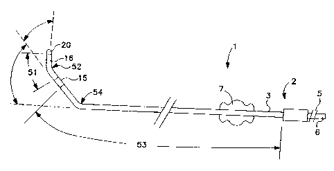
FIG. 1 is a plan view of the lead of the present
invention.
FIG. 2 is a fragmented detail of the construction
of the lead body.
FIG. 3 is a detailed view of the distal end of the
lead shown in FIG. 1.
FIG. 4 is a cross sectional view of the distal end
of the lead shown in FIG. 3.
FIG. 5 is a cross sectional view of the distal end
of the lead shown in FIG. 3 having a stylet inserted through
the center lumen and the lead straightened.
FIG. 6 is a partial sectional view of the distal
end of the lead shown in FIG. 3 having been inserted into a
guide catheter, thereby causing the distal end to become
relatively less bent.
CA 02249611 1998-09-22
WO 97/40883 PCT/US97/U4174
It should be understood the drawings are not necessarily to scale.
DETAILED DESCRIPTION OF THE PREFERRED EMBODIMENTS
Turning now to FIG. 1, which is a plan view of the lead of the present
invention. As
seen lead 1 consists essentially of three portions, connector assembly 2, lead
body 3,
S and distal electrode assembly 4. Connector assembly 2 is constructed to meet
the
industry standard IS-1 Bi, although other types of connectors could be used,
depending on the type of lead (e.g. unipolar) and its use (e.g. temporary.) As
seen,
connector assembly 2 has sealing rings 5 and connector pin 6, all of the type
known
in the art.
An anchoring sleeve 7 may also be provided for suturing the lead to body
tissue. Anchoring sleeve 7 and connector assembly 2 are preferably fabricated
from
silicone, although they may also be constructed of any other suitable bio-
compatible
material known in the art, such as polyurethane.
Connector pin 6 preferably has a lumen therethrough which corresponds to a
lumen within the lead, as discussed below, to permit the introduction of a
stylet into
the lead and thereby impart stiffness.
As best seen in FIG. 2, lead body 3 consists of two coiled conductors and two
insulating sleeves. In particular, inner conductor 11 is disposed within and
electrically
insulated by inner sleeve 14. Outer conductor 12 is positioned concentric
about inner
sleeve 14 and inner conductor 11. Outer sleeve 13 is further positioned
concentric
over inner sleeve 14, inner conductor 1 l, and outer conductor 12. Sleeves are
preferably constructed from polyurethane, although they may be constructed
from any
other bio-compatible material known in the art, such as silicone. Conductors
are
preferably multifilar coils and preferably are constructed from a body
compatible
alloy, such as MP35N.
Lead body 3 has essentially two sections. First section 51 extends between
ring electrode 15 and tip electrode 20. Second section 53 extends from
connector
assembly 2 to ring electrode 15. Second section 53 is less stiff than first
section 51.
As seen in FIG. 1, first bend 52 located along first section 51, while second
bend 54
is located along second section 53.
CA 02249611 1998-09-22
WO 97/40883 PCT/US97/04174
6
Turning now to FIG. 3 which shows a detailed view of the distal end of the
lead shown in FIG. 1. As seen, first section 51 of lead body 3 is pre-shaped
to be
canted between 15 and 90 degrees, with 45 degrees preferred, relative to the
prior
second section 53 of lead body. Pre-shape cant or bend 52 is provided to this
section
of lead body 3 through the TR (tip to ring) spacer 16 covering the conductor
in this
section of the lead body 3. This canting or pre-shape bend near the electrode
tip, in
conjunction with the pre-shape bend 54 within second section 54 of the lead
body 3
permits the electrode tip to come in very close contact with the coronary
sinus upper
wall and, thus, with the left atrium inferolateral wall. As also seen, has
second
section 52 has a pre-shape bend 54 of between 15 and 90 degrees, with 45
degrees
preferred, relative to the more proximal section of lead body 3. Second pre-
shape
bend 54 (see in FIG. 1 ) is provided to lead body 3 through inner sleeve 14,
outer
sleeve 13 and conductor 12. Second pre-shape bend 54, however, is of a small
enough
bias such that introduction of a straight stylet into the center lumen of the
lead body 3
1 S or the insertion of lead body 3 into a guide catheter (both discussed
below) may cause
the bend to be straightened. Although depicted as being within the same plane,
it
should be understood the above-described pre-shape bends may also be in
different
planes.
FIG. 4 is a cross sectional view of the distal end of the lead shown in FIG.
3.
As seen, outer sleeve 13 covers outer coil 12. Outer sleeve 13 and outer coil
12 are
fashioned to provide the pre-bend to this section of lead body 3, discussed
above.
Outer coil 12 couples with ring electrode 15. Ring electrode 15 is preferably
a
polished platinum alloy, although other materials may also be used. Butted
against
the distal end of ring electrode 1 S is TR (tip to ring) spacer 16. TR spacer
16 covers,
in part, inner coil 17. Inner conductor is crimped into shank 18. Shank 18, in
turn, is
distally crimped into tip coil 19. Shank 18 is preferably an electrical
conductor, such
as MP35N as is tip coil 19. Tip coil 19 is electrically couple to tip
electrode 20.
Although tip electrode 20 is preferably positioned at the distal end of lead
body 3, it
- may also be positioned off set on lead body 3, such that it is positioned
along only one
side of lead body 3. Tip electrode 20 is preferable a hemispherical porous
platinized
CA 02249611 2003-12-16
6672-677
7
electrode, such as the~Medtronic CapSure SP. although other types of
electrodes may
be used. In addition, it should be understood that other materials other than
platinum
may be used for both tip electrode 20 and ring electrode 15 including any
conductive
material from the class of materials consisting essentially of platinum,
palladium,
titanium, tantalum, rhodium, iridium, carbon, vitreous carbon and alloys,
oxides or
nitrides of such metals. Located within a hollow of tip electrode 20 is a
monolithic
controlled release device ("MCIZD") 21. In the preferred embodiment, MCRD is
loaded with a drug or pharmaceutical agent, such as the sodium salt of
dexamethasone
phosphate, to provide therapeutic dosage to the tissue immediately adjacent
tip
electrode.
One important aspect of the present invention is that the tip electrode 20 is
biased relative to the lead body 3, but which may be straightened merely with
a stylet.
As best seen in FIG. 5, stylet 25 through the center lumen of lead body 3
causes the
lead body 3 to become relatively straight.
Another important aspect of the present invention is that the lead body 3 has
varying degrees of flexibility along its length. In particular, fit section 51
of lead
body 3 between ring electrode 15 and tip electrode 20 has a first degree of
flexibility
while second section 53 of lead body 3 between connector assembly 2 and ring
electrode 15 has a second degree of flexibility. The first degree of
flexibility is less
than the second degree of flexibility.
Still another important aspect of the present invention is the location of the
bends along the lead body 3. In particular, the first or more distal bend
located
between connector assembly 2 and ring electrode 1 S is preferably located 1.15
inches
from the tip electrode 20, although it may conceivably be located anywhere
between
0.75 to 2 inches from the tip electrode 20. The second or more proximal bend
located
between ring electrode 15 and tip electrode 20 is preferably located 0.25
inches from
the tip electrode 20, although it may conceivably be located anywhere between
0.10 to
0.40 inches from the tip electrode 20.
The above are important aspects because, taken together, they are intended
to anchor or wedge the lead into position within the coronary sinus. This more
distal
*Trade-mark
CA 02249611 1998-09-22
WO 97/40883 PCT/US97/04I74
8
bend, moreover, is intended to angle the tip electrode 20 towards the tissue
to be
stimulated. Both bends, however, occur over a uniform area.
FIG. 6 is a partial sectional view of the distal end of the lead shown in FIG.
3
having been inserted into a guide catheter 35, thereby causing the distal end
to
become relatively less bent. As seen guide catheter 35 is used to deliver lead
1 to the
desired location within the body. Guide catheter 35 may be any acceptable,
guide
catheter 35 preferably having a stiffness which is greater than the stiffness
of either
bend along lead body 3. Guide catheter 35, moreover, may be either
substantially
straight along its length of have one or more bends. Overall, the ability of
lead 1 to be
relatively straightened within guide catheter 35 so as to be precisely
delivered into a
location within the body is another important aspect of the present invention.
It is to he understood that the present invention is not limited to use only
in
pacing leads, and may be employed in the construction of may of various type
of
therapeutic and diagnostic devices, including defibrillation leads, intended
to be
i 5 disposed within the coronary sinus. In fact, for the purposes of this
specification and
claims, the term "lead" is used herein in its broadest sense and includes any
stimulation lead or sensing lead, a combination thereof or any other elongated
member, such as a catheter, which may usefully be introduced into a body. For
purposes of illustration only, however, the present invention has been
described in the
context of transvenous pacing lead.
Although a specific embodiment of the invention has been disclosed, this is
done for purposes of illustration and is not intended to be limiting with
regard to the
scope of the invention. It is contemplated various substitutions, alterations
and/or
modifications may be made to the disclosed embodiment without departing from
the
spirit and scope of the invention. Such modifications may include substituting
elements or components which perform substantially the same function in
substantially the same way to achieve substantially the same result for those
described
herein.