Note : Les descriptions sont présentées dans la langue officielle dans laquelle elles ont été soumises.
CA 02263086 1999-02-26
.. ~ ~ F ZM0339 FOUR COMPARTMENT KNEE
BACKGROUND OF TI4E INVENTION
The present invention relates to knee prostheses for replacing the articular
surfaces
of a diseased or injured human knee. More particularly, the present invention
relates to a
knee prosthesis having an extended range of flexion.
Disease and trauma affecting the articular surfaces of the knee joint are
commonly
effectively treated by surgically replacing the articulating ends of the femur
and tibia with
prosthetic femoral and tibial implants, referred to as total knee replacements
(TKR). These
implants are made of materials that exhibit a low coefficient of friction as
they articulate
against one another so as to restore normal, pain free, knee function. Modern
TKR's are tri-
compartmental designs. That is, they replace three separate articulating
surfaces within the
knee joint; namely the patello-femoral joint and the lateral and medial
inferior tibio-femoral
joints. These implants are designed to articulate from a position of slight
hyperextension to
approximately 115 to 130 degrees of flexion. Such a tricompartmental design
can meet the
needs of most TKR patients even though the healthy human knee is capable of a
range of
motion (ROM) approaching 170 degrees. However, there are some TKR patients who
have
particular need to obtain very high flexion in their knee joint, usually as a
result of cultural
considerations. For many in the orient, and for some in the west, a TKR which
permits a
patient to achieve a ROM in excess of 150 degrees is desirable to allow deep
kneeling,
squatting, and sitting on the floor with the legs tucked underneath.
SUMMARY OF THE INVENTION
In order to meet such a liigh flexion requirement, the present invention
provides a
fourth articulating compartment, namely the superior posterior femoral
condyles. All prior
TKR designs ignore the superior posterior condyles. The articulating surface
of the posterior
condyles of prior TKR's continue their natural curves until the posterior
condylar surface
meets the interior posterior wall of the TKR fixation surface. Where the two
surfaces meet,
an edge is formed. For simply aesthetic reasons, the posterior superior edge
of standard
TKR's may have a small fillet. If such a TKR is able to articulate beyond 130
degrees at all,
CA 02263086 1999-02-26
-2-
then the edge directly articulates against the tibial articulating surface
which is usually made
of ultra high molecular weight polyethylene (UHMWPE). Such a condition is
contraindicated
as it will lead to extremely small contact areas between the articulating
components and could
lead to exceptionally high wear rates. Such a condition could ultimately lead
to the
destruction and failure of the TKR. In the present invention, provision is
made to add an
additional articulating surface to each of the superior posterior femoral
condyles so that at
very high flexion angles, a proper articulation is maintained. Articulation
along the superior
posterior condylar surface of the present invention is intended. Thus, the
superior posterior
condyles represent a fourth compartment of articulation.
The superior posterior articulating surface is achieved by first increasing
the thickness
of the superior posterior condylar portion of the TKR femoral component to
widen the
superior posterior edge of the posterior condyle. Second, the newly created
surface at the
superior posterior condyle is smoothly rounded to provide an articular surface
with no sharp
changes in the surface contours. In one embodiment, the fourth articular
compartment of this
invention is provided in a one piece femoral design. In another embodiment, it
is provided
as a modular addition to an existing prior art femoral component. In another
embodiment,
the fourth compartment is combined with a posterior stabilized (PS) TKR design
that includes
a tibial post and cooperating femoral cam characterized by low engagement of
the cam on the
spine.
BRIEF DESCRIPTION OF THE DRAWINGS
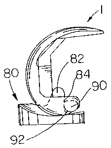
FIG. 1 is a side plan view of a femoral knee implant according to the present
invention.
FIG. 2 is a side plan view of an alternative embodiment of the femoral knee
implant
according to the present invention.
FIG. 3 is a side plan view of an alternative embodiment of the femoral knee
implant
according to the present invention.
CA 02263086 1999-02-26
~
-3-
FIG. 4 is a front plan view of an articular surface module according to the
present
invention.
FIG. 5 is a side plan view of the articular surface module of FIG. 4.
FIG. 6 is a top plan view of the articular surface module of FIG. 4 shown
mounted on
a femoral knee implant;
FIG. 7 is a side plan view of the articular surface module of FIG. 4 shown
mounted on
a femoral knee implant;
FIGS. 8-14 are side plan views of the femoral knee implant of FIG. 1
articulating with
a tibial component of the present invention between 90 degrees and 160 degrees
of flexion.
FIG. 15 is a side view of an alternative embodiment of the femoral knee
implant
according to the present invention.
FIGS. 16-22 are side plan views of the femoral knee implant of FIG. 15
articulating
with a tibial component of the present invention between 90 degrees and 160
degrees of
flexion.
DETAILED DESCRTPTION OF THE TNVENTION
FIGS. 1, 2, 3, 7 and 15 show embodiments of the femoral knee component of the
present invention oriented at zero degrees of flexion. Unless otherwise noted,
the geometric
relationships of this invention are descriptive of a femoral knee implant in
this orientation.
FIG. 1 depicts an exemplary one-piece femoral knee implant 1 according to the
present invention. The irnplant 1 includes arcuate medial 2 and lateral (not
shown) condyles
joined together at their anterior aspects to form a patellar flange 4. Each of
the medial 2 and
lateral condyles includes a distal condyle 5, a posterior condyle 6, and a
superior condyle 7.
The patellar flange 4, the distal condyles 5, the posterior condyles 6, and
the superior
condyles 7 define a smooth articular surface extending around the exterior of
the implant 1.
The interior of the implant 1 is defined by a box 9. The box 9 includes an
anterior box surface
10, a distal box surface 11 and a posterior box surface 12. The anterior 10
and distal 11 box
surfaces are blended by an anterior chamfer surface 13. The distal 1 1 and
posterior 12 box
CA 02263086 1999-02-26
(. ~. ..
-4-
surfaces are blended by a posterior chamfer surface 14. The four compartment
knee of the
present invention accommodates flexion in the range of 165 degrees.
In order to provide the superior condyles 7 of the present invention, the
superior
aspect of the posterior condyles 6 is extended toward the anterior flange 4 to
allow the
articular surface to extend further around and back anteriorly than with prior
femoral
implants. Extending the superior aspect of the posterior condyle can be done
in several ways.
As shown in FIG. 1, the entire posterior condyle is thickened such that the
posterior box
surface 12 is further from the posterior condyle 6 exterior surface and nearer
the anterior box
surface 10. This widens the superior aspect of the posterior condyle so that
the articular
surface can be extended to form the superior condyle 7. Alternatively,
posterior condyle 6
can be shortened by removing material from the superior aspect where the
condyle begins to
taper which will have the effect of leaving a thicker superior aspect that can
be shaped into
a superior condyle. Yet another alternative is to change the angle that the
posterior box
surface 12 makes with the distal box surface 11. By making the included angle
between these
two surface smaller, the superior aspect of the posterior condyle is made
wider to provide for
a superior condyle 7.
Taking this angle change further leads to the embodiment of FIG. 2. Here, the
angle
between the posterior box surface 16 and the distal box surface 18 has been
made less than
90 degrees to provide ample width for a superior condyle 20. The dashed line
22 depicts the
angle of the posterior box surface of a typical prior art femoral component.
In order for the
femoral component to be easily implantable, posterior box surfaces 16 and the
anterior box
surface 24 must be parallel or slightly diverging toward the box opening.
Therefore it may
be necessary, as shown in FIG. 2, where the posterior box surface has been
angled inwardly,
to angle the anterior box surface 24 outwardly. The dashed line 26 depicts the
angle of the
anterior box surface of a typical prior art femoral component.
FIG. 3 illustrates another alternative embodiment for moving the superior
aspect of
the posterior condyle 28 anteriorly. In this enlbodiment, the entire box;
including the
posterior surface 30, distal surface 32, anterior surface 34 and chamfers 36
and 38; is rotated
CA 02263086 1999-02-26
-5-
about a medial-lateral axis thus shortening the anterior condyle 40 and
extending the posterior
condyle 28 anteriorly and slightly superiorly. A superior condyle 42 can then
be formed at
the superior aspect of the posterior condyle 28. The dashed lines 44 depict
the box and
articular surfaces of a typical prior art femoral component before the box is
rotated.
In prior art implants the distal box surface 27 (dashed) is parallel to the
tangent 3 1 of
the distal condyles at their most prominent point. This helps a surgeon orient
the femoral
component at full extension. In the embodiment of FIG. 3, the box is rotated
so that the distal
surface 32 is angled relative to the tangent 31.
FIGS. 4-7 depict an alternative modular embodiment of the invention. The use
of a
modular add-on allows a conventional implant to be adapted for four
compartment
articulation. The implant 50 includes arcuate medial 52 and lateral 53
condyles joined
together at their anterior aspects to form a patellar flange 54. Each of the
medial 52 and
lateral 53 condyles is made up of a distal condyle 55 and a posterior condyle
56. The patellar
flange 54, the distal condyles 55 and the posterior condyles 56 define a
smooth articular
surface extending around the exterior of the implant 50. The articular surface
terminates at
the apexes 58 of the posterior condyles 56. The terminal portion of the
articular surface is
defined by the radius R. The interior of the implant 1 is defined by a box 59.
The box 59
includes an anterior box surface 60, a distal box surface 61 and a posterior
box surface 62.
The anterior 60 and distal 61 box surfaces are blended by an anterior chamfer
surface 63. The
distal 61 and posterior 62 box surfaces are blended by a posterior chamfer
surface 64.
FIGS. 4 and 5 depict an articular surface module 65. The module 65 includes a
front
surface 66, a back surface 67, a bottom surface 68, side surfaces 69, and a
top surface 70.
The back 67 and bottom 68 of the module 65 are shaped to seat against the
posterior box
surface 62 and posterior chamfer surface 64 respectively. The top surface 70
has an articular
shape matching the articular surface of the implant 50 near the apexes 58.
When the back 67
and bottom 68 of the module 65 are seated in the implant box 59, the top 70 of
the module
forms an extension of the articular surface, or a superior fourtli
compartment, as shown in
FIGS.6 and 7. The extended articular surface blends functionally with the
articular surface
CA 02263086 1999-02-26
. ~;..
-6-
to allow additional articulation of the femur relative to the tibia. Thus, a
smooth transition
is provided from articulation on the implant to articulation on the module. In
the embodiment
shown in FIG. 7, the module 65 extends the radius R. A module is used
similarly on both the
medial and lateral posterior condyles. A through hole 71 in the module 65 and
corresponding
threaded holes in the posterior condyles allow the module 65 to be securely
attached to the
implant 50. Other well known means of attachment may also be used such as
cement or clips.
The femoral component of the present invention accommodates deep flexion
through
the use of a fourth articular region. Other femoral features help to maximize
the potential of
this improved articular surface design. FIGS. 8-14 illustrate the femoral
component 1 of FIG.
1 articulating with a tibial component 80. The tibial component 80 includes a
spine 82 having
an articular surface 84. The femoral component 1 includes a cam 90 having an
articular
surface 92. In flexion, the cam articular surface 92 bears on the spine
articular surface 84.
This spine/cam interaction creates a center for rotation of the femoral
component relative to
the tibial component and prevents anterior subluxation of the femoral
component relative to
the tibial component. The distance from the spine/cam contact to the top of
the spine is called
the "jump heiglit" and is a measure of the subluxation resistance of a
particular spine/cam
combination because the cam would have to jump over the spine for subluxation
to occur.
In extreme flexion, such as that for which the present invention is designed,
jump height is of
increased concern. Likewise, bending of the spine is a concern due to
increased loads during
activities such as squatting. In many prior art implant designs, the cam is
located relatively
low compared to the top of the distal condyles. If these prior art knees are
flexed deeply, the
cam begins to ride up the spine and the jump height can be significantly
shortened leading to
an increased possibility of subluxation and an increased possibility of
bending the spine
because of the greater bending moment. In the present invention a high cam
placemerit is
used similar to the design of the NexGeng Complete Knee Solution manufactured
and sold
by Zimmer, Inc. By combining high cam placement with a fourth articular
compartment, the
extreme flexion potential of the knee is enlianced. Extreme flexion is
facilitated while
maintaining a safe level of subluxation resistance. As shown in FIGS. S- 14,
the jump height
CA 02263086 1999-02-26
. ~ '
-7-
increases from 90 degrees, FIG. 8, to approximately 130 degrees, FIG. 12.
Beyond 130
degrees, the cam rises only slightly, thus maintaining a large jump height
even in deep flexion.
The embodiment of FIG. 15 further enhances the jump height of the spine cam
articulation. The exemplary cam in FIGS. 1 and 8-14 is cylindrical at its
functional
articulating surface. It is placed far superiorly between the superior
posterior condyles to
increase jump height in flexion. To further enhance jump height, the cam in
FIGS. 15-22 is
made non-cylindrical, being made up of blended circles or other geometries. An
exemplary
non-cylindrical cam is shown in FIG. 15. The cam 100 includes a relatively
flat portion 101,
a first spine contact portion 102 having a first radius defining a circle, and
a second spine
contact portion 104 having a second radius defining a circle. The first spine
contact portion
102 is an arc of the circle defined by the first radius. The second spine
contact portion 104
is an arc of the circle defined by the second radius. The second spine contact
portion extends
further posteriorly than the perimeter of the circle defined by the first
radius. In the
embodiment shown in FIG. 15, the first and second spine contact portions form
an ovoid
articular surface 102, 104. Because the cam radius extends posteriorly, the
second spine
contact point is lower relative to the spine than it would otherwise be. The
posterior
extension of the cam 100 causes it to reach downwardly and contact the spine
lower at
higher angles of flexion as shown in FIGS. 16-20. The second contact portion
104 causes the
femur to roll back in deep flexion to prevent the femoral bone, where it exits
the posterior
box, from impinging on the tibial articular surface. The top 108 of the cam
100 completes
the cam profile.
The cani 100 alternatively includes a third spine contact portion 106, also
shown in
FIG. 15, having a third radius defining a circle. The alternative third spine
contact portion
projects beyond the condyles in order to maintain the proper femoral position
relative to the
tibia in deep flexion. The radius of the third portion 106, when present,
forms the posterior
most cam surface and the end of the cam articular surface.
/ CA 02263086 1999-02-26
1f ~
-g-
One way to achieve the described relationships between the spine contacting
portions
is to increase the radius of the cam 100 posteriorly from the first spine
contact portion 102
to the second spine contact portion 104. The third spine contacting portion
106 would be
made smaller than the second spine contacting portion 104 and would articulate
as shown in
FIGS. 21 and 22. Another way to achieve the inventive relationships is to
offset the centers
of the first and second radii in the anterior/posterior direction. Depending
on the particular
radius values and offset chosen, additional radii may be necessary to smoothly
blend the first
and second spine contacting surfaces.
It will be understood by those skilled in the art that the foregoing has
described a
preferred embodiment of the present invention and that variations in design
and construction
may be made to the preferred embodiment without departing from the spirit and
scope of the
invention defined by the appended claims.
