Note : Les descriptions sont présentées dans la langue officielle dans laquelle elles ont été soumises.
CA 02266580 2005-01-05
50336-79
1
DESCRIPTION
THREE-DIMENSIONAL INTRALUMINAL ULTRASOUND IMAGE RECONSTRUCI70N
Field of the Invention
This invention relates to methods and apparatus for ultrasound
imaging within a body cavity or lumen, and more particularly relates to
methods
and apparatus which enhance the accuracy of three-dimensional (3D) image
reconstruction where the 3D image is reconstructed from a series of images
taken
over a nonlinear path.
Background of the Invention
Intravascular ultrasound imaging is now a common technique used
to determine the position and characteristics of stenotic lesions in the
arteries of a
patient. Presently, 3D images of a region of a vessel are generated by
acquiring
image data from an ultrasound transducer during pull-back: of the transducer
within a region of interest, and then stacking the sequence of 2D images thus
acquired to generate a 3D image. In procedures commonly used, the 2D images
are stacked equidistantly along a straight centerline, assuming uniform
velocity of
pull-back. If pull-back velocity is nonuniform, then the position of each
image is
registered, and images are stacked at registered positions. Although this
non-uniform pull-back does not lead to 3D distortion, it may result in gaps in
the
image due to nonuniform spacing. These methods assume that the transducer
follows a linear path during pull-back, whereas in reality the path is often
curved
in 3D space. Thus, by making this assumption, substantial error may be
introduced into the 3D reconstruction, resulting in an image having
significant
distortion in the 3D representation of reality. This error results because
each
CA 02266580 2005-01-05
50336-79
2
image is assumed to lie in a plane which is parallel to the plane of each
adjacent
image, whereas the image planes are, in reality, angled relative to one
another.
At least one attempt has recently been made to correct for this error
by generating a 3D catheter centerline, and then stacking 2D images according
to
the geometry of the catheter centerline. See Slager et al., The Thorax Center
Journal 7/3:36-37 (1995); Roelandt et al., Circulation 2Q(2):1044-1055 (1994);
Slager et al., Journal of the American College of Cardiology, 2,.5:144A
(1995);
and Laban et al., Thorax Center, University Hospital, Rotterdam, Netherlands,
"ANGUS: A new Approach to Three-dimensional Reconstruction of Coronary
Vessels by Combined Use of Angiography and Intravascular Ultrasound".
According to the Slager method, use is
made of a catheter having radiopaque markers, and the catheter centerline is
reconstructed from data obtained through bi-plane fluoroscopy before and/or
during catheter pull-back. The use of fluoroscopy as a technique for the
determination of a 3D centerline is, however, not without certain drawbacks
and
side effects. Thus, a need exists for apparatus and methods to determine pull-
back trajectory without using fluoroscopy, so as to permit accurate 3D
reconstruction from sequential ultrasound images.
Summary of the Invention
We have discovered methods and apparatus for imaging an organ,
lumen, or other internal structure within a body to obtain accurate 3D
reconstruction of the organ, lumen, or other internal structure. The methods
and
apparatus will find applicability to the coronary arteries, arteries
generally, and
more generally the vascular system, as well as to imaging anatomic spaces
within
organs, such as the cavities of the heart, including the atria and ventricles.
It will
also be understood that the methods and apparatus will find applicability to
imaging within the esophagus (e.g., transesophegeal echocardiography), the
urethra, the uterus, etc.
CA 02266580 1999-03-19
WO 98/11823 PCT/US97/16455
3
The apparatus of the invention includes both an ultrasound imaging
catheter system and a catheter tracking system. The ultrasound imaging system
generally is provided in the form of a conventional intraluminal catheter
having
ultrasound imaging capabilities. For details on the general design and
construction
of such catheters, the reader is directed to Yock, U.S. Patent Nos. 4,794,931,
5,000,185, and 5,313,949; Sieben et al., U.S. Patent Nos. 5,243,988, and
5,353,798; Crowley et al., U.S. Patent No. 4,951,677; Pomeranz, U.S. Patent
No. 5,095,911, Griffith et al., U.S. Patent No. 4,841,977, Maroney et al.,
U.S.
Patent No. 5,373,849, Bom et al., U.S. Patent No. 5,176,141, Lancee et al.,
U.S.
Patent No. 5,240,003, Lancee et al., U.S. Patent No. 5,375,602, Gardineer et
al.,
U.S. Patent No. 5,373,845, Seward et al., Mayo Clinic Proceedings
71(7):629-635 (1996), Packer et al., Cardiostim Conference 833 (1994),
"Ultrasound Cardioscopy," Eur.J.C.P.E. 4(2):193 (June 1994), Eberle et al.,
U.S. Patent No. 5,453,575, Eberle et at., U.S. Patent No. 5,368,037, Eberle et
al., U.S. Patent No. 5,183,048, Eberle et at., U.S. Patent No. 5,167,233,
Eberle
et al., U.S. Patent No. 4,917,097, Eberle et al., U.S. Patent No. 5,135,486,
and
other references well known in the art relating to intraluminal ultrasound
devices
and modalities. The catheter will typically have proximal and distal regions,
and
will include an imaging tip located in the distal region. Such catheters have
an
ability to obtain echographic images of the area surrounding the imaging tip
when
located in a region of interest inside the body of a patient. The catheter,
and its
associated electronic circuitry, will also be capable of defining the position
of the
catheter axis with respect to each echographic data set obtained in the region
of
interest.
The catheter tracking system generally includes at least one
ultrasound transducer mounted adjacent the imaging tip of the catheter, the
signal
of which is used to track the location and/or angulation of the imaging tip
during
movement. In the reception mode, the signal used to track location of the
imaging
tip is electric, while in the emission mode the signal will be acoustic. The
movement is usually pull-back, but also including lateral movement in all six
degrees of freedom ((x,y,z) and three angles). In the remaining disclosure we
CA 02266580 2005-01-05
50336-79
4
shall typically discuss pull-back alone, but it will be understood that all
other
forms of movement-are contemplated including tip deflection within a steerable
catheter. The tracking transducer operates in two modes. In the reception
mode,
the signal used to track location of the imaging tip is electric, while in the
emission mode the signal will be acoustic. In another embodiment, a pair of
transducers mark the location of the imaging tip during pull-back. The pair of
closely spaced transducers define a line which approximates the tangent to the
curve defined by the catheter at that point. Thus, angulation of the catheter
is
determined by finding the line through the positions of at least two
transducers.
adjacent the imaging tip as an approximation of the catheter tangent. Where
only
a single transducer marks the location of the catheter tip during pull-back,
the
catheter tangent is approximated by the Iine defined by two sequential
positions of
the marker transducer during pull-back.
The catheter tracking system further includes a number of
transducers located away from the intraluminal ultrasound (ILUS) catheter,
generally two or more, more preferably three or more, more preferably four or
more. These transducers form a reference frame, and they may be located
internally and/or externally of the patient. The tracking system further
includes
electronic circuitry for activating certain transducers to generate ultrasound
signals
for reception by certain other ultrasound transducers. The system also
includes
circuitry for measuring elapsed time between generation of the ultrasound
signals
and reception by respective other ultrasound transducers. Moreover, the
tracking
system will include electronic circuitry for calculating positions of the
frame
transducers and catheter tip transducers relative to each other using known
velocity of ultrasound waves and triangulation, and using measured elapsed
times.
This tracking system allows the user to determine the 3D coordinates (x, y, z)
of
each marker transducer at successive times during catheter pull-back. An
example of a catheter tracking system, and its method of use to determine 3D
coordinates (x, y, z) of a moving point, are described in Smith et al., U.S.
Patent
No. 5,515.853.
CA 02266580 2005-01-05
50336-79
The methods of the invention will generally include a step of
positioning the ILUS catheter imaging tip within the patient at a region of
interest.
The ILUS catheter may then be operated to obtain a series of echographic
images
during catheter pull-back. In a preferred embodiment, the pull-back is an
5 ECG-gated pull-back as disclosed in Roelandt et al., Circulation 9-Q(2):1044-
1055
(1994) and Laban et al., Thorax Center, University Hospital, Rotterdam,
Netherlands, "ANGUS: A new Approach to Three-dimensional Reconstruction of
Coronary Vessels by Combined Use of Angiography and Intravascular
Ultrasound." During acquisition of each echographic data set, the position of
the
catheter axis with respect to the data set is determined and recorded. This
step is
done by the imaging catheter. Moreover, the 3D coordinates of the catheter tip
are also determined and recorded for each echographic data set. This step is
done
by the tracking system. The recorded positions of the catheter tip are used to
calculate a catheter pull-back trajectory in 3D space, taking into account a
correction for the imaging transducer not being positioned at exactly the
location
of the one or more marker transducers. The echographic images are stacked
around the catheter trajectory. This step is the 3D reconstruction. The origin
of a
first echographic data set is placed at the first recorded position, and then
each
subsequent image is positioned at its respective distance from the first
image.
During this positioning step, it is preferable to align the recorded catheter
axis
with the pull-back trajectory so that each image data set has the proper angle
of
orientation relative to the pull-back trajectory.
CA 02266580 2007-10-29
50336-79
5a
In accordance with an aspect of the present
invention, there is provided a method for imaging an organ,
lumen, or other internal structure to obtain three-
dimensional reconstruction of a region of interest of the
organ, lumen, or other internal structure, comprising the
use of an imaging system, which has been positioned within
the region of interest which imaging system includes a
catheter imaging tip; and a catheter tracking system
comprising at least one tracking element which has been
mounted adjacent to the catheter imaging tip, and a
plurality of reference frame elements located away from the
catheter imaging tip; said method comprising the steps of:
obtaining a plurality of images of the surrounding of the
catheter imaging tip during movement of the catheter imaging
tip using the imaging system when the catheter imaging tip
is positioned within the region of interest within the
organ, lumen, or other internal structure; recording,
relative to each image obtained during movement, the
position of a tangent to the catheter at the imaging tip,
said tangent providing the local catheter axis; recording
the X, Y, Z coordinates of the image origin in the catheter
tip during movement using the catheter tracking system; and
stacking the plurality of images around a curved three-
dimensional catheter movement trajectory by positioning the
origin of a first image at a first recorded position, and
positioning subsequent images at their respective distances
from the first image and perpendicular to the catheter axis
at each recorded position along the curve.
In accordance with another aspect of the present
invention, there is provided a catheter imaging system for
imaging an organ, lumen, or other internal structure to
obtain accurate three-dimensional reconstruction of a region
of interest of the organ, lumen, or other internal
CA 02266580 2007-10-29
50336-79
5b
structure, said system comprising: a catheter having a
proximal region, a distal region, and an imaging tip
operably disposed within the distal region of the catheter;
a catheter tracking system comprising at least one tracking
element mounted adjacent to the catheter imaging tip, and a
plurality of reference frame elements located away from the
catheter imaging tip; an automated longitudinal position
translator operably coupled to the imaging tip; and a system
coupled to the imaging tip, the tracking system, and the
automated longitudinal position translator; wherein, the
system is configured to automatically pull back the imaging
tip, obtain a plurality of images with the imaging tip,
track the position coordinates of the imaging tip, and stack
the plurality of images around a three-dimensional catheter
movement trajectory.
Brief Description of Drawings
Fig. 1 depicts a human heart as a site for use of
the method and apparatus disclosed herein;
Fig. 2 depicts an exploded view of a region of the
coronary arteries having an ILUS catheter positioned in a
region of interest;
Fig. 3 depicts a pull-back of an ILUS catheter
having one omni-directional tracking transducer;
CA 02266580 1999-03-19
WO 98/11823 PCT/US97/16455
6
Fig. 4 depicts lateral movement which may occur during pull-back
of the ILUS catheter shown in Fig. 3;
Fig. 5 depicts a pull-back of an ILUS catheter having two omni-
directional tracking transducers;
Fig. 6A depicts the ILUS catheter of Fig. 5 positioned within a
linear region of a vessel;
Fig. 6B depicts the ILUS catheter of Fig. 5 positioned within a
highly curved region of a vessel;
Fig. 7 depicts a patient having a catheter tracking system positioned
for use and an automated pull-back device mounted on the ILUS catheter;
Fig. 8 depicts a schematic block diagram of an optical coherence
domain reflectometer; and
Fig. 9 depicts an embodiment of an optical coherence tomography
catheter module.
Detailed Description
The methods disclosed herein are applicable to ultrasound imaging
of the coronary arteries as depicted in Fig. 1, or to any body cavity where
the
image is to be obtained over a region where the position trajectory of the
catheter
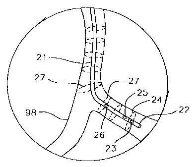
may vary. With reference to Fig. 1, heart 99 includes coronary arteries 98
which
follow a tortuous path along the surface of the heart, having curvatures in
many
locations. Fig. 2 shows an exploded view of curvature 97, having an ILUS
catheter disposed within a region of interest therein. Catheter 21 has distal
end 22
and a proximal end (not shown), and is generally designed in accordance with
imaging catheters known in the art. The catheter thus includes an intraluminal
ultrasound imaging system capable of obtaining echographic images of the
surrounding of catheter tip 22. The imaging system includes transducer 23 and
its
associated electronics for displaying an echographic data set, e.g., obtained
by
rotating transducer 23 over a 360-degree path 24 about distal tip 22 of
catheter 21
to scan the vessel interior, or by a sector scan which makes a 60 or 90 degree
scan
of the vessel interior. In an alternative embodiment, transducer 23 is
replaced by
CA 02266580 1999-03-19
WO 98/11823 PCTIUS97/16455
7
a phased array of transducers as disclosed in Griffith et al., U.S. Patent No.
4,841,977. Scanning of the vessel interior is repeated many times during pull-
back to obtain a plurality of echographic data sets taken at a sequence of
positions
27 within vessel 98.
In one embodiment, each echographic data set obtained during
pull-back comprises a transverse or cross-sectional image of the vessel at the
point of the image, as shown in Fig. 2. In another embodiment, each image
represents essentially a conical plane having its top at the position of the
imaging
transducer. In this case, the top angle of the conical plane is usually large,
and
typically 150-170 degrees. In another embodiment, each echographic data set
represents essentially a 3D cloud having its origin at the position of the
ultrasound
imaging transducer.
The apparatus herein further includes a catheter tracking system
which enables real-time determination of the 3D coordinates and angulation of
the
catheter imaging tip during use within a body. The tracking system includes at
least one transducer 25 mounted on the catheter and typically adjacent the
imaging
tip 23, as shown in Fig. 2. Transducer 25 can be mounted either proximal or
distal to scanning transducer 23. However, a distal mount may lead to image
artifacts because of transducer leads which cross the image plane. In another
embodiment, catheter 21 includes two tracking transducers 25 and 26 spaced by
a
short distance. Optimum spacing is determined by considering two competing
parameters; accuracy of angulation measurement and percent error due to limits
on
resolution. Tracking system resolution is the limiting factor in determining
how
close the two transducers can be placed. It is desired to have the two
transducers
placed as close as possible in order to optimize the accuracy of the
angulation.
However, because the currently available resolution permits determination of
position to within about 1 mm, in order to minimize the percent error due to
the
limits on resolution, it is best to space the tracking transducers by greater
than 5
mm, more preferably greater than 7 mm, more preferably greater than 9 mm, and
most preferably at or greater than about 10 mm. In any case, it is best to
limit the
spacing of the tracking transducers to no greater than about 15 min or less,
more
CA 02266580 1999-03-19
WO 98/11823 PCT/US97/16455
8
preferably no greater than about 12 mm or less, more preferably no greater
than
about 10 mm or less, with about a 10 mm spacing being most preferred.
It is desirable to be able to calculate the tangent to the catheter
centerline or to the trajectory of the imaging transducer at each point where
echographic data is acquired. In one embodiment, where two or more tracking
transducers are employed, the tangent is approximated by the line defined by
the
two or more points determined by the location of the tracking transducers. In
another embodiment, where only one tracking transducer is employed, the
tangent
is approximated by the line through two points determined by the successive
locations of the tracking transducer at two positions during pull-back. The
tangent, or angulation, once determined, provides the proper angle of
placement of
each echographic data set since each data set may be placed so that the
recorded
catheter axis for each data set coincides with-the tangent at the point where
the
echographic data set was recorded. Numerous averaging techniques known in the
art can be used to smooth large deviations in the obtained trajectory caused
by
stochastic errors or resolution deficiency.
The catheter tracking system also includes a number of transducers
located away from the ILUS catheter which collectively define a reference
frame.
It is desired to have at least three transducers away from the catheter and in
the
reference frame so that the tracking transducer and the reference frame
transducers
are not all included in one plane. The design and use of a catheter tracking
system
is fully discussed in Smith et al., U.S. Patent No. 5,515,853.
An ILUS catheter and a tracking system as disclosed herein is
shown deployed in a patient in Fig. 7. The tracking system includes reference
frame transducers disposed within chest harness 101. Catheter 21 enters the
patient through the femoral artery and is positioned within a region of
interest,
e.g., within the coronary arteries. The proximal end of catheter 21 includes
handle 102 which is coupled to an automatic pull-back device (APD) 103.
Processing system 104 receives electronic input from the reference frame
transducers through wires 105, from the tracking transducers through wires
106,
CA 02266580 2005-01-05
50336-79
9
from the ILUS catheter imaging system through wires 107, and from the APD
through wires 108.
In use, the ILUS catheter is deployed according to procedures well
known in the art. For imaging in the coronary artery, the catheter is inserted
through an incision in the femoral artery (Fig. 7) and is advanced upwardly
through the descending aorta, typically with assistance from a guiding
catheter,
until it crosses the aortic arch and reaches the coronary arteries. Where the
region
of interest lies within a different vessel or organ, the means for entry will
obviously differ, but will follow established techniques known in the art. The
ILUS catheter is positioned so that the imaging tip lies within a region of
interest
inside the body of a patient, as depicted in Fig. 2. The catheter imaging
system is
then carried through a pull-back sequence, optionally using an automated
pull-back device as disclosed by Webler et al., U.S. Patent Nos: 5,361,768 and
5,485,846, and Ream, U.S. Patent Nos. 5,827,313 and 5,957,941.
During pull-back, a series of echographic data sets is
obtained, each of which will provide the necessary input to produce an image
which can be displayed according to processes and using electronic equipment
well
known in the art.
At the time each image is captured, the position of the catheter axis
with respect to each echographic data set obtained during pull-back is defined
by
the position of the transducer, and is therefore known. During pull-back of
the
catheter imaging tip, it is also desired to record the position (x, y, z) and
the time
at each interval for which data is recorded, according to the method of U.S.
Patent
No. 5,515,853. From this information, it is possible to calculate the velocity
of
catheter tip movement and to record this information as well. Moreover, it is
desired to determine and record the angulation in 31) space of the imaging tip
for
each image using the coordinates (x, y, z) of the one or more-tracking
transducers
as discussed above.
The coordinates (x, y, z) of each point of image acquisition along
the catheter pull-back path are then used in conjunction with the time data to
CA 02266580 2005-01-05
50336-79
.
reconstruct a 3D image by stacking the echographic images around the catheter
pull-back trajectory. The origin of the first image is placed at a first
recorded
position. The next image is then positioned at its respective distance from
the first
image, and this process is repeated until all images within the region of
interest
5 have been placed along the pull-back trajectory. Each echographic data set
is
adjusted so that the catheter axis recorded for that data set is aligned with
the
catheter pull-back trajectory. In this manner, for cross-sectional scanning,
each
echographic data set is oriented at a substantially 90-degree angle to the
longitudinal axis of the ILUS catheter. Linear interpolation is performed
between
10 digitized adjacent image sets, resulting in a volumetric (voxel) data set,
as
described in Evans et al., Circulation 9x:567-576 (1996).
The voxel data set can be resliced, creating a new series of 2D frames.
3D images can be created by processing the data with algorithms developed
specifically for voxel-based image display (Sonoview, Pura Labs). The
resulting
images are displayed on a workstation for display and analysis.
Where the echographic data sets are 3D clouds, each cloud may
overlap at its boundaries with the adjacent image cloud. Thus, it may be
desired
to make certain adjustments in the echographic data to eliminate distortions
which
may occur at the boundaries between overlapping images. Moreover, error may
occur due to what is known as "sock rotation," which refers to the rotational
orientation of each echographic data set relative to a fixed reference. We
adjust for
this error by using anatomic landmarks within the vessel or cavity being
imaged,
so that the rotational orientation of each data set is adjusted relative to
the
placement of that landmark.
In another embodiment, adjustment may be made for image rotation
caused by torsion of the catheter during pull-back due to bends in different
planes.
This typically shows up as image rotation which may distort a reconstructed
image
if not corrected for. This distortion is referred to as "twist," and arises
because
the images are rotated to a different degree around their origin along their
path
during pull-back. This phenomenon, and a solution to correct for it, is
described
in Laban et al., Thorax Center, University Hospital, Rotterdam, Netherlands,
CA 02266580 2005-01-05
50336-79
11
"ANGUS: A new Approach to Three-dimensional Reconstruction of Coronary
Vessels by Combined Use of Angiography and Intravascular Ultrasound," and will
not be further discussed here in the interest of brevity.
In another embodiment, the catheter tip position is recorded during
pull-back by using an electromagnetic position and orientation determining
system
as described by Aretz et al., International Journal of Cardiac Imaging ¾:231-
237
(1991), Acker et al., U.S. Patent No. 5,558,091.
Example- 1: 3D Reconstruction Using Catheter with One Omni-Directional
Tracking Transducer
Stacking of images to obtain 3D reconstruction is accomplished
using a catheter with one tracking transducer by calculating an approximate
tangent to the pull-back trajectory using two successive points for the
tracking
transducer, as depicted in Fig. 3. Transverse images 52 and 53 are obtained
during pull-back 57 along trajectory 51. At a first point in time, t=t1, the
tracking transducer lies at position 1, corresponding to point 54. The image
plane
52 is displaced by distance 56 (s) from position 1, and this displacement s
corre-
sponds to the distance between a distal tracking transducer and a slightly
proximal
scanning transducer. At a second point in time, t=t2, the tracking transducer
lies
at position 2, corresponding to point 55. The image plane 53 is again
displaced by
distance s from position 2.
Thus, at position 1 (t=t,), the tip coordinates are given by (x1, yl,
z1), while at position 2 (t=t2), the tip coordinates are given by (x2, y2,
z2). The
direction and the length of the vector between position 1 and position 2 is
given by
the vector
(x2-x1, Y2-Y1, z2-z1)
CA 02266580 1999-03-19
WO 98/11823 PCT/US97/16455
12
The direction and the length of the vector between position 1 and the image
plane
52 is given by the vector
S
(x2-x1, Y2-Y,, z2-Z1)
(x2-x')2 + (Y2-Y1)2 + (Z2-z1)2
The normal vector on image plane 52 is
(X2-XI' y2-y1, z2-z 1)
(n1, n2, n3) =
(x2-x 1)2 + (Y2-Y 1)2 + (z2-Z, )2
This vector positions image plane 52 at a 90 angle to the tangent, the
direction of
which is approximated by the direction of the vector between position 1 and
position 2. The mathematical description of image plan 52 is therefore defined
by
s(x2-x1)
n,x + n2y + n3z = n, X1 + +
(x2-x,)2 + (y2-y,)2 + (z2-z,)2
S (y2-y, )
n2 y, +
(x2-x1)2 + (Y2-Y1)2 + (z2-z1)2
s(z2-z1)
n3 z, +
(x2-x1)2 + (Y2-Y1)2) + (z2-z1)2
Therefore, in reconstruction, the coordinates of the origin are given
by
CA 02266580 1999-03-19
WO 98/11823 PCTIUS97/16455
13
X + s (x2-x1)
(x2-x1)2 + (Y2-Y1)2 + (z2-z1)2
s (y2-y1)
y1+
(x2-x1)2 + (Y2-Y1)2 + (z2-z,)2
z s (z2-z 1)
1 + (x2-x1)2 + (y2-yl)2 + (z2-zi)2
and the angulation of the plane is defined by its normal vector (n1, n2, n3).
Image
plane 53 is defined analogously after definition of a position 3, by
substitution
1---2 and 2-3 in (x1, y,, z1) and (x2, y2, z2).
It will be understood that this system and method of calculation has
a potential for significant error if the pull-back is not done in line with
the
catheter axis at the catheter tip location. This error is therefore likely to
occur
where there is substantial lateral movement during pull-back, such as would
occur
in an anatomic cavity within an organ (e.g., the chambers of the heart). This
error is illustrated in Fig. 4 where it can be seen that, for a pull-back
starting at
position 54. the entire trajectory is displaced by lateral movement 57'. Thus,
while the real image plane should be calculated as shown by plane 52, an
assumed
image plane 58 is generated instead, resulting in a substantial angulation
error 59
between the real and assumed image planes. Moreover, this angulation error
causes error 60 to occur in positioning the origin of the image.
Example 2: 3D Reconstruction Using Catheter With Two Omni-Directional
Tracking Transducers
Stacking of images to obtain 3D reconstruction is accomplished
using a catheter with two tracking transducers by calculating an approximate
tangent to the pull-back trajectory using the line between the two tracking
trans-
CA 02266580 1999-03-19
WO 98/11823 PCT/US97/16455
14
ducers at the point in time when echographic image acquisition occurs, as
depicted
in Fig. 5. Transverse image 52 is obtained at time t = t, during pull-back
along
trajectory 51. At t=t,, the first tracking transducer (transducer 1) lies at
point 54'
(position 1) while the second tracking transducer (transducer 2) lies at point
55'
(position 2), separated by distance f corresponding to gap 61. Image plane 52
is
displaced by distance 56 (s) from position 1, as described in Example 1.
Thus, at t,, the tip coordinates for transducer 1 are given by (x,, y,,
z,), while the tip coordinates for transducer 2 are given by (x2, y2, z2). The
distance between transducers 1 and 2 is known and measured to be
Q = (x2-x1)2 + (y2-Y,)2 + (z2-z,)2
and the image plane is positioned at distance s from transducer 1.
The normal vector on image plane 52 is
(n' n2 n3) (x2-x1, Y2-y1, Z2-Z,)
(x2-x,)2 + (YZ-Y,)2 + (Z2-z1)2
This vector positions image plane 52 at a substantially 90 angle to the
tangent,
the direction of which is approximated by the direction of the vector between
transducers 1 and 2.
The mathematical description of image plane 52 is therefore defined
by
CA 02266580 1999-03-19
WO 98/11823 PCT/US97/16455
s(x2-x1)
nix + n2y + n3z = n1 x1 + +
(x2-x1)2 + (Y2-Y1)2 + (z2-z1)2
s(y2-y1)
n2yl+ +
(x2-x1)2 + (Y2-Y 1)2 + (z2-z1)2
s(z2-z1)
n3 z1+
V (x2-x1)2 + (Y2-Y1)2 + (z2-Z1)2
Therefore, in reconstruction, the coordinates of the origin are given
by
s(x2-x1)
xl +
(x2-x1)2 + (Y2-Y1)2 + (z2-z1)2
s(y2-y1)
yJ+
(x2-x1)2 + (Y2-Y1)2 + (z2-z1)2
S (z2-z1)
zl +
(x2-x1)2 + (Y2-Y1)2 + (z2-z1)2
and the angulation of the plane is defined by its normal vector (n1, n2, n3).
During
pull-back, this determination is repeated at t = t2 (= tl + At), at t = t3 (=
t2 +
5 At), etc.
It will be understood that this technique assumes that transducers 1
and 2 lie on a straight line with image plane 52 oriented perpendicularly, as
shown
in Fig. 6A. This assumption may lead to significant error where the catheter
has a
high degree of curvature between transducers 1 and 2 at any point of
echographic
10 data acquisition. This error is illustrated in Fig. 6B where it can be seen
that, for
an image acquired at t,, the tangent is approximated by line 61, while the
true
CA 02266580 2005-01-05
50336-79
16
tangent 62 has a significant displacement therefrom. Thus, while the real
image
plane should be calculated as shown by plane 52, an assumed image plane 58 is
generated instead, resulting in angulation error 59 between the real and
assumed
image planes. Moreover, this angulation error causes error 60 to occur in
positioning the origin of the image.
On the other hand, the use of two tracking transducers prevents
error associated with lateral movement which occurs if the pull-back is not
done
in-line with the catheter axis at the catheter tip location. This is because
the
tangent to the pull-back trajectory is calculated from coordinates recorded at
a.
single point in time, whereas error due to lateral movement occurs when two
successive points for a single tracking transducer are used to calculate the
tangent.
In another embodiment the invention makes use of optical
coherence tomography (OCT) to image tissue within a body instead of
intraluminal
ultrasound. OCT is described generally in Swanson et al., U.S. Patent No.
5,321,501. Referring to Fig. 8, an optical
coherence domain reflectometer (OCDR) 200 is shown. The OCDR includes a
short coherence length (broad spectral bandwidth) optical source 201 coupled
to an
optical coupler 202. The other input to coupler 202 is laser 204 generating an
optically visible output which is applied to the coupler through a fiber optic
path
205. Laser 204 does not contribute to the normal operation of the system and
is
utilized only to provide a source of visible light for proper alignment with a
sample, when the light from diode 201 is in the infrared region and thus not
visible. Further details on the construction and operation of the optical
coherence
domain reflectometer are given in Swanson et al., U.S. Patent No. 5,321,501.
Fig. 9 illustrates a catheter module which may be utilized for
imaging tubular structures 250 such as blood vessels, the esophagus, or the
like.
Fiber 251 is embedded in inner rotating sheath 252 which is rotatably mounted
within an outer sheath 253. Inner sheath 252 has lens 2:54 at the end of fiber
251
and angled mirrored surface 255. The probe is scanned longitudinally 257 along
vessel wall 250, while inner sheath 252 is rotated to scan the vessel wall in
a
second dimension. Scanning in the depth dimension to provide a
CA 02266580 1999-03-19
WO 98/11823 PCTIUS97/16455
17
three-dimensional scan may be achieved by one of the techniques described in
Swanson et al., U.S. Patent No. 5,321,501.
Although the foregoing invention has, for purposes of clarity of
understanding, been described in some detail by way of illustration and
example,
it will be obvious that certain changes and modifications may be practiced
which
will still fall within the scope of the appended claims.