Note : Les descriptions sont présentées dans la langue officielle dans laquelle elles ont été soumises.
CA 02268956 1999-03-10
WO 99/18847 PCTIUS98I21710
IMAGING DISEASED TISSUE USING AUTOFLUORESCENCE
BACKGROUND OF THE INVENTION
S A. Field of the Invention
The present invention relates to an apparatus and method for imaging bodily
tissue
by using autofluorescence. More particularly, the present invention relates to
an endoscopic
apparatus and method for imaging and sampling bodily tissue using
autofluorescence
techniques.
B. Description of Related Art
Cervical cancer often begins as a precancerous lesion on the cervix (i.e., the
outer
end of the uterus) and is called cervical intraepithelial neoplasia (CIN). The
lesion can
deepen over a period of years and if left untreated can become an invasive
cancer. A Pap
smear test is currently a common method of providing a type of screening for
cervical
cancer. The test involves taking a sample of cells from the cervix, and
sending the sample
to a laboratory to be analyzed. Test results usually take two or three weeks
to complete.
If the laboratory analysis determines that abnormal cells are detected from a
first Pap
test, a follow up test it typically performed. A second abnormal Pap smear
will often
pmmpt a colposcopic examination wherein the cervix is examined usually with a
low-power
stereo microscope. During colposcopy, suspect abnormal tissue is often
biopsied and again
sent to a laboratory for analysis. Because patients must often wait another
two to three
weeks for these results, a heightened period of anxiety and fear for the women
and their
families is created. Often, the first and second abnormal Pap smear result
from false
positive test results. Therefore, oftentimes, when a tissue sample has been
biopsied, the
sampled tissue was incorrectly determined to be cancerous and did not need to
be removed.
CA 02268956 1999-03-10
WO 99/18847 PCT/US98/21710
Spectroscopic autofluorescence, a minimally invasive procedure for analysis of
cervical
cytological, has been used to decrease a number of the problems normally
associated with
Pap smear tests.
Spectroscopic methods for differentiating cervical neoplasia from normal
cervical
tissue in vivo can be used to detect abnormal cells on the outside of the
cervix. Typically, a
fluorescence spectroscope has optical fibers at the end of a small probe which
illuminate
areas of the cervix. Suspect tissue is exposed to ultraviolet and visible
Laser or lamp Light,
causing substances naturally present in the tissue to fluoresce. The specific
wavelength or
signature of the light absorbed and emitted by cervical tissue is analyzed.
The fluorescence
spectra is then measured and compared at different intensities and wavelengths
since
abnormal or cancerous tissues consistently display different results from
normal or non-
cancerous tissue. Typically, a computer algorithm analyzes the fluorescence
spectrum and
assesses the degree of cell abnormality.
Generally, there are two types of fluorescence measurement techniques: the
first
being emission spectroscopy and the second being excitation spectropscopy. In
emission
spectroscopy, the exciting light is kept at a fixed wavelength and the emitted
fluorescent
intensity is measured as a function of the emitted wavelength. In excitation
spectroscopy,
the emission wavelength is kept fixed and the fluorescence intensity is
measured as a
function of the excitation wavelength.
Both emission and excitation spectra measurements have limitations. For
example,
both types of spectra measurements analyze only a single parameter to
determine cell
abnormality. The nature of the human tissue, however, is such that the
application of any
one single method produces a large amount of data, most of which is extraneous
to the
intended measurement. A primary reason for this situation is that tissues
contain an
extensive and diverse assortment of fluorescent species. Many of the species
are present in
2
CA 02268956 1999-03-10
WO 99/18847 PCT/US98I21710
high concentrations and have excitation bands distributed throughout the
ultraviolet and the
visible spectra regions. _
Another limitation is that the emission band of one fluorophore may overlap
the
excitation band of another fluorophone, consequently leading to energy
transfer between the
emission and excitation bands. Consequently, emissions from one fluorophore
could
possibly excite another fluorophore. The net effect is that optically exciting
a tissue sample
at almost any wavelength in the ultraviolet or visible wavelength regions
causes tissue
autofluorescence over a broad spectral range. As these emissions are typically
composed of
contributions from multiple fluorophores, utilizing a single analytical
parameter makes the
autofluoresence spectrum complex and problematic to solve. Consequently, a
robust
discrimination between tissue states is often difficult to obtain.
Another limitation of typical fluorescence measurement techniques is that they
cannot be readily combined with an apparatus or method for taking a biopsy. In
other
words, once an abnormal tissue area is detected, samples from this particular
suspect area
1 S cannot be simply, quickly and accurately taken. With current devices
utilizing fluorescent
measurement techniques, after locating the abnormal area, the endoscope must
be
withdrawn from the patient. Once the endoscope is withdraw, the Pap smear
specimen can
then be taken as a blind sample. Typically, there is no correlation between
where on the
cervix the sample is taken and where the suspect tissue was identified. Taking
a blind
sample, therefore, often results in samples being taken from normal areas or
perhaps even
areas which have not been previously investigated. Usually, the sample is also
taken by
relatively imprecise sampling devices such as brushes, scraper devices or the
like.
Once the sample has been taken and withdrawn from the body, the specimen is
typically smeared onto a microscope slide. This is often done by the physician
performing
the test. The slide is then submitted to a remote laboratory for
cytopathological microscopic
3
CA 02268956 1999-03-10
WO 99/18847 PCT/US98/21710
examination. Pertinent patient data must be sent along with the slide
including the medical
history, day in menstrual cycle, family history and other known risk factors.
Gathering and
collating these patient data, which are critical to the proper evaluation of a
specimen, is a
time-consuming, expensive, inefficient and labor-intensive process. The
laboratory
administrative personnel who gather such data are also responsible for
manually recording
the results of the Pap smear tests and ensuring that both the slide and
paperwork provided to
the cytotechnologist relate to the same patient. As the complexity of testing,
analyzing,
handling and transporting the Pap smear samples increases, the probability for
a false
positive, a false negative or sample contamination increases.
The typical Pap smear test has a number of other disadvantages. For example,
in
alinost every instance where a slide specimen is produced, the slide is
forwarded to a
laboratory. No preliminary analysis to eliminate possible unnecessary
laboratory testing is
conducted. This increases the cost of performing a Pap smear since their is no
preliminary
detenx2ination as to the possibility of normality or absence of abnormality.
Moreover,
because the sampling is taken "blind", there is typically no assurance as to
whether the
suspect abnormal cells have in fact been sampled. Oftentimes, only after
having forwarded
the sample to the testing facility and waiting two to three weeks is it
eventually determined
that another sample must be taken. Incidents of poor sample or slide
preparation are also
common because of the large amount of human interface with each specimen
slide.
Moreover, because slides are often sent to a location remote, there is an
increased risk that
the sample may become lost, broken or contaminated. The complexity of
maintaining a
secure and sterile transporting medium further increases the cost of sample
transport. In
addition, there is a psychological disadvantage in having to wait up to two
weeks or longer
for the test results to either confirm or rebut a primary abnormal reading.
4
CA 02268956 2003-11-14
62396-1017
SUI~iARY OF THE INVENTION
The present invention provides a diagnostic tool
to assist an operator in diagnosing cancerous tissue in a
human gynecological tract, the diagnostic tool comprising,
in combination: an endoscope having a proximal end and a
distal end, wherein the endoscope directs a first light from
the proximal end of the endoscope to tissue near the distal
end of the endoscope, the first light arriving at the tissue
and exciting autofluorescence in the tissue, a second light
thereby being produced, the second light comprising light
reflected from the tissue and light emitted from the tissue,
the emitted light representing a spatial distribution of the
autofluorescence, wherein the distal end of the endoscope
receives the emitted light, and wherein the endoscope
directs the emitted light from the distal end to the
proximal end of the endoscope; a position locator at the
distal end of the endoscope, wherein the position locator
generates coordinates representing where the distal end of
the endoscope is in relation to the gynecological tract; a
plurality of imaging detectors located at the proximal end
of the endoscope for receiving the emitted light from the
tissue, wherein the imaging detectors are responsive to the
emitted light and produce an autofluorescence image of the
tissue comprising a plurality of pixels each representing a
respective visible portion of the tissue; a computing
device, wherein the computing device conducts an analysis of
the plurality of pixels, the analysis producing diagnostic
characterizations of the respective visible portions of the
tissue, wherein the computing device presents the
characterizations to the operator as a derived image and
associates the characterizations with the coordinates from
5
CA 02268956 2003-11-14
62396-1017
the position locator, whereby the coordinates and the
associated characterizations of the tissue may be recorded
in response to instructions by the operator; and means for
steering the distal end of the endoscope within the
gynecological tract, whereby the operator may steer the
distal end of the endoscope to a position in the
gynecological tract corresponding to recorded coordinates.
These and many other features and advantages of
the invention will become more apparent from the following
detailed description of the preferred embodiments of the
invention.
5a
CA 02268956 1999-03-10
WO 99/18847 PCT/US98/21710
BRIEF DESCRIPTION OF THE DRAWINGS
FIG. 1 is a schematic view of an imaging video endoscopic system incorporating
a
preferred embodiment of the present invention.
FIG. 2 is a schematic view of the endoscope shown in FIG. 1.
FIG. 3 is a data input block flowchart for generating a preprocessed image.
FIG. 4 illustrates a ratio block flowchart for generating a composite ratio
image from
the data input blocks generated by the flowchart shown in FIG. 3.
FIG. S is a schematic view of an alternative embodiment of the endoscope shown
in
FIG. 1.
FIG. fi is a schematic view of an imaging video endoscope system incorporating
another preferred embodiment of the present invention.
6
CA 02268956 1999-03-10
WO 99/18847 PCT/US98/21710
DETAILED DESCRIPTION OF THE PREFERRED EMBODIMENTS
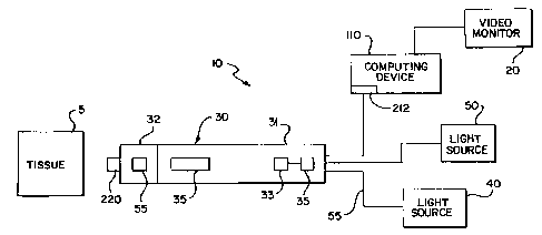
FIG. 1 illustrates a video endoscopic system 10 incorporating a preferred
embodiment of the present invention. The system 10 is utilized as a diagnostic
tool for
imaging tissue 5. Preferably, the tissue 5 being investigated by the
endoscopic system 10 is
the tissue of a cervix. Alternatively, the endoscopic system 10 is a
diagnostic tool used to
generate an image of any in vivo tissue where use of minimally invasive
procedures are
advantageous. The endoscopic system 10 utilizes the autofluorescence and
reflectance
properties of tissues to discriminate between normal and abnormal tissues.
The system 10 includes an endoscope 30, a video monitor 20, a data collection
device or computing device 110 and a light source 40. Alternatively, a second
light source
50 is provided and is preferably a white light source. The imaging endoscopic
system 10 is
suitably sized and shaped for cervical inspection. The endoscope 30, having a
distal end 32
and proximal end 31, includes a light processing unit 33, distal optics 35,
and imaging
detectors 45. Preferably, the light processing unit 33 is either a wavelength
division
multiplexes or a beam splitter. As will be further discussed with reference to
FIG. 2, the
endoscope 30 also has a sampling device 220.
The endoscope 30 functions as an imaging reflectance fluorometer or as a
reflectance spectrometer depending upon whether the light processing unit 33
is a
wavelength division multiplexes or a light processing unit beamsplitter,
respectively. The
light processing unit 33 accepts light in a spectral region which is generated
by the light
source 40. The light generated by the light source 40 is communicated to the
light
processing unit by way of fiber optic cable 55. Alternatively, the endoscope
includes both a
wavelength division multiplexes and a beam splitter may be concatenated to
provide both
functional modes simultaneously.
7
CA 02268956 1999-03-10
WO 99118847 PCT/US98/21710
The light generated by the light source 40 or 50 is communicated to the light
processing unit 33 by way of fiber optic cable 55. The light emitted by light
source 40, 50
may encompass a continuum of wavelengths within the ultraviolet, visible and
near infrared
spectral regions or one or more wavelengths or groups of wavelengths within
one or more
of these regions. The light processing unit 33, which is used for fluorescence
or reflectance
measurements, respectively, accepts light of the appropriate spectral
distribution from either
the first light source 40 or the second light source 50 by way of the fiber
optic cable 55.
The light is directed from the light processing unit 33 through the endoscope
30 to the distal
end 32 where the light illuminates the tissue 5. The endoscope 30 includes
optical fibers
which can transmit light in both directions between the distal end 32 and the
proximal end
31.
The light emerging from the distal end 32 of the endoscope 30 is reflected by
and
excites autofluorescence in the tissue 5. The light reflected by and the
autofluorescent
emissions from the tissue 5 is collected by the distal optics 35 contained
within the distal
end 32 of the endoscope 30. Preferably, this light is collected by the distal
optics 35 and
transmitted to the proximal end 31 of the scope in a spatially resolved manner
where it is
routed to the light processing unit 33. The light processing unit 33 separates
the light
reflected from or emitted by the tissue S from the light 41 being directed
from either light
source 40 or 50 to the proximal end 31 of the scope. The light processing unit
further
separates light emitted from the tissue 5 from Iight reflected by the tissue
5. The light
emerging from the light processing unit 33 is routed to one or more imaging
detectors 45.
Each of the imaging detectors 45 or flourescence detectors 45 is responsive to
light in a
different spectral band. Each detector 45 generates a separate image
representative of the
specific spectral band associated with that detector 45.
s
CA 02268956 1999-03-10
WO 99/18847 PCT/US98/21710
Each generated image is communicated to a computing device 110 for
manipulating
and storing the images. Preferably, the computing device 110 is in
communication with-a
video monitor 20 wherein the images, either individually or as a composite of
a plurality of
images can, after the appropriate manipulations, be examined for possible
tissue
abnormalities.
In a combined image, due to the difference in the wavelengths of
autofluorescence
between normal and abnormal tissues, various regions of the tissue 5 having
abnormalities
are visible. In the preferred embodiment where a cervix is being examined, the
composite
image facilitates localization of any abnormalities on the surface of the
cervix. The
computing device 110 can alternatively perform image processing and image
analysis
techniques such as edge enhancement and segmentation which can be applied to
these
images either singly or in combination thereby facilitating abnormality
detection and
interpretation. Feature recognition permits electronically establishing visual
reference
points on the examined tissue and relates to the location of features of
interest to these
reference points. Furthermore, this capability allows joining multiple
contiguous fields of
view to produce a panoramic display. Once an area of abnormality in the tissue
5 is
detected, the scope 30 may then take a sample of this area.
Information relating to the state of the tissue S can be obtained through
various
methods. For example, in one method of differentiating between normal and
abnormal
tissues, the intensities of flavinoid autofluorescence at several selected
wavelengths is
measured. The ratios between these emission intensities for normal and
abnormal tissues
vary in a characteristic manner. By varying the light transmitted by the light
processing
device 33 and therefore the excitation wavelengths, various other cellular
constituents such
as porphyries can be made to autofluorescence in a diagnostically useful
manner, and
similarly characteristic ratios can be computed. Computing such a ratio on a
pixel by pixel
9
CA 02268956 1999-03-10
WO 99/18847 PCTNS98/21710
basis from a suitably selected pair of images can produce a derived
ratiometric image in
which the differences between normal and abnormal tissue are accentuated.
These derived
images may be further processed by methods such as edge enhancement and
segmentation
to further accentuate any differentiation.
It is generally known that one can differentiate between normal and abnormal
tissues
by exciting autofluorescence by illuminating the tissue with light in one
wavelength band,
measuring the intensity of the light emitted in one or more wavelength bands,
computing
ratios between those emitted intensities, and discriminating between normality
and
abnormality on the basis of these ratios. However, since tissue
autofluorescence is
comprised of emissive contributions from a multiplicity of fluorophores and,
even under
ideal conditions, the emissions from a single fluorophore tends to be
spectrally broad, the
autofluorescence spectrum of tissue tends to be relatively undifferentiated
with few
pronounced features. Furthermore, the fluorescent emission intensities at
multiple
wavelengths under a single excitation condition are highly correlated.
Therefore, the
information gained by computing intensity ratios between multiple pairs of
emission
wavelengths represents only an incremental improvement over that obtained from
computing the ratio between a single pair of emission wavelengths.
In a preferred embodiment, the system 10 utilizes information obtained at a
multiplicity of emission wavelengths generated at a multiplicity of excitation
wavelengths,
each wavelength combination selected to, in and of itself, to maximize
discriminatory
power between normal and abnormal tissues. Therefore, instead of relying upon
a single
measurement to obtain the desired differentiation, system 10 utilizes multiple
independent
measurements that are combined to obtain substantially improved
discrimination.
Furthermore, as described below, system 10 allows the results obtained through
the use of
multiple independent measurement techniques to be combined to further improve
the
to
CA 02268956 1999-03-10
WO 99118847 PCT/US98/21710
robustness of the discrimination between normal and abnormal tissues. The use
of
statistically based classification functions and mufti-dimensional pattern
matching
techniques to effect this merging and interpretation on the multiplicity of
independent data
sets furthers this goal.
Preferably, additional information is derived and interpreted by acquiring the
autofluorescence signals in a time resolved manner. The relaxation times and
fluorescent
lifetimes of different fluorophores, which are determined from time resolved
measurements,
differ substantially between fluorophores. The various relaxation times can
therefore
provide an indication of a fluorphore's identity. These parameters are
frequently influenced
by the environment surrounding the fluorophore in ways that, in turn, reflect
the normality
or abnormality of the surrounding tissue.
In another preferred embodiment, reflectance spectrometry provides yet another
means of probing tissue status as changes in the tissue status are often
evidenced by the
changes in colored constituents of the tissue. Although normally practiced in
the visual
spectral region, reflectance spectrometry can be extended into the near infra
red as well as
ultraviolet. Reflectance spectrometry is extended to a depth where the
incident light
penetrates the tissue 5 sufficient enough such that additional tissue
information can be
obtained. This information could include such characteristics as the
concentrations of
certain metabolites and the degree of blood oxygenation. Raman scattering
could also be
used because of increased incident light penetration and the fact that there
is a relative
scarcity of fluorophores having excitation bands in the near infra red.
The previously discussed methods can be used individually as a means of
detecting,
and in some cases interpreting differences in tissue status. Unfortunately,
the nature of
human tissue is such that the application of any one of these methods produces
an
overabundance of data, most of which is extraneous to the intended
measurement. The
11
CA 02268956 1999-03-10
WO 99/18847 PCT/US98/217I0
primary contributor to this situation is that tissues contain an extensive and
diverse
assortment of fluorescent species. Many of the fluorescent species are present
in high
concentrations and have excitation bands distributed throughout the
ultraviolet and most of
the visible spectral regions.
As previously discussed, the emission band of one fluorophore may overlap the
excitation band of another fluorophore thereby leading to energy transfer
between the two.
Consequently, there is a distinct possibility that emissions from one
fluorophore may excite
another. The net effect is that optically exciting a tissue sample having a
wavelength in the
ultraviolet or visible wavelength region will cause the tissue to
autofluorescence over a
broad spectral range. As these emissions typically are composed of
contributions from
multiple fluorophores, the autofluorescence spectrum is complex and difficult
to resolve.
This, in turn, makes it difficult to obtain a robust discrimination between
tissue states
through the use of a single method.
A preferred embodiment of the present invention resolves these problems by
applying methods that have been developed for applications such as the mapping
of natural
resources and military reconnaissance. These "multispectraI" methods in effect
"fuse" or
combine the outputs of multiple sensing modalities to obtain a result that is
considerably
more robust than the results obtained using any one modality independently.
For example, the classical approach to determining tissue autofluorescence is
to treat
the tissue as a single homogenous entity, excite it at one wavelength and
measure the
emissions at another wavelength. This sort of measurement is critically
dependent upon
using a stable, well-calibrated instrument and upon having negligible, or at
least relatively
low extraneous background fluorescence at the emitted wavelength. In contrast
to the
classical approach, robust determinations are preferably made by measuring the
fluorescence at two or more emission wavelengths. The use of multiple emission
12
*rB
CA 02268956 1999-03-10
WO 99/18847 PCT/US98/217I0
wavelengths performs internal consistency checks. Calculating ratios between
the emission
intensities provides another means for discriminating between changes of
interest from
background noise.
Because the classical approach treats the tissue 5 as a single homogeneous
entity, the
classical approach does not provide the spatial resolution needed to determine
whether an
abnormality is localized or whether an abnormality is widely distributed.
Generating the
fluorescence measurements described above at multiple discrete points on an
image
provides the spatial information needed to determine the location of any
abnormalities.
In an alternative embodiment, multiple excitation wavelengths are used and
other
techniques are applied such as time resolved spectrometry. Each additional
parameter
applied supplies unique information that can be used to discriminate between
tissue states.
However, the multiple parameters provide redundant or extraneous information.
Adding
derived parameters such as intensity ratios to the data set can define or
reflect significant
tissue characteristics that can, in turn, facilitate interpretation.
The preferred approach to interpreting this voluminous mass of data is
"preponderance of evidence" wherein each data set is interpreted by the
computing device
110 independently of any other data set. A majority vote method is applied to
the collection
of conclusions derived from the data sets. An alternative, more sophisticated
approach
applies a multivariate classifier or discriminate function that combines
information from the
various data sets in a prescribed, and statistical manner. A single composite
result is then
obtained. Such methods are usually applied at each point or pixel of an image
in a spatially
resolved image. Incorporating multiple lines of evidence into a single
determination works
to filter out extraneous and redundant information while improving the
robustness of the
final determination.
13
CA 02268956 1999-03-10
WO 99/18847 PCTIUS98/21710
In another preferred embodiment, more advanced techniques such as contour
following are used. For example, if the data sets are viewed as a stack of
image planes each
of which collected under different defined conditions, a contour through the
stack describes
the changes or evolution of the signal level at each pixel as a function of
the measurement
parameters. The shape or shapes of the contours provide additional
discrimination between
different possible interpretations.
The endoscopic system 10 of FIG. 1 acquires data under a multiplicity of
conditions
as previously described. The computing device 110 evaluates the image data
acquired
under these conditions. The generated data is interpreted, not in isolation of
each individual
generated parameter, but in its entirety as a composite whole. This preferred
composite
analysis improves the accuracy and robustness of the final determination as to
the normality
or abnormality of the tissue 5. Due to the large amounts of data involved in
this preferred
composite type of approach, it may be desirable to apply fuzzy logic.
Alternatively, a
neural network or other "self teaching" method can be used for the
interpretation of either
the entire data set or to subsets thereof.
In addition to the individual images taken under multiple conditions, the
second
light source 50 is used to generate a "white light" image by directly using
broad band
illumination. Alternatively, the white light image is synthesized from
multiple narrow band
images.
In addition to its potential relating to its diagnostic utility, the white
light image
allows identifying visual positional reference features within the field of
view of the
endoscope. In a preferred embodiment, the white light image is overlaid with
markers. The
markers designate certain reference features, locations or regions of the
tissue that analytical
methods such as those described above have identified as being abnormal or
suspect. Such
reference features facilitate the ability of an examining physician to return
at a later time to
14
CA 02268956 1999-03-10
WO 99/18847 PCT/~3598/21710
a previously investigated location of the cervix. For example, if the
physician performs a
preliminary test to evaluate a sample recently taken and subsequently
determines that, for
one reason or another, a follow-up sample is required, the location from where
the initial
sample was taken can be quickly and accurately identified.
Preferably, two types of interpreting schemes are used to interpret the
multiplicity of
data sets generated by the system 10. Before these interpreting schemes are
described,
however, the processing of the generated data sets will be discussed.
FIG. 3 illustrates a data input block flow chart 300 which shows how a
preferred
embodiment of the present invention generates a preprocessed image 310. First,
image data
305 is acquired at the image acquiring step 305. The image data relates to a
particular
excitation wavelength N and a particular emission wavelength M which together
define an
initial image NM. Once the image data and therefore the initial image NM is
acquired,
shading correction is applied during a shading correction step 307. After the
image has
been corrected for shading, curvature correction is applied during a curvature
correction
step 309. The preprocessed image NM is then defined as an image block NM.
Various
image blocks can be similarly generated for each excitation wavelength N and
each
emission wavelength M pair. Preferably, a plurality of image blocks NM are
generated and
configured as data input blocks, which in turn are used to generate a
composite image. A
resulting preprocessed image NM 310 is then used generate a plurality of ratio
blocks as
shown in FIG. 4.
FIG. 4 illustrates a ratio block flow chart 400 for generating a composite
ratio image
from the data input blocks 320 generated in FIG. 3. First, the ratio block
flowchart 400
generates a plurality of intensity ratios 410 during the intensity ratio step.
An intensity ratio
415 is computed for varying data input blocks 320. For example, a first
intensity ratio Ratio
NM12 415 is computed for the data input blocks NM1 and NM2. A second intensity
ratio
CA 02268956 1999-03-10
WO 99/18847 PCTIUS98121710
Ratio NM23 417 is computed for the data input blocks NM2 and NM3. This process
is
repeated for each different emission wavelength M. Preferably, an intensity
ratio is
computed for each pixel of the resulting image. The generation of intensity
ratios are
repeated for each pair of excitation wavelengths N and emission wavelengths of
interest M.
Preferably, at least two emission wavelengths M for each excitation wavelength
are
generated. The resulting matrix of generated intensity ratios is then used to
generate a ratio
image which is then interpreted to analyze the state of the tissue.
In a preferred embodiment, a first approach to interpreting the ratio image
applies a
first and a second threshold value to each pixel in the ratio image. The ratio
image is then
segmented, categorized or defined into various regions. Preferably, the pixels
of the ratio
image are segmented into either normal, suspect or abnormal regions.
Preferably, the first
and the second threshold values are empirically derived based upon the
statistical
distribution of ratio values in a large number of reference images. For
example, an upper
(Tu) and a lower (Tb) threshold are defined and are used for image
interpretation.
For example, if a ratio value at a given pixel position is defined as R, then
three
scenarios are possible. First, if the ratio value R is greater than the upper
threshold Tu, then
the pixel is classified as being abnormal. Second, if the ratio value R is
greater than the
lower threshold Tb but less than the upper threshold Tu, then the pixel is
classified as being
suspect. Third, if ratio value R is less than the lower threshold Tb, then the
pixel is
classified as being normal. A resulting image can then be generated and
analyzed according
to pixel classification.
In another preferred embodiment, a segmentation algorithm is applied to
interpret
the ratio image. First, a search is made of the entire ratio image for Local
minima.
Alternatively, a search is made of the entire ratio image for local maxima. In
the case where
a search for Ioca1 minima is made, at each local minimum found, a temporary
threshold
16
*rB
CA 02268956 1999-03-10
WO 99/18847 PCT/US98/21710
value is defined and equated to the ratio value at the minimum plus one. The
ratio values at
all neighboring pixel locations are then examined. If the value at the
neighboring pixel
being examined is less than or equal to the temporary threshold value, the
neighboring pixel
is tagged with the identifier for the minimum with which it is now associated.
If the pixel
S has been previously tagged, the designation is not changed. This cycle is
repeated until all
pixels making up the ratio image are "tagged." All pixels having the same tag
value are
considered to belong to the same region, whether that region is defined as
normal, abnormal
or suspect.
Preferably, a conventional matched filter is applied to detect particular
features or
feature shapes within the initial preprocessed image or the ratio image or
alternatively in the
threshold renditions of these images. Filtering, usually in combination with
dilation and
erosion, smoothes the boundaries between regions and removes noise from the
resulting
images.
The preprocessed images generated as described in FIG. 3 or the ratio image
generated as described with reference to FIG. 4 can also be interpreted using
a linear or
statistical classifier. Preferably, the classifiers are of the form: Figure of
Merit = F(I1, I2,
I3, .... In) where I represents pixel values in the preprocessed image or the
ratio image.
Preferably, the function "F" is determined by the multivariate statistical
analysis of a large
population of reference images. Standard statistical tests for significance
determine which
images or pixels are used in interpreting measurements made on any particular
type of
tissue image. The multivariate statistical analysis effectively determines how
much weight
should be given to each of the remaining parameters.
FIG. 2 illustrates the details of the endoscope 30 shown in FIG. 1. The
endoscope
may be rigid or flexible. Endoscope 30 includes a sheathing member 205, a
distal end or
25 endoscope tip 32, and a sampling device 220 located at the distal end or
tip 32. The
17
CA 02268956 1999-03-10
WO 99/18847 PCT/US98I21710
sampling device 220 is used to extract samples from the tissue 5 under
investigation.
Preferably, the sampling device 220 is manipulated by a physician while
performing
autofluorescence imaging as previously discussed. The sampling device 220 is
also
steerable. By steerable, it is meant that the sampling device 220 is
rotatable, pivotable and
retractable within the endoscope sheathing 205 in such a manner that the
sheathing 205 does
not need to be manipulated or controlled.
Preferably, the sampling device 220 is configured as a brush 225 having a
plurality
of sampling members 226. To take a sample of tissue S, the sampling device 220
is
manipulated and steered to a location adjacent the suspect tissue 5. The brush
225 rotates
preferably in a counter-clockwise direction. The sampling device 220 or more
preferably
the brush 225 extends towards the tissue 5 such that the sampling members 226
come into
contact with the tissue S. The rotating sampling members 225 securely remove
an outer cell
layer of the examined tissue 5.
Alternatively, the sampling device 220 includes a sampling ribbon, an unwind
reel
and a rewind reel. Before any samples have been taken, the entire sampling
ribbon resides
on the unwind reel. As samples are taken, a predetermined length of the
sampling ribbon is
unwound from the unwind reel onto a rewind reel. During sampling, a portion of
the ribbon
comes into contact with the tissue 5 thereby securing a tissue sample. As
subsequent
samples are taken, the rewind reel takes up the sampling ribbon segment
containing the
sampled tissue. Another sample can then be taken. Once the entire sampling
ribbon is
completely transferred from the unwind to the rewind reel, the sampled tissue
stored on the
ribbon of the rewind reel can be taken out of the endoscope 30 and tested. The
sampling
ribbon can then be tested at the physician's facility and then sent to a
laboratory for further
testing.
18
CA 02268956 1999-03-10
WO 99118847 PCT/US98/21710
During the imaging procedure previously described with respect to FIG. 1, the
sampling device 220 remains in a retracted state. In this retracted state, the
sampling device
220 remains inside the sheathing 205 of the endoscope 200 and decreases any
interference
the sampling device 220 may create during imaging. Once the physician
operating the scope
30 determines that a sample of the investigated tissue 5 should be taken, the
sampling
device 220 is extended beyond the distal end of the scope 30, towards the
tissue 5. After a
sample is taken, the sampling device 220 can then be retracted back within the
distal end
206 of the endoscope 200. Further imaging or sampling can then take place.
Preferably, the sampling device 220 removes a plurality of tissue samples from
the
tissue 5 in a sequential order. This enables the operator of the device to
collect a set of
samples from either the same suspect location or alternatively from a variety
of different
areas. The ability to take a plurality of tissue samples during one minimally
invasive
procedure results in a number of benefits. For example, where a plurality of
samples are
taken from the same or different location, specimens will generally have an
enhanced
probability of containing abnormal cells. Moreover, having a collection of
samples taken
from an abnormal tissue area increases the probability that the samples
contain a portion of
the suspect tissue which initially gave rise to the determination that a
sample should be
collected. Having a set of samples that are not taken blindly also enables the
physician to
revisit and perform further investigation of various, previously investigated
suspect areas.
Preferably, once the sampling device 220 extracts a sample, the sample is
drawn into
a captive unit 260. Preferably, the captive unit 260 is a sterile container
such as a sleeve,
capsule, or like device. More preferably, the endoscope 30 contains a
plurality of captive
units 260 such that each time a tissue sample is taken, the sample is placed
in its own,
separate sterile captive device 260. The captive device 260 can be provided
with an
identification means such as a label, print-out, log or other similar type of
identifier. The
19
CA 02268956 1999-03-10
WO 99/18847 PCT/US98121710
captive device 260 is preferably detachable from the sampling device 220 and
therefore
detachable from the endoscope 30. Therefore, the captive units 260 can be used
as storage
or shipping containers. The storage container simplifies the transporting,
marking and
identifying various aspects of the sample.
Preferably, the captive units 260 are disposable. After final testing of a
sample, a
captive unit 260 originally containing the sample can therefore be disposed
of.
Alternatively, the captive unit 260 is reusable such that it can be repeatedly
sterilized and
reused.
The extracted samples contained within the captive units 260 can be initially
examined at the physician's location and then subsequently sent to a
laboratory for further
testing. Preferably, the physician performs an initial test on the sample. The
initial test can
be used to determine whether the samples are indeed samples taken from the
investigated
suspect tissue. The initial test also enables the physician to make a
relatively quick
determination as to whether any additional samples of the patient are
required. Local
testing also enables the physician to determine relatively quickly whether the
tissue is
actually abnormal. This is an important consideration since it has been
documented that
over ninety percent of Pap smear tests sent to labs for testing result in a
negative result.
Consequently, by providing the physician with a preliminary screening test for
determining
whether the sample contains abnormalities provides a number of advantages.
For example, the patient is not required to go through an additional two or
three
weeks of anxiety waiting for the results of the test. The samples removed by
way of the
previously discussed method can also be examined by any standard method. The
initial test
is performed by the physician by taking a portion of the sample tissue and
smearing it on a
slide. This slide can then be analyzed at the same location where the sample
was taken. If
this preliminary test or screen performed by the physician results in an
abnormal reading,
CA 02268956 1999-03-10
w0 99118847 PCTIUS98/21710
the entire captive unit can then be transported to a laboratory where the
standard analysis
can be conducted.
By being able to take a plurality of samples, a "map" of cervix sampling
locations is
generated. By mapping previously sampled locations and by using the previously
discussed
visual reference points, these same sampling locations can be revisited during
subsequent
follow-up examinations. In addition, by taking a plurality of tissue samples
and thereby
enhancing the probability of detecting and collecting abnormal cells, the
proposed system
also provides a cost effective and efficient means for follow up testing based
on an
abnormal Pap smear.
One preferred means of performing an immediate examination of a sample is to
subject the sample to fluorimetric measurements similar to those described
above. In this
preferred method, an aliquot of the sample taken by the sampling device may be
tested in its
current state (i.e., "as is"). Alternatively, the sample is suspended within a
captive unit in an
appropriate fluid medium. In this alternative embodiment, the sampling device
places each
sample into a captive unit. The unit holds both the extracted sample and a
fluid medium.
This method eliminates a number of the problematic areas normally associated
with the
preparation, transportation, and testing of Pap test samples. Moreover, this
method reduces
the number of samples that are sent out for costly analysis to an off site
investigation. In
addition, because the number of Pap smear false positive test results will be
decreased,
anxiety of women waiting for follow up test results will be decreased.
As the conditions sunrounding the proposed methods of Pap test re-testing are
better
defined and controlled than those used for the in-situ identification of
sampling regions, a
positive second test provides a high probability of confirming that the
sampled tissue was
abnormal. This information can then be used as a means of verifying that the
intended
21
CA 02268956 1999-03-10
WO 99/18847 PCTIUS98/21710
sample was indeed taken and could be used to determine which samples should be
subjected
to a more vigorous analysis.
Return to FIG. 1, the present system 10 may utilize both intrinsic and
extrinsic
testing. Therefore, although it is not essential for using the system 10, one
alternative
embodiment of the present invention utilizes photodynamic therapy. During such
extrinsic
testing, a photodynamic agent or photodynamic drug is applied to tissue to be
examined.
The drug will then generally be incorporated into atypical or cancerous cells.
In some circumstances, the system 10 utilizes a drug to label atypical or
cancerous
cells. However, the system 10 may also use other suitable probes that assist
in the
identification of atypical or cancerous cells. Applicants note, however, that
the use of
probes may often provide advantages.
With photodynamic therapy ("PDT"), a PDT drug is a probe that can label
abnormal
tissue in the examined area. This labeling occurs when the photodynamic drug
is
metabolically incorporated into an atypical or cancerous cell in a
substantially higher (or
simply different) concentration than typical or non-cancerous cells.
Tests that rely upon the metabolical incorporation of a drug into a cell
provides a
number of advantages over tissue analysis with autoflouresence alone. For
example, by
metabolically incorporating a drug into a cell or tissue region, atypical
cells or atypical
tissue regions can be made to flouresce. Induced fluoresence, caused by
excitation with an
illumination or light source, results in increased sensitivity and increased
specificity,
compared with visual examination of the tissue.
The photodynamic therapy drug or agent ("agent") may be administered in a
number
of different methods. Typically, the agent will require to be applied a
predetermined time
period before the subject area can be investigated. For instance, often after
application of an
agent, the agent may take 60 minutes or longer to be metabolically
incorporated into in vivo
22
CA 02268956 1999-03-10
WO 99118847 PCT/US98/21710
tissue. This somewhat lengthy incorporation time period may result, for
example, in a
patient having to wait an hour or more before an in-vivo examination may
commence.
To reduce or eliminate the need for such waiting period while in the
physician's
office (e.g., an Obstetrician/Gynecologist's practice), the agent may be
administered before
the patient arnves for the examination. For instance, the drug may be self-
administered by
the patient. Self administration can occur by way of a number of different
methods. For
example, a tampon or cervical sponge may be used. Prior to the date and time
of the
examination, the patient may be sent a tampon or cervical sponge containing
the agent. The
patient can then insert the tampon or sponge into her gynecological tract a
predetermined
period of time prior to an examination. Alternatively, where high agent
concentrations may
be necessary, the tending physician may require or suggest that the agent be
applied by the
physician or other experienced administrator prior to the examination.
Accordingly, the agent may be applied a sufficient amount of time before
cervical
examination. In this way, the agent gradually reaches an adequate level of
metabolic
incorporation. Once an adequate metabolic level is reached, the agent can
thereby provide
sufficient sensitivity for discrimination between normal and abnormal tissue.
Such a procedure for applying the testing agent may also reduce the amount of
time
that a woman must wait in the examination room or waiting room. Additionally,
this
application method has psychological advantages. For example, the woman may
apply the
agent in the privacy and comforts of her own home. Furthermore, the
concentration levels
of the agent, as well as the tissue application sites of the agent, can be
monitored and
maintained for safety and testing efficiency.
Preferably, once the agent has been applied and has had a suffcient amount of
time
to induce fluorescence, which time is generally correlated to the degree of
tissue
atypicallity, the fluorescing areas may be examined by introducing an
endoscope 30 into the
23
CA 02268956 1999-03-10
WO 99/18847 PCTNS98/21710
gynecological tract. The endoscope 30 may be either flexible or rigid. In
either case, the
endoscope allows a physician or other operator to {1) image the tissue, (2)
induce
florescence, (3) provide image data to an analytical instrument, including a
computer
system, and (4) collect site-specific tissue samples.
The computer system can include the computing device 110 and video monitor 20
as
shown in the diagnostic tool illustrated in FIG. 1. Alternatively, the
computer system can
comprise the system shown in FIG. 6. The computer system 204 of FIG. 6
includes an
endoscope 214, an optical fiber cable 224, a ratio unit 234, amplifiers 234,
and computer
unit 244. The ratio unit 234 performs the wavelength analysis as previously
described. The
computer unit 244 includes a monitor 245. The endoscope 214 shown in FIG. 6
may be the
endoscope 30 shown in FIG. 1 or in FIG. 5.
Photodynamic therapy ("PDT") offers a number of advantages over other methods.
Drugs may be more sensitive and more specific to atypical cells or cancerous
cells. Thus,
for example, the use of PDT drugs may result in the earlier detection of
lesions in the tissue.
This results from an increase in sensitivity to atypical cells. Earlier
detection and
consequently an earlier treatment of cancerous tissue may be possible.
In addition, by increasing the sensitivity to atypical cells, and if the test
results in a
high degree of specificity, unnecessary Pap tests may be avoided. Therefore,
the costs and
associated inconveniences of Pap testing may also be avoided in the clearly
negative case.
Use of PDT agents also provides psychological advantages to patients
undergoing
Pap smear testing. For example, a Pap test result typically takes two weeks to
process at a
remote, clinical diagnostic cytopathology laboratory. By avoiding the need for
a Pap test,
the patient is not burdened, for example, with the unpleasant prospect of
waiting two weeks
after an initial tissue investigation to learn whether or not she has been
diagnosed as having
cancer. Many patients may prefer not having to wait for weeks in order to
learn the results
24
CA 02268956 1999-03-10
WO 99/18847 PCT/US98/21710
of their Pap tests. Another advantage of PDT agents is that the PDT drugs are
generally not
accumulated by the human body since the drugs are generally either
metabolically
decomposed or are excreted by way of the natural human body fiznctions..
As will be explained, the system 10 may obtain greater sensitivity and
specificity by
capturing either (substantially) point or regional measurements of examined
tissue. The
investigating physician may therefore obtain precise data on atypical or
cancerous cells and
the examined tissue's location.
Furthermore, the system 10 or 204 utilizes a single wavelength of light to
monitor
florescence, searching for a high (or low) reflection of a particular
wavelength. Such
reflection is correlated to atypical or cancerous tissue. Alternatively, the
system analyzes
two or more wavelengths of reflected light to determine a ratio of the
different wavelength
reflections. The ratio unit 234 shown in system 204 may perform such function.
By
analyzing two or more wavelengths, the present invention deternlines whether a
particular
tissue is atypical or cancerous.
In the exemplary embodiment shown in FIG. 6, the computing device 244 receives
the florescence signals representing reflected light from within the
gynecological tract. The
computer computes the ratio of two different wavelengths flouresced by the
illuminated
tissue under investigation by the endoscope 214. The ratio is computed to
determine
whether a particular area of tissue under consideration in the gynecological
tract is atypical
or cancerous. In one preferred embodiment, the computer computes and analyzes
the ratio
of two different wavelengths where the compared wavelengths are (a) 400 to 500
micrometers and (b) 300 to 400 micrometers. Such an analysis compares the
level of (a)
Flavinoids to (b) Collagen in the examined tissue.
In an alternative embodiment of the present invention, the system performs a
more
complex analysis of the reflections of a plurality of wavelengths. For
example, a function
CA 02268956 2003-11-14
62396-1017
weighted by the strength of reflection of one or more
wavelengths, together with one or more different ratios of
the levels reflections of different wavelengths, might be
used to obtain a result with an even higher correlation to
tissue atypicallity in the gynecological tract.
The present invention may be used with a variety
of photodynamic drugs. One such photodynamic drug is
distributed under the trademark, Levulan (5-aminolevulinic
acid), and is manufactured by Dusa Pharmaceuticals, Inc.,
("Dusa") of Toronto, Ontario. The drug is currently being
examined for use in detecting for bladder cancer.
Dusa has announced that it has filed an
Investigational New Drug application with the U.S. Food and
Drug Administration for beginning a Phase I/II multicenter
clinical trial. Use of the drug is described generally, for
example, in J.C. Kennedy et al., "Photodynamic Therapy (PDT)
and Photodiagnosis (PD) Using Endogenous Photosensitization
Induced by 5-Aminolevulinic Acid (ALA): Mechanisms and
Clinical Results", Journal of Clinical Laser Medicine &
Surgery, Vol. 14, No. 5, 1996, pp. 289-304, and E.W. Jeffes,
"Photodynamic Therapy of Actinic Keratosis With Topical
5-Aminolevulinic Acid", Arch Dermatol, Vol 133, June 1997,
pp. 727-732.
When applied to tissue in low concentrations,
cells treated with a PDT drug fluoresce. This allows for
the detection (photodiagnosis) of atypical or cancerous
cells. When applied in high concentrations, the drug can
kill atypical cells. The articles by Kennedy et al. listed
above suggest that a topical solution with a 20%
concentration of Levulan, applied to non-melanoma skin and
26
CA 02268956 2003-11-14
62396-1017
head and neck cancers, may be used for photodiagnosis.
Jeffes et al. suggests that topically based solutions with
PDT concentrations of 10 to 30% may be used to treat face
and scalp lesions.
26a
CA 02268956 1999-03-10
WO 99/18847 PCTNS98/2I710
Applicants note that a PDT, such as Levulan, may also be utilized successfully
with
the present system 10 shown in FIG. 1 or the system 204 shown in FIG. 6. More
particularly, Levulan may be used for detection and treatment of atypical or
cancerous cells
in the gynecological tract. In low concentrations, the drug may be used for
applications
such as for the detection, visualization, localization, screening, and
diagnosis of atypical and
cancerous cells.
Aside from such diagnostic applications, Applicants believe that PDT drugs may
also be used for therapeutic applications in the gynecological tract. For
example, by
applying the drug in generally high concentrations, the drug can treat lesions
in the
gynecological tract. Therefore, in one embodiment, the PDT drug is used both
to assist with
diagnosis of atypical or cancerous tissue in the gynecological tract, as well
as the treatment
of that tissue.
As previously discussed, the photodynamic therapy agents can be applied via a
number of different methods. FIG. 5 illustrates an alternative embodiment for
applying a
photodynamic probe and shows an endoscope examining tissue 5. The endoscope
505
shown in FIG. 5 may be used for applying photodynamic agents, as well as
assisting with an
examination of tissue fluorescence. As shown in FIG. 5, the endoscope includes
a sheathing
member 505, an application mechanism 525, and a catheter 530. Typically, the
photodynamic agent is applied via the endoscope 505 having an application
mechanism
525. The application mechanism may be, for example, a brush, sponge, or
similar
application device. Brushes for such purposes ("cytobrushes") are readily
available from a
variety of distributors, such as, for example, Andwin Scientific of Canoga
Park, California;
Globe Scientific of Paramus, New Jersey; IMEB Inc. of San Marcos, California;
Medical
Packaging Corp. of Camarillo, California; Medscand (U.S.A.) Inc. of Hollywood,
Florida;
Shandon Lipshaw, of Pittsburgh, Pennsylvania; and Surgipath Medical Industries
of
27
CA 02268956 1999-03-10
WO 99/18847 PCT/US98/21710
Richmond, Illinois. The endoscope 305 may be used to assist in locating a
suspect area of
tissue 5 and then used to apply an agent once the suspect tissue located or is
identified.
Regardless of the mechanism used for diagnosis and treatment of atypical or
cancerous tissue, the specific areas of the vaginal canal screened and treated
may be
monitored and recorded by a data collection device. Accordingly, the operator
may have,
for example, documentation that the entire relevant area has been previously
reviewed,
along with a record of any fluorescence detection. A record of how such tissue
areas were
treated can also be saved..
By monitoring and recording the area where fluoresence was noted during a
previous examination, a physician may know more easily later where
supplemental
treatment should occur. Such supplemental treatment could include the
application of a
highly concentrated solution of a PDT drug. Similarly, by monitoring and
recording the
location of an agent application, for example, a physician may document
treatment of the
area. A physician may therefore, during a supplemental investigation, return
an
investigating device, such as the endoscopes shown in FIG. 1 or FIG. 5, to the
same suspect
tissue area for follow-up treatment.
In an alternative embodiment, the endoscope 535 is provided with a single
diagnostic and therapeutic device. In this embodiment, the drug used with the
present
system 10 is applied via the single device. The single device is used both to
collect tissue
samples and is used to apply the agent. The endoscope 535 may be either is
flexible or
rigid. Whether flexible or rigid, however, a preferred embodiment utilizes a
steerable
endoscope. The device is remotely controlled from one end of the endoscope (or
the
catheter 530) after inserting the other end into the endo-cervical canal.
The application mechanism 525 may contain a brush that can be used for
collection
of suspect tissue, as well as, for example, a brush or sponge to topically
apply the agent.
28
CA 02268956 1999-03-10
WO 99118847 PCTIUS98/21710
Again, the agent may be applied in varying concentrations. In low
concentrations, the agent
stimulates fluorescence of atypical or cancerous sell. In a high
concentration, the agent kills
atypical or cancerous cells. The endoscope may comprise a number of different
types of
tips to facilitate a variety of different applications for the same endoscope.
In still another alternative embodiment of the present invention, the
endoscope
comprises a plurality of endoscopic tubes. One of the tubes may contain the
fiber optics
coupled to a light source. Other tubes could contain a catheter. The tissue
sample could
then be removed from the patient by way of one of the endoscopic tubes. In
this manner,
the sample or multiple tissue samples could be taken from the same patient.
In the system shown in FIG. 1, the endoscope 10 is preferably coupled to the
computing device 110. The computing device 100 monitors and records both ( 1 )
the
diagnostic procedures taken by a physician when examining the gynecological
tract for
luminescent tissue, and (2) the therapeutic procedures taken by a physician in
apply a higher
concentration of the agent to tissue deemed atypical or cancerous. The
endoscope supplies
data to the device 110 for recording these procedures taken by the physician.
In this
manner, an archive is established regarding the tissue areas that the
physician analyzed for
luminances and which areas were detected as being atypical as atypical or
cancerous. Such
data may then be recalled during supplemental investigations, thereby
directing the
physician to the same areas, so that a treatment drug may be applied to the
appropriate
tissue. The system shown in FIG. 6 operates in a similar manner.
In an alternative embodiment, the system 10 includes a direction-indicator.
For
example, the computing device 110 shown in FIG. 1 includes a direction-
indicator 212.
The direction indicator directs the person operating the endoscope how to
manipulate the
sterible endoscope 30 so as to return the distal end or tip 32 of the
endoscope 30 to a
particular tissue area.. This area may be an area that was previously noted by
the physician
29
CA 02268956 1999-03-10
WO 99/18847 PCT/US98I21710
as luminesing, thereby identifying atypical or cancerous cells. The system
shown in FIG. 6
can operate in a similar manner. -
Alternatively, the tissue area may have been previously noted as an area that
had
been treated (with, for example, a high concentration solution of the PDT
drug) and the
direction indicator 212 assists the physician in returning to the same area
for further
treatment to confirm that the treatment was successful.
Therefore, a data collection device, such as the computing device, records the
location of the tissue that fluoresces during an initial examination.
Furthermore, recordation
provides the investigating physician documentation as to the tract location.
Recordation
also verifies that the physician investigated all suspect areas identified via
the agent.
In an alternative embodiment, the endoscope 30 contains a position locator or
position mechanism 55. (FIG. 1) The position mechanism provides for
determining the
absolute and/or relative XYZ location coordinates of the endoscope 30. This
information
can then be used by the data collection device for medical recording
documentation
1 S localization during screening and diagnostics and relocation of endoscope
and drug delivery
systems for patient therapy.
The position mechanism 55 may be, for example, a position locator with a Radio
Frequency emitter. The emitter may be located at the distal end 32 of the
endoscope. The
distal end or endoscope tip 32 may be replaceable.
The electrical power for the system may be bundled with the fiber optics of
the
endoscope. Preferably, the position mechanism is a XYZ spatial coordinate
sensor. The
position mechanism allows the endoscope 30 and related sensor to determine the
absolute
and/or relative location of the endoscope distal end within the canal. By
noting the position
of the endoscope tip while the tip is adjacent particular areas within the
canal (such as areas
that luminese or areas which are undergoing treatment), the coordinates may be
recalled by
*rB
CA 02268956 1999-03-10
WO 99/18847 PCT/US98/21710
the physician at a later time. This allows the physician to return the
endoscope tip to the
same area at a later for further analysis and treatment.
The position mechanism 55 provides a number of advantages. For example, it
provides a reference point to return to within the gynecological tract for
further
investigation and reapplication of a drug. This allows the investigating
physician to return
to applied area to determine if the suspect cells are still atypical.
In still another alternative embodiment, the endoscope contains a sampling
device.
As previously discussed with respect to FIG. 2, the sampling device can
contain a plurality
of tissue samples taken from suspect or non-suspect areas. These samples may,
for
example, be extracted through a catheter of an endoscope such as the catheter
530 shown in
FIG. 5.
Preferably, the sampling device places the tissue in a sample container having
a
plurality of compartments, divisions, sections, or the like. The compartments
facilitate the
isolation of various tissue samples. Therefore, a number of different samples
from the same
or different patients may be taken without getting the samples mixed with one
another.
The container also includes a log for identifying from whom the sample was
taken.
The log could also identify the exact location from where within the patient
the sample was
taken. These various samples may then be individually analyzed.
Thus, the present system allows a physician to (1) image the tissue, (2)
induce
fluorescence, (3) provide image data to an analytical instrument, including a
computer
system, and (4) collect site-specific tissue samples in a way that is
accurate, repeatable, and
efficient. The system for diagnosis and treatment does not significantly
increase the degree
of invasiveness. The system also reduces the time for examining and taking of
tissue
samples, as compared to conventional Pap tests.
31
CA 02268956 1999-03-10
WO 99118847 PCT/US98/2171a
Since the tip 32 of the endoscope may be sterilized or may be replaced, the
tip 32
can be adapted for use even in a practitioner's office. The system supports
examination of
the entire cervix and vagina, and is usable for the large majority of patients
undergoing Pap
tests. The system is relatively low cost and is easy to use by a single
operator. The operator
may confirm visually that all atypical or cancerous areas have been noted or
sampled. The
equipment used may be mass produced and is compatible with most of the
relevant agents.
Further, the monitoring and recordation device allows users to image, detect,
localize, quantitatively characterize, and visualize atypical or cancerous
tissues from the
human gynecological tract. Further, the system documents and records the areas
reviewed
by the user when diagnosing the patient. The system also records and documents
the
actions taken in treating the patient with, for example, a PDT drug.
While the invention has been described in conjunction with the presently
preferred
embodiments of the invention, persons of skill in the art will appreciate that
variations may
be made without departure from the scope and spirit of the invention. This
true scope and
spirit is defined by the appended claims, as interpreted in light of the
foregoing.
32