Note : Les descriptions sont présentées dans la langue officielle dans laquelle elles ont été soumises.
CA 02280286 1999-08-13
1
Process for improving the image quality of video imaging
processes in the field of dentistry, accessories for a
dental camera, support fixture.
Technical area of the invention
The invention concerns a method to improve the image
quality of video imaging processes in the field of
dentistry, as well as accessories to a dental camera, in
particular for intra-oral mapping images, as well as a
support fixture.
State of the art
Mapping cameras have been applied in the field of
dentistry for several years. They serve to produce videos
of individual teeth or groups of teeth in order to map the
tooth through the use of these images via a processor which
generally serves to calculate the reconstruction of the
tooth which, in turn, is shaped from a ceramic material by
an integrated milling device. These inlays, onlays,
maskings, full or partial crowns which are milled
automatically and in a highly precise fashion receive - if
needed - a final touching up and are subsequently glued
virtually invisibly with a special plastic onto the
remaining tooth. The advantages of this automated full-
ceramic reconstruction consist in the material properties of
the ceramic, the time required to produce the ceramic body,
and the precise fit between the prepared tooth and the
ceramic body. Furthermore, tooth impressions and temporary
arrangements are avoided and the manufacturing process is
significantly shortened. One problem during the mapping
process of a tooth is the imaging duration during which the
lens of the camera must remain in still position. The
imaging duration is listed at 133/1000 seconds or 1/7 of a
second for the leading product on the market offered by
Siemens, the Cerec 2. The dentist can hold the camera still
CA 02280286 1999-08-13
2
during the imaging duration only if he supports the optic
with both hands on the teeth or the jaw, as recommended by
the manufacturer. This manual support approach is not even
feasible for some positions.
For the reproduction of an existing bite surface, it
can be mapped by the camera prior to the preparation of the
tooth. After the preparation, the program calculating the
ceramic insert requires a second image. The ceramic insert
is then calculated from both images so that on one hand, it
will fit exactly onto the prepared area, and, on the other
hand, it assumes the appropriate outer surface contour. The
two images must be congruent to each other for the processor
to be able to merge or correlate the two images so that the
ceramic body can be appropriately manufactured. The dentist
requires a significant amount of skill to match the position
of the lens during the second image exactly with the lens
position, which was used during the first image. A check of
the positioning is possible, by superimposing the two images
onto a screen, whereby, two relevant contour lines of the
first image are to be aligned with the second image. It is
easy for the distance between the lens and the tooth or the
angular position of the lens, relative to the tooth, to be
off in some direction causing the images to be mismatched.
To correct the position of the lens, in case the images do
not match in one way or another, requires some experience.
To avoid a blurring of the images with the lens in the
desired position provides for an additional challenge.
Objective of the invention
The objective of this invention is to improve the
quality of the mapping process and to simplify the
generation of two congruent- to-each-other images.
CA 02280286 1999-08-13
3
Description of the invention
To improve the quality of the image and to simplify the
generation of two congruent-to-each-other images, using
video imaging processes developed for use in dentistry, the
optical unit of the camera is = in accordance to this
invention - supported through the aid of a support fixture,
which is positioned in relation to the part of the jaw which
is to be mapped or simply recorded. Because of this support
fixture, the lens of the camera can be held in position with
one hand. The support fixture reduces the risk of causing a
blurring of the image during the duration of the imaging
event. Furthermore, the support fixture has the advantage
that its position relative to the lens is constant so that a
repeated placement of the support fixture in the same
locality causes the lens also to be placed in the same
location in a repeatable fashion. The support fixture can
also be tailored to accommodate unique situations in the
mouth of the patient in order to enhance its hold in the
area where it is positioned.
Although it is imaginable for the support fixture to be
positioned in the treatment room and maintain the part which
is to be recorded stationary relative to the support
fixture, it is advantageous to place the support fixture
simply next to the part to be imaged, for example, on top of
a tooth, a part of prostheses, or on top of the gum. That
way, the support fixture will move together with any
movement of the jaw, without affecting the position of the
lens relative to the part to be imaged.
It is advantageous to be able to move the lens in the
support fixture in order to obtain an optimum image detail.
The ability to move the lens in the support fixture makes it
possible to choose the optimum support points in the mouth
while obtaining an optimum image detail. The support
points, therefore, do not need to be placed at a certain
distance to the object, which is to be imaged.
CA 02280286 1999-08-13
4
Because of the ability to select various support
arrangements of the support fixture such as, for example,
one-sided support or support on both sides of the tooth to
be imaged, support by means by means of a plastic material,
pre-made or molded support beam, etc., the individual
anatomical situation in the mouth of the patient, as well as
the preferences of the dentist, can be accommodated.
This invention suggests a support fixture for the lens
of a dental camera, as an accessory to the camera equipment,
for the purpose of providing support to the lens on the jaw
of the mouth. This support fixture comprises a retaining
piece to locate the lens of a camera, and a support piece,
which aids in the placement of the support fixture i.e. on a
tooth or the gums, in the area adjacent to the tooth, which
is to be imaged. This improves the quality of the images
since the lens, relative to the part to be imaged, can be
held more stationary as compared to supporting the lens by
hand. The support fixture also eases the fine-tuning of the
camera alignment by means of the image details on the screen
and, therefore, eases the generation of congruent mapping
images. The retaining piece and support piece ensure a
constant relationship between the object and the lens during
the entire imaging period.
The retaining piece, which is holding the lens in
place, is preferably made in form of a ring or a clasp in
order to ease the placement or removal of the support
fixture from a conventional lens. The ring or clasp
encompasses the contour of the lens housing. The housing is
movable within the clasp or the ring and extractable from
same.
The support fixture should further comprise a frame,
within which the optical window of a lens, which is
assembled to the support fixture, is placed. This frame is,
in at least one dimension, namely in direction of the motion
of the support fixture relative to the lens, larger than the
outer dimension of the optical window, so that the optical
window can be moved within the frame, without having the
CA 02280286 1999-08-13
image detail cropped by the frame. The frame thereby
connects a front and rear support element or a front support
element with the retainer piece of the support fixture.
The support fixture is preferably made from a material
5 that can be altered by the individual using this instrument.
This gives the dentist the flexibility to modify the support
fixture to adapt to the individual circumstances.
It would be functionally practical to make the support
fixture from a synthetic material, which can repeatably be
sterilized, can withstand disinfectant solutions and is W
resistant, thereby meeting the hygienic requirements of a
dental office and allowing it to be used repeatedly.
This invention further relates to a set of support
fixtures comprising a plurality of such support fixtures
with various support element configurations, i_e., a support
element only in front, support elements in the front and the
back, each using the same or different support elements, a
support element only in the back, a support element in form
of a support beam, a support leg, or a support element with
a cut-out to accommodate a plastic, hardening material
serving the function of a support. Such a set of support
fixtures allows the dentist to use different support
fixtures to suit different requirements.
This invention furthermore relates to a set of support
fixtures with a plurality of support fixtures for the
individual treatment of the support elements by the
individual using the instrument. It is feasible to have
different support fixtures available. A plurality of
identical support fixtures is preferred.
Brief description of the figures:
The following applies:
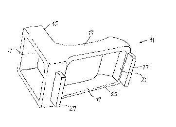
Fig. 1 a three-dimensional depiction of a support
f fixture ,
CA 02280286 1999-08-13
6
Fig. 2 a three-dimensional depiction of the same
support fixture placed on a lens,
Fig. 3 a lens placed with the support fixture placed
in position to record an image in the molar tooth area,
Fig. 4 a lens placed with the support fixture placed
in position to record an image in the frontal tooth area,
Fig. 5 a support fixture with simply a frontal
support element,
Fig. 6 a support fixture with a wedge-shaped rear
support element,
Fig. 7 a support fixture with a rear support element
made from a plastic, hardening material,
Fig. 8 a support fixture with an individually-made
support beam.
Descriptions of the embodiments:
The support fixture 11 depicted in Figures 1 and 2 is
an accessory of a mapping camera and is capable of being
attached and removed from the lens housing 13. The oblong-
shaped lens housing 13 comprises at the front end of the
housing a lens window 23, through which the video images can
be projected. The support fixture 11 is movably attached
behind the lens window 23 onto the lens housing 13 by means
of a ring 15. Said ring 15 could comprise a gap 17 at one
place, as shown in dashed line in Fig. 1. The ring 15 or
the clasp comprising the gap facilitates the clamping of the
support fixture 11 against the lens housing 13.
Two parallel arms 19 extend from the retainer piece,
that is the ring 15 or the clasp, to a connecting piece 21
linking the two arms together, whereby, said connecting
piece is positioned at a distance to ring 15. In assembled
condition, the arms 19 envelope the lens housing 13 on both
sides of the lens window. Ring 15, arms 19 and the
connecting piece 21 form together a frame 25, which, if
attached to the lens housing 13, encompasses the lens window
23. The size of the opening of frame 25 between the ring 15
CA 02280286 1999-08-13
7
and the connecting piece 21 is larger than the respective
length of the lens window 23. Because of the clamping
connection between the ring 15 and the lens housing 13, the
support fixture 11 can be moved relative to the lens 13
directionally along a line parallel to arms 19, which is
also the same direction along which the size of the opening
of the frame 25 is substantially larger than the lens window
23.
A suitable displacement distance lies between 5 and
10 mm. A support beam 27, 27' is attached to ring 15 and
connecting piece 21. Ring 15, arms 19, connecting piece 21
and support beams 27, 27' are made in one piece from a
synthetic material. The synthetic material can be shaped by
the dentist, dental technician, or by the laboratory
assistant with his or her traditional tools. It is also
capable of being sterilized and de-germinated by means of UV
radiation without rapidly affecting the material properties.
The support beams 27, 27' are depicted in Fig. 1 and 2
in their fundamental shapes, which allow different
derivations. These derivations can be finished parts, or
they can be individually fabricated by modifying the
fundamental shape by the individual using the support
fixture 11. The fundamental shape of the support beam 27,
27' is approximately 3 to 4 mm high, 12 to 20 mm long and 2
to 3 mm wide.
For applications in the molar area (ref. Fig. 3), the
support beams 27, 27' are reduced to 1.5 mm, depending on
the circumstances. This can also be done on only part of
the support beam 27, 27' contacting the tooth 29. Thereby,
even the shallowest areas of object 31 to be imaged - in
Fig. 3 a pin for a crown - are still in focus for the
camera.
For applications in the frontal tooth area (ref. Fig.
4), support beam 27' preferably remains in tact in terms of
its height and length and is resting against gum 33 above
(or below in case of lower frontal teeth) tooth 35 to be
mapped. The rear support beam 27 is thereby not needed.
CA 02280286 1999-08-13
8
Figures 5 through 8 illustrate various shapes of the
support elements 27, 27'. In Fig. 5, the rear support 27 is
missing. This support fixture lla is therefore suitable for
the frontal tooth area. Figure 6 displays the support
fixture llb with the rear support 27b sharpened in a wedge-
like manner. Point 37 can be allowed to rest in the area
between two teeth, while the front support, here the support
beam 27'b in its fundamental shape, can be placed against a
tooth.
Figure 7 illustrates an important exemplification llc
of the support fixture. In place of the rear support beam
27, retention cavities 39 are designed into frame 15.
Thermoplastically or chemically hardening, plastic matter 41
can be pressed into these retention cavities 39. With this
matter 41 applied, the support fixture llc can be placed
into position for the imaging process. In doing so, the
matter conforms to the outer surface area of the tooth onto
which it is seated and starts to harden. The hardened
matter 41 can serve as a precise guide and support for the
support fixture should a second image be required to be
taken from the same location. The mold of the tooth surface
can be placed without any trouble exactly in the same place.
The elasticity of the hardened matter 41 will allow a slight
correction of the position, if needed. Such matter can be
placed in the front or in the rear, or in the front and in
the rear. The hardening, plastic material which can be used
for this purpose, are materials which are commonly used for
dental molds such as silicone, waxes, or synthetic
materials.
Figure 8 displays a support fixture lld with a support
beam 27, 27' which has been adapted to the unique
circumstances of the patient. This embodiment lld
exemplifies that the support beams can be adapted to the
specific requirements also in the transverse direction, and
this can occur during the treatment of the patient. Figure
8 shows furthermore the ability of choosing the image detail
by laterally moving the lens 13 inside the support fixture
CA 02280286 1999-08-13
9
lld. Figure 8 shows lens 13 moved towards the retainer
piece, that is the ring 15, unlike the lens shown in Fig. 7.
Since the commonly-used lens housings 13 are slightly
tapered towards the front, the retainer piece 15 has to be
able to clamp on different cross-sectional areas of the lens
housing 13. To do so, the inside of ring 15 comprises a
subtly protruding friction pad 43, which is located
approximately in the center of each of the four sides of the
rectangular cross-section 43. This allows some clearance in
the corner areas between the ring 15 and the lens 13. If
the cross-sectional area of the lens is larger, the friction
pads 43 are compressed and the corner areas of ring 15 are
positioned closer to the lens housing 13. A minimal
clearance between housing 13 and ring 15 allows this subtle
deformation of ring 15. There are additional friction pads
43' on each of the arms 19 in order to stabilize the arms
and the front support element 27'.
In summary, it can be stated that this support fixture
improves the image quality of dental mapping cameras, while
shortening the time required to position the camera and
while improving the congruency of two images taken from the
same position. The support fixture is mounted in a movable
fashion on the lens housing and is positioned, i.e., on
teeth that are adjacent to the area which is to be mapped.
The support elements can be adapted to suit the individual
circumstances with dental tools and dental materials.
