Sélection de la langue

Search

Sommaire du brevet 2283317 

Énoncé de désistement de responsabilité concernant l'information provenant de tiers

Une partie des informations de ce site Web a été fournie par des sources externes. Le gouvernement du Canada n'assume aucune responsabilité concernant la précision, l'actualité ou la fiabilité des informations fournies par les sources externes. Les utilisateurs qui désirent employer cette information devraient consulter directement la source des informations. Le contenu fourni par les sources externes n'est pas assujetti aux exigences sur les langues officielles, la protection des renseignements personnels et l'accessibilité.

Disponibilité de l'Abrégé et des Revendications

L'apparition de différences dans le texte et l'image des Revendications et de l'Abrégé dépend du moment auquel le document est publié. Les textes des Revendications et de l'Abrégé sont affichés :

  • lorsque la demande peut être examinée par le public;
  • lorsque le brevet est émis (délivrance).
(12) Brevet: (11) CA 2283317
(54) Titre français: SYSTEME ET DISPOSITIF DE REFERENCE PERMETTANT DE DIRIGER UN FAISCEAU EN RADIOTHERAPIE
(54) Titre anglais: ARRANGEMENT AND REFERENCE MEANS TO DIRECT A BEAM IN RADIATION THERAPY
Statut: Périmé et au-delà du délai pour l’annulation
Données bibliographiques
(51) Classification internationale des brevets (CIB):
  • A61N 5/10 (2006.01)
  • A61B 6/12 (2006.01)
  • A61M 25/01 (2006.01)
  • A61M 25/098 (2006.01)
(72) Inventeurs :
  • WIDMARK, ANDERS (Suède)
  • BERGSTROM, PER (Suède)
  • LOFROTH, PER-OLOV (Suède)
(73) Titulaires :
  • BEAMPOINT AB
(71) Demandeurs :
  • BEAMPOINT AB (Suède)
(74) Agent: NORTON ROSE FULBRIGHT CANADA LLP/S.E.N.C.R.L., S.R.L.
(74) Co-agent:
(45) Délivré: 2003-03-11
(86) Date de dépôt PCT: 1998-03-17
(87) Mise à la disponibilité du public: 1998-09-24
Requête d'examen: 2000-05-01
Licence disponible: S.O.
Cédé au domaine public: S.O.
(25) Langue des documents déposés: Anglais

Traité de coopération en matière de brevets (PCT): Oui
(86) Numéro de la demande PCT: PCT/SE1998/000481
(87) Numéro de publication internationale PCT: SE1998000481
(85) Entrée nationale: 1999-09-08

(30) Données de priorité de la demande:
Numéro de la demande Pays / territoire Date
9700984-9 (Suède) 1997-03-18

Abrégés

Abrégé français

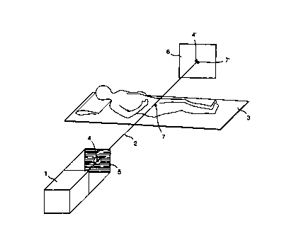
La présente invention concerne un système et un procédé de référence utilisés pour pointer avec précision un faisceau de rayons au cours du traitement d'une tumeur cancéreuse interne affectant un patient. Ce procédé de traitement comprend au moins un marqueur de référence introduit dans le corps du patient dans une position définie par rapport à la tumeur cancéreuse. On définit la géométrie de la tumeur et on planifie le traitement par rapport au marqueur de référence au moyen d'un tomodensitomètre ou par tomographie. Le patient, par un dispositif connu, est placé en position pour le traitement. Un point dans la section transversale du faisceau est défini par un viseur, et le patient, à l'aide d'au moins un marqueur de référence dans la position définie, est d'abord traité par rayons, les positions du ou des marqueurs de référence et du viseur étant lues à l'aide du faisceau qui irradie le patient, et les positions sont ajustées les unes par rapport aux autres de façon à s'assurer que le faisceau de traitement est dirigé avec précision sur la tumeur cancéreuse avant que les doses restantes de rayons soient administrées au patient. Le dispositif de cette invention comprend un équipement de traitement (1) connu qui émet un faisceau (2) de traitement d'une section transversale définie (5) et un dispositif (3) qui supporte le patient. Le dispositif de l'invention se distingue par au moins un marqueur de référence (7) qui est introduit dans le corps du patient par rapport auquel est déterminée la position de la tumeur cancéreuse; un viseur (4) placé dans la trajectoire du faisceau de façon à définir un point dans la section transversale du faisceau; un lecteur de faisceau (6) placé de façon à recevoir le faisceau qui a irradié le patient et visualiser la position d'au moins un marqueur de référence (7') et la position du viseur (4'). On détermine ainsi la position du faisceau par rapport à celle de la tumeur cancéreuse.


Abrégé anglais


The present invention concerns an arrangement and a reference method for the
precision aiming of a radiation beam for treating an internal cancer tumour in
a patient. The method of treatment includes that least one reference marker
introduced into the patient in a defined position in relation to the cancer
tumour, whose geometry is determined and the treatment planned in relation to
the reference marker by means of computer tomography/tomography, that the
patient is by known means brought into position for the treatment, that a
defined point in the cross section of the beam is defined by a sight, that the
patient with at least one said reference marker in the said defined position
is initially treated with radiation, that the positions of the reference
marker or the reference markers and the sight in relation to one another are
read with the help of the beam that passes through the patient, and that the
positions in relation to one another are adjusted to ensure that the treatment
beam is directed precisely at the cancer tumour before the remaining doses of
radiation are given to the patient. The device according to the invention
includes known treatment equipment (1) for emitting a treatment beam (2) with
a defined cross section (5) and a device (3) for supporting the patient,
whereby the characteristics of the device include that at least one reference
marker (7) is introduced into the patient in relation to which the position of
the cancer tumour is determined, that a sight (4) is arranged in the path of
the beam to define a point in the cross section of the beam, that a device to
read the beam (6) is arranged to receive the beam that has passed through the
patient and visualise the position of at least one said reference marker (7')
and the position of the sight (4'), whereby the position of the beam in
relation to the cancer tumour is determined.

Revendications

Note : Les revendications sont présentées dans la langue officielle dans laquelle elles ont été soumises.


Embodiments of the invention in which an exclusive property or privilege is
claimed are
defined as follows:
1. ~Arrangement for the precision aiming of a treatment beam against an
internal cancer
tumour in a patient, including a known treatment equipment for emitting a
treatment beam
with a defined cross-section and a device for supporting the patient,
characterised in that at
least one reference marker is introduced into the patient in relation to which
the position of
the cancer tumour is defined, that a sight is arranged in the path of the beam
to define a point
in the cross-section of the beam, that a device to read the beam is arranged
to receive the
beam that has passed through the patient and to visualise the position of at
least one said
reference marker and the position. of the sight, whereby the position of the
beam in relation to
the cancer tumour is determined.
2. ~Arrangement according to claim 1, characterised in that the reading device
includes a
surface for the incident beam that has passed through the patient, that the
incident surface
includes a layer sensitive to the incident beam, whereby a sight arranged in
the beam and a
reference marker of high density can be visualised by projections.
3. ~Arrangement according to claims 1 or 2, characterised in that the reading
device is an
X-ray film.
4. ~Arrangement according to claims 1 or 2, characterised in that the reading
device
includes a receiving unit, a digital image processing system and a monitor for
visualising the
cross-section of the treatment beam.
5. ~Arrangement according to claim 4, characterised in that the reading device
uses what
is known as a portal imaging system.
6. ~Arrangement according to any of claims 1 to 5, characterised in that a
device is
arranged to adjust the positions of the patient and the treatment equipment
relative to one
another following an initial radiation dose.
7. ~Arrangement according to claim 6, characterised in that the adjustment
device is
arranged in the device that supports the patient.

8. Reference device carrying reference markers for use in the device according
to claim
1, characterised in that the reference device includes several reference
markers in a material
with high density, that the reference markers are arranged at defined
distances from each
other, that the reference device also includes a position fixing-device for
the reproducible set-
up of the reference markers in a defined position relative to the cancer
tumour.
9. Reference device according to claim 8, characterised in that the reference
device
includes a catheter having a position fixing-device at its free end, that the
position fixing-
device is an inflatable balloon, and that at least one marker is arranged at a
defined position
along the catheter.
10. Reference device according to any of claims 8 or 9, characterised in that
the
reference markers are produced from a material of high density to enable
visualisation in a
high energy beam.
11. Reference device according to claim 10, characterised that the reference
markers are
made of one or more materials from a group consisting of lead, tantalum and
tungsten.
12. Reference device according to any of claims 10 or 11, characterised in
that the
catheter has a channel for manoeuvring the inflatable balloon, a channel to
allow urination,
and that the reference markers are arranged within the wall of the catheter.

Description

Note : Les descriptions sont présentées dans la langue officielle dans laquelle elles ont été soumises.

CA 02283317 1999-09-08
WO 98/41282 PCT/SE98/00481
Arrangement and reference means to direct a beam in radiation therapy
The present invention concerns an arrangement and a reference means for the
precision
aiming of a radiation beam for treating an internal cancer tumour.
During the last 40 years, radiation therapy of localised prostate cancer has
been used as
one of a number of treatment methods. The levels of radiation considered
suitable for treatment
fall within the interval 65-70 Gy. However, the occurrence of viable cancer
cells even after such a
level of treatment has been demonstrated. One way of dealing with these
remaining cells is to
increase the level of treatment, known as scaling up the dose, initially to
the interval of 76-80 Gy
or more.
Because of the lack of precision of current techniques, radiation treatment
also carries
with it an increased risk of affecting organs adjacent to the tumour. In the
area around the
prostate, this means primarily an increased risk for side effects seen as
damage to the rectum and
the urinary bladder as these are normally found in the radiation field. The
risk for damage is
especially pronounced with high doses of radiation.
1 S The preparations for the radiation treatment of prostate cancer can be
described briefly in
the following way. The patient undergoes computer tomography to determine the
geometry and
location of the prostate. The position of the prostate is shown by markings on
the skin of the
patient. These markings then constitute the directional points for the beams
used in treatment.
Even if the geometry of the prostate is known, problems can arise during
radiation
treatment because the position of the prostate is not constant in relation to
the pelvis or, for that
matter, to the skin where the markings have been made. The patient's
subcutaneous fat means
that the markings can be displaced in relation to the pelvis, and different
muscle contractions, such
as those in the area around the prostate, can displace the prostate up to one
centimetre in relation
to the pelvis. Furthermore, variation in the contents of the intestine and the
bladder can affect the
position of the prostate. When added together, this has the effect that the
field of radiation must
have a margin of 1.5 - 2 cm around the determined position of the prostate,
which means that the
rectum and bladder can be subjected to radiation, thus limiting the
possibility of giving very high
doses of radiation.
Even though the problem has initially been described in connection with the
treatment of
prostate cancer, it should be emphasised that many aspects of the problem are
common to the
treatment of cancer tumours in other organs, which is why the invention also
has applications

CA 02283317 1999-09-08
WO 98/41282 PCT/SE98/00481
2
within these areas. This is especially true for tumours of the cervix, urinary
bladder, stomach,
intestinal tract, pancreas, mouth, throat, etc. Embodiments of the invention
have further
applications within general internal radiation treatment of cancer, as will be
made evident below.
During radiation therapy, a treatment beam is emitted from equipment used for
the
treatment. Depending on the shape of the object to be treated, a cross-section
of the treatment
beam is chosen so that the treatment is restricted to a specified treatment
area. If the treatment
area is to be reduced to a size approximating the size of the object to be
treated, it is of great
importance that the beam strikes its target, the tumour, with great precision.
One objective of the present invention is to achieve the precise aiming of the
direction of a
treatment beam during the radiation treatment of internal cancer tumours so
that the margins of
the treatment beam can be reduced, thereby minimising the effect on adjacent
organs and tissue.
This objective is achieved with an arrangement and a reference device fast
mentioned
above and that have the features that are evident from the characteristics of
the enclosed
independent claims.
The advantages and features of the invention are evident from the non-
independent claims
and the following description.
By using the invention, it is possible to describe the geometry of an organ
based on one or
more reference points that always occupy the same position with reference to
the organ. In
addition, the possibility to repeatedly direct the treatment beam with
precision in relation to at
least one said reference point so that the beam always strikes the cancer
tumour with precision, is
also achieved. As such, the beam does not need as large a safety margin as has
been required until
now, but can be assigned a profile that is more or less exactly identical with
that of the object to
be treated.
Before each treatment occasion, it is thus possible to check, adjust and
verify that the
cancer tumour really is in the path of the treatment beam.
Additional features and advantages of the invention will be made evident by
the following
detailed description of one preferred embodiment of the invention, which
constitutes one example
and as such does not limit the area of protection of the invention. To
simplify understanding, the
text contains references to the enclosed drawings, in which equivalent or
similar parts are assigned
the same designation.
T ~

CA 02283317 1999-09-08
WO 98/41282 PCT/SE98/00481
Fig. 1 shows schematically an arrangement according to one embodiment of the
present
invention.
Fig. 2 shows schematically a reference device according to one embodiment of
the present
invention.
The manner in which we intend to use the arrangement is as follows. For the
precision
aiming of the direction of a treatment beam 2 against an internal cancer
tumour of a patient, at
least one reference marker 7 is introduced into the patient at a defined
position in relation to the
cancer tumour and, if possible, into the tumour itself The geometry of the
tumour is determined
and the treatment is planned with reference to the reference marker 7. For the
geometrical
determination of the tumour, computer tomography/tomography, magnetic X-rays,
or other
suitable radiological techniques can be used with advantage, and the reference
markers) can, for
example, be composed of a liquid contrast agent, lead, tantalum, tungsten or
other material that is
visible when computer tomography/tomography, magnetic X-rays, or other similar
radiological
techniques are used.
In addition, when the actual treatment is begun, the patient is brought into a
treatment
position and a set point in the cross-section of the beam is defined by a
sight 4
The geometrical determination and description of the cancer tumour is
performed based
on one or more reference markers 7 and, on the basis of that information, the
cross-section of the
treatment beam is built up and the position of the sight in the beam's cross-
section is moved so
that when the incidence of the beam corresponds with the cancer tumour, the
positions of the
sight and the reference markers coincide, even if they are located on separate
elevations.
With at least one said reference marker in the defined position, the patient
receives the first
initial treatment, after which the relative positions of the reference marker
or markers and the
sight to one another are read with the help of, for example, the beam that
passes through the
patient. The relative positions can be adjusted later as needed to ensure the
precision of the
direction of the beam towards the cancer tumour before the remaining doses in
the treatment are
given to the patient. By allowing the beam to project the position of the
sight 4' and the
positions) of the markers) 7' on a sensitive surface 6, their relative
positions to one another can
be visualised.
It is desirable to keep the initial radiation dose low as radiation that
misses the target does
more harm than good. Experiments have shown that a dose equivalent to 0.03 Gy
has been

CA 02283317 1999-09-08
WO 98/41282 PCTlSE98/00481
4
sufl-icient to visualise the reference marker and the sight on film. It is
desirable to use a dose level
that is less than 5% of the radiation dose used in treatment, preferably less
than 2%.
When treating prostate cancer, the reference marker can be introduced through
the urinary
tract by means of a catheter 10, as shown in fig. 2. The reference markers
need not be the same
when determining the geometry of the cancer tumour and when treating it. The
key factor is that
the reference markers are located in known positions and in the same position
in relation to the
cancer tumour. To achieve this, the catheter 10 can be introduced through the
urinary tract so that
its free end enters the bladder 11 where an attachment arranged at the free
end of the catheter, in
this case a balloon device 12, is inflated inside the bladder to fix the
position of the catheter in
relation to the urinary tract, which is in turn essentially fixed in position
relative to the prostate
13. The catheter is withdrawn slightly, which pulls the inflated balloon 12
against the floor of the
bladder where it joins with the urinary tract. The actual reference marker 7
or reference markers 7
can be permanently positioned in the catheter or can be able to be introduced
into the catheter in a
defined position therein.
In addition to the said inflatable balloon, adjustable fold-out flaps that
increase the local
circumference, hooks or clips of different types or other similar locating
devices suitable for
reproducible location are also suggested as locating devices for the catheter
or other carrier of the
reference markers.
An arrangement according to the invention for performing the task includes
what is, in
fact, known treatment equipment 1 for emitting a treatment beam 2 and a device
3 for supporting
the patient, whereby the patient has at least one reference marker 7 in
relation to which the
position of the cancer tumour is determined, a sight 4 arranged in the path of
the beam to define a
point in the cross-section of the beam, a device 6 to detect the beam arranged
to receive the beam
passing through the patient and visualise the position of at least one said
reference marker 7' and
the position of the sight 4', whereby the position of the beam in relation to
the cancer tumour is
determined.
The reading device 6 can be an X-ray film in a cassette suitable for high
energy radiation.
The reading device 6 can even be one without film and instead include a
receiving unit, a
digital image processing system and a monitor for visualising the cross-
section of the treatment
beam. It can be advantageous if the reading device uses what is known as a
portal imaging
system.
r i

CA 02283317 1999-09-08
WO 98/41282 PCT/SE98/00481
When a deviation from the desired agreement between the beam and the cancer
tumour is
registered, the device for adjusting the relative positions of the patient and
the treatment
equipment to one another is activated, either manually or automatically. This
can occur, for
example, by a change in the elevation or position of the device supporting the
patient or by
adjustment of the treatment equipment.
It is advantageous if the reference marker 7 or reference markers 7 are
included in a
reference device. The reference device then has at least one reference marker
7 in a material with
high density and a position fixing-device.
The reference device can include several reference markers arranged at defined
distances
from each other. A marker can, for example, take the form of a dot, a line, an
arc, a cross, a
circle, etc.
The position-fixing device results in the markers) taking up a defined
position in relation
to the cancer tumour, preferably a reproducible set-up of the reference
markers in a defined
position in relation to the cancer tumour.
In one embodiment, the reference device includes a catheter 10 that has at its
free end a
reproducibly inflatable balloon, plus markers in the form of spheres arranged
at defined positions
along the catheter. The spheres are produced from a material of high density
to enable
visualisation in a high energy beam, preferably lead, tantalum and/or
tungsten.
The catheter shown in fig. 2 includes tubing 10 with several spheres 7
arranged in the
internal channel of the tubing. The markers can naturally be arranged within
the wall as spheres,
rings, lines arcs, etc., to maintain an internal passage for, for example,
urination. In this case, the
use of several reference markers means that the catheter can be used by
different patients with
different anatomical measurements. For example, the third sphere can be used
as a reference by
one patient, whereas with another patient, the fourth sphere may be better
placed to function as
the reference point. A smaller channel passes through the tubing to the
inflatable balloon 12 so
that this can be expanded.
For certain treatments, it is not necessary to direct the beam at a point.
Instead, a Iine can
be judged to provide sufficient accuracy. For example, with a catheter with a
wire or with several
spheres arranged at a shorter distance from each other, the arc or line that
the urinary tract forms
through the prostate can be visualised and used to direct the beam.

CA 02283317 1999-09-08
WO 98141282 PCT/SE98/00481
6
Even if the text above to a large extent deals with the invention's
application for the
treatment of prostate cancer, it should be remembered that the invention also
has uses with other
types of cancer tumour.
In further applications of the invention, the reference marker can be
introduced to, and
kept in place by, surrounding body tissues, for example, in a cancer tumour.
For example, a marker provided with a clip or other attachment device can be
arranged in
a defined position in relation to a cancer tumour by means of, for example,
surgery, peep-hole
surgery or by the marker being introduced through one of the body's openings.
The marker can
then be kept in position throughout the whole cycle of treatment, from
determining the position of
the tumour to completed treatment, after which it can be removed by an
appropriate means, such
as the means by which it was introduced or, if made of material suitable for
the purpose, be left in
the body.
In those cases where a carrier is arranged to carry several reference markers
with known
positions relative to one another, it can be appropriate to arrange these in a
pattern so that it is
possible to determine where along the carrier the beam is directed. For
example, the distance
between the reference markers or their shape at a known position can deviate
in a way that is easy
to identify. The reference markers in a catheter can, for example, be arranged
one cm apart,
except for one position along the catheter where one marker is missing. In
this way, the position
of the beam along the carrier can be determined with increased certainty.
____________
r r r

Dessin représentatif
Une figure unique qui représente un dessin illustrant l'invention.
États administratifs

2024-08-01 : Dans le cadre de la transition vers les Brevets de nouvelle génération (BNG), la base de données sur les brevets canadiens (BDBC) contient désormais un Historique d'événement plus détaillé, qui reproduit le Journal des événements de notre nouvelle solution interne.

Veuillez noter que les événements débutant par « Inactive : » se réfèrent à des événements qui ne sont plus utilisés dans notre nouvelle solution interne.

Pour une meilleure compréhension de l'état de la demande ou brevet qui figure sur cette page, la rubrique Mise en garde , et les descriptions de Brevet , Historique d'événement , Taxes périodiques et Historique des paiements devraient être consultées.

Historique d'événement

Description Date
Le délai pour l'annulation est expiré 2011-03-17
Lettre envoyée 2010-03-17
Inactive : TME en retard traitée 2009-04-27
Inactive : Demande ad hoc documentée 2009-04-16
Inactive : Paiement - Taxe insuffisante 2009-04-15
Lettre envoyée 2009-03-17
Inactive : Lettre officielle 2006-05-25
Inactive : Paiement correctif - art.78.6 Loi 2006-05-12
Inactive : CIB de MCD 2006-03-12
Accordé par délivrance 2003-03-11
Inactive : Page couverture publiée 2003-03-10
Inactive : Grandeur de l'entité changée 2003-01-06
Préoctroi 2002-12-17
Inactive : Taxe finale reçue 2002-12-17
Lettre envoyée 2002-08-28
Inactive : Transfert individuel 2002-07-08
month 2002-06-20
Un avis d'acceptation est envoyé 2002-06-20
Lettre envoyée 2002-06-20
Inactive : Approuvée aux fins d'acceptation (AFA) 2002-06-10
Modification reçue - modification volontaire 2002-04-12
Modification reçue - modification volontaire 2002-03-28
Inactive : Dem. de l'examinateur par.30(2) Règles 2001-11-29
Lettre envoyée 2000-05-24
Requête d'examen reçue 2000-05-01
Exigences pour une requête d'examen - jugée conforme 2000-05-01
Toutes les exigences pour l'examen - jugée conforme 2000-05-01
Inactive : Page couverture publiée 1999-11-15
Inactive : CIB en 1re position 1999-11-02
Inactive : CIB attribuée 1999-11-02
Inactive : Notice - Entrée phase nat. - Pas de RE 1999-10-14
Demande reçue - PCT 1999-10-08
Demande publiée (accessible au public) 1998-09-24

Historique d'abandonnement

Il n'y a pas d'historique d'abandonnement

Taxes périodiques

Le dernier paiement a été reçu le 2002-02-18

Avis : Si le paiement en totalité n'a pas été reçu au plus tard à la date indiquée, une taxe supplémentaire peut être imposée, soit une des taxes suivantes :

  • taxe de rétablissement ;
  • taxe pour paiement en souffrance ; ou
  • taxe additionnelle pour le renversement d'une péremption réputée.

Les taxes sur les brevets sont ajustées au 1er janvier de chaque année. Les montants ci-dessus sont les montants actuels s'ils sont reçus au plus tard le 31 décembre de l'année en cours.
Veuillez vous référer à la page web des taxes sur les brevets de l'OPIC pour voir tous les montants actuels des taxes.

Historique des taxes

Type de taxes Anniversaire Échéance Date payée
Taxe nationale de base - petite 1999-09-08
TM (demande, 2e anniv.) - petite 02 2000-03-17 1999-09-08
Requête d'examen - petite 2000-05-01
TM (demande, 3e anniv.) - petite 03 2001-03-19 2001-03-14
TM (demande, 4e anniv.) - petite 04 2002-03-18 2002-02-18
Enregistrement d'un document 2002-07-08
Taxe finale - générale 2002-12-17
TM (brevet, 5e anniv.) - générale 2003-03-17 2003-03-05
TM (brevet, 6e anniv.) - générale 2004-03-17 2003-12-22
TM (brevet, 7e anniv.) - générale 2005-03-17 2005-03-11
TM (brevet, 8e anniv.) - générale 2006-03-17 2006-03-08
2006-05-12
TM (brevet, 9e anniv.) - générale 2007-03-19 2007-03-16
TM (brevet, 10e anniv.) - générale 2008-03-17 2008-02-25
Annulation de la péremption réputée 2009-03-17 2009-03-20
TM (brevet, 11e anniv.) - générale 2009-03-17 2009-03-20
Titulaires au dossier

Les titulaires actuels et antérieures au dossier sont affichés en ordre alphabétique.

Titulaires actuels au dossier
BEAMPOINT AB
Titulaires antérieures au dossier
ANDERS WIDMARK
PER BERGSTROM
PER-OLOV LOFROTH
Les propriétaires antérieurs qui ne figurent pas dans la liste des « Propriétaires au dossier » apparaîtront dans d'autres documents au dossier.
Documents

Pour visionner les fichiers sélectionnés, entrer le code reCAPTCHA :


Pour visualiser une image, cliquer sur un lien dans la colonne description du document (Temporairement non-disponible). Pour télécharger l'image (les images), cliquer l'une ou plusieurs cases à cocher dans la première colonne et ensuite cliquer sur le bouton "Télécharger sélection en format PDF (archive Zip)" ou le bouton "Télécharger sélection (en un fichier PDF fusionné)".

Liste des documents de brevet publiés et non publiés sur la BDBC .

Si vous avez des difficultés à accéder au contenu, veuillez communiquer avec le Centre de services à la clientèle au 1-866-997-1936, ou envoyer un courriel au Centre de service à la clientèle de l'OPIC.


Description du
Document 
Date
(yyyy-mm-dd) 
Nombre de pages   Taille de l'image (Ko) 
Page couverture 2003-02-03 2 61
Revendications 2002-03-27 2 74
Abrégé 1999-09-07 1 76
Description 1999-09-07 6 340
Revendications 1999-09-07 2 84
Dessins 1999-09-07 2 26
Dessin représentatif 1999-11-14 1 6
Page couverture 1999-11-14 2 99
Revendications 2002-04-11 2 82
Avis d'entree dans la phase nationale 1999-10-13 1 193
Accusé de réception de la requête d'examen 2000-05-23 1 178
Avis du commissaire - Demande jugée acceptable 2002-06-19 1 164
Courtoisie - Certificat d'enregistrement (document(s) connexe(s)) 2002-08-27 1 112
Avis de paiement insuffisant pour taxe (anglais) 2009-04-14 1 91
Avis concernant la taxe de maintien 2009-04-14 1 171
Quittance d'un paiement en retard 2009-05-11 1 164
Avis concernant la taxe de maintien 2010-04-27 1 170
Correspondance 2002-12-16 2 48
PCT 1999-09-07 10 402
Correspondance 2006-05-24 1 17