Note : Les descriptions sont présentées dans la langue officielle dans laquelle elles ont été soumises.
CA 02287107 1999-10-25
CANADA
APPLICANT: Medizintechnik GmbH
TITLE: DEVICE FOR REMOVING PATHOLOGICAL CENTERS IN HUMAN
AND VETRINARY MEDICINE
CA 02287107 1999-10-25
DEVICE FOR RE1~IOVING PATHOLOGICAL
CENTERS IN HU~ItANS AND ANIMALS
BACKGROUND OF THE INVENTION
J
1. Field of the Invention
The present invention relates to a device for removing pathological centers
useful
for humans and animals. The device includes a supply capillary having a supply
channel for a
pressurized flow and a discharge capillary with a discharge channel , which is
open to the
pathological center, for suction flow.
Such devices are used in human and veterinary medicine, wherein pathological
centers are understood as including bodily impairments and deficiencies in
general.
2. Description of the Related Art
Pathological or etiological centers of the aforedescribed type can be found in
the brain
and in the central nen~ous system, including the eyes, and in those body
structures which calls for
a particularly mild form of invasive surgery due to the concentration of nerve
and blood vessel tissue
or other conditions. According to the present teachings, specialists treat or
remove such pathological
centers by surgical or micro-surgical means. These means are characterized by
an applied force
which is entirely or at least predominantly directed in a forward direction.
These means include, for
?0 example, scalpels of any kind, coagulators and lasers of any kind,
ultrasound aspirators, and the like.
An pathological center or degenerative impairment of the aforedescribed type
is. for example. an lens
of the eye which may be impaired by a cataract and therefore may have to be
removed and replaced
by an artificial lens.
CA 02287107 1999-10-25
The first incision in the eve is a tunnel incision and the anterior capsule is
opened
with a flawless, preferably circular capsolorhexis. .-~n instrument is then
pushed into the diseased
lens through this opening. The lens is then fractured, preferably by
ultrasound, initially into small
fragments which are subsequently suctioned off After all fragments is have
been removed and the
chamber of the eye has been cleaned, an artificial lens is inserted into the
chamber of the eye through
the channel of a special instrument. The lens relaxes when the instrument is
pulled out and again
assumes its original lens shape. Finally the artificial lens is oriented and
secured in place.
Although this surgical procedure since has become routine, complications may
still
occur. For example, the residual fragments of the diseased lens can still not
be completely removed
from the chamber of the eye, since some of the peripheral fragments of the
diseased cell residues are
obscured from the view of the surgeon and may therefore remain in place. This
creates a risk that
the cataracts return. The cell residues can only be partially mobilized by
manually injecting a fluid.
In addition, the ultrasound energy produces excess heat which heats the
corneal tissue and causes
a loss of endothetical cells.
This surgical procedure also requires a variety of different surgical tools
and a
considerable number of independent time-consuming steps. This increases the
costs of the surgical
procedure. The large number of surgical steps and the large number of surgical
tools also are
demanding on the surgeon. As a result, the success of such a surgical
procedure depends to a large
extent on the surgeon's qualifications.
?0 It is therefore an object of the invention to develop a method of the
aforedescribed
type which is less demanding on the surgeon, which can be performed in less
time, and which is
more gentle on the healthy tissue.
3
CA 02287107 1999-10-25
SUMMARY OF THE INVENTION
It is another object of the invention to provide a multi-functional device for
carrying out the method.
The object is solved by providing a device for removing pathological centers
which includes a supply capillary with a supply channel for a pressurized flow
and a discharge
capillary with a discharge channel, which is open to the pathological center,
for a suction flow,
the supply channel of the supply capillary has a throttle located at the
proximal end of the supply
channel and is operatively connected with the discharge channel of the
discharge capillary.
The invention eliminates the aforedescribed disadvantages of the present state
of
the art.
In addition, the method and the device have many applications and can be used
at
many locations where tissue has to be removed in order to be replaced or
tested, independent if
surgery is performed on the open body or in a body cavity. Advantageously, the
surgical
procedure is of high-quality and very venue on healthy tissue. This is mainly
due to the fact that
with the method of the invention, the hydraulic jet no longer exerts a forward-
directed force and
therefore eliminates pressure build-up and possible turbulence in the body
cavity. In particular.
the retrograde effective direction of the hydraulic jet protects the healthy
tissue. Furthermore, the
volume of the removed tissue can be compensated which is advantageous with
very small body
cavities, for example in neurosurgery and ophthalmology.
As an additional advantage, the device is multi-functional and combines
several
functional elements. This protects the healthy tissue and simplifies and
shortens the surgical
procedure.
4
CA 02287107 1999-10-25
The invention will be described hereinafter in more detail with reference to
an
embodiment.
Other objects and features of the present invention will become apparent from
the following detailed description considered in conjunction with the
accompanying drawings.
It is to be understood, however, that the drawings are intended solely for
purposes of
illustration and not as a definition of the limits of the invention, for which
reference should be
made to the appended claims.
CA 02287107 1999-10-25
BRIEF DESCRIPTION OF THE DRAWINGS
In the drawings, wherein like reference numerals delineate similar elements
throughout the several views:
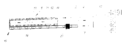
FIG. 1: schematically a simplified diagram of a hydro-jet device, and
FIG. 2: a device according to the invention.
6
CA 02287107 1999-10-25
DETAILED DESCRIPTION OF THE PRESENTLY PREFERRED EMBODIMENTS
Referring to FIG. 1. the hvdro-jet device essentially includes a device 1
according
to the invention for removing pathological centers. The device 1 is connected
via a supply line 2
with a pressurized flow generator 3 and via a discharge line 4 with a reduced
pressure flow
generator ~. The pressurized flow generator 3 is associated with a pulse
generator 6 which can
be connected in addition. The device 1 also includes a second supply line 7
which is connected
with a hydraulic pump 8 and with an auxiliary unit 9 which can be
alternatively connected. The
supply lines 2, 7 and the discharge line 4 pass through a control unit 10
which is operated
manually by the surgeon
The device 1 for removing pathological centers preferably consists, as
indicated
with particularity in FIG. 2, of an outer supply capillary 11 which is
penetrated by an inner
discharge capillary 12. The inner discharge capillary 12 includes a discharge
channel 13 and is
connected with the discharge line 4 of the reduced pressure flow generator 5.
Both capillaries 11
and 12 are located on a common axis. Through proper selection of the inside
diameter of the
supply capillary 11 and of the outside diameter of the discharge capillary 12,
an annular supply
channel 14 with a defined unobstructed width is created which is connected to
the supply line 2
of the pressurized flow generator 3. The defined unobstructed width is
determined by the desired
ratio between the cross-sectional surfaces of the annular supply channel 14
and the discharge
channel 13 of the discharge capillary 12. The supply capillary 11 and the
discharge capillary 12
are located at the proximal end of the device 1 and form a special hydro-
membrane nozzle 15.
The front side of the outer supply capillary 11 is covered by a front ring 16,
and the inner
discharge capillary 12 is recessed lengthwise with respect to the outer supply
capillary 11 by a
7
CA 02287107 1999-10-25
predetermined amount. The difference a length between the supply capillary 1 1
and the
discharge capillary 12 is greater than the wall thickness of the front ring
16, so that a radial
annular throttle gap 17 is produced between the supply capillary 1 1 and the
discharge capillary
12. For producing a desired throttle effect, the cross-sectional surface of
the radial annular
throttle gap 17 is smaller than the cross-sectional surface of the annular
supply channel 14. The
front ring 16 has a central nozzle opening 18, wherein the diameter of the
nozzle opening 18 is
matched within a predetermined tolerance range to the in a diameter of the
discharge capillary
12
The device 1 for removing an pathological center also includes a separate
supply
capillary 19. A pressure sensor 20 is arranged inside the supply capillary 19
and connected either
to the supply line 7 of the hydraulic pump 8 or to the auxiliary unit 9. The
supply capillary 19 is
preferably rigidly connected to the outer supply capillary 11 or formed as a
separate volume-
compensating capillary. The length of the supply capillary 19 is identical to
the length of the
supply capillary 11.
For removing a clouded eye lens and replacing the clouded eye lens by an
artificial lens, the eye is first incised in a conventional manner with a
tunnel incision. The front
capsule is then opened using a flawless, preferably circular capsulorhexis.
The supply capillary
11 for the pressurized flow generator 3 and the volume-compensating supply
capillary 18 of the
device 1 is pushed into the diseased lens through this opening. Subsequently,
the surgeon
?0 enables a hydraulic supply flow in the direction of the device 1 by
activating the pressurized flow
generator 3. The flow and pressure parameters of the pressurized flow
generator 3 can be preset
at the control unit 10. At the same time, the surgeon activates the reduced
pressure flow
8
CA 02287107 1999-10-25
generator 3 to produce a discharge tlow at the preset flow <md pressure
settings and flowing in
direction of the reduced pressure flow generator ~. :~s a result, a
pressurized hydraulic flow
passes from the pressure flow generator 3 through the supply line 2 into the
annular supply
channel 14, then passes through the radial annular throttle gap 17 and exits
into free space. At
this point. the hydraulic flow is entrained by the discharge flow of the
reduced pressure flow
generator ~ and transported through the discharge channel 13 of the discharge
capillary 12 and
the discharge line 4 in a retrograde direction to the reduced pressure flow
generator ~. Different
flow velocities between the supply flow and the discharge flow can be
generated by suitably
setting the flow and pressure parameters at the control unit 10 of the two
flows. This produces a
reduced pressure region at the hydro-membrane nozzle 1 S. The reduced pressure
region
generates at the mouth of the inner discharge capillary 12 a conical hydro-
membrane which
produces attracting forces acting on the immediate surroundings of the hydro-
membrane nozzle
15. The strength of these attracting forces can be set and adjusted at the
control unit 10.
The attracting forces dislodge the diseased tissue of the eye lens from the
healthy
tissue of the neighboring parts of the eye and, at the same time, comminute
the dislodged lens
fragments and transport the dislodged lens fragments to the mouth of the hydro-
membrane
nozzle 15. Smaller lens residues enter the discharge channel 13 unimpededly,
whereas larger
lens fragments impinge on the front ring 16 of the outer supply capillary 11,
where they are
further comminuted to a size suitable to be transported. The removal action
produced by the
attracting forces can be amplified by pulsating the supply flow through
activation of the pulse
generator 6.
9
CA 02287107 1999-10-25
To compensate for the volume loss within the lens chamber. the hvdro-pump 8 is
activated for supplying the eve chamber with a quantity of fluid that is
equivalent to the volume
of the diseased lens tissue which has been removed. This volume compensation
can be
performed continuously using the pressure sensor 20 or can be controlled
manually.
Alternatively, the volume can be compensated. for example, by using an
auxiliary unit 9 in the
form of a water jet device which produces a water jet flowing through the
supply capillary 19 or
a laser beam to perform additional measures in conjunction with the surgical
procedure.
Advantageously, a heating device 21 may be embedded in the supply line 2 for
suitably adjusting the temperature of the hydraulic flow.
Thus, while there have been shown and described and pointed out fundamental
novel features of the invention as applied to a preferred embodiment thereof,
it will be
understood that various omissions and substitutions and changes in the form
and details of the
devices illustrated, and in their operation, may be made by those skilled in
the art without
departing from the spirit of the invention. For example, it is expressly
intended that all
combinations of those elements and/or method steps which perform substantially
the same
function in substantially the same way to achieve the same results are within
the scope of the
invention. Substitutions of elements from one described embodiment to another
are also fully
intended and contemplated. It is also to be understood that the drawings are
not necessarily
drawn to scale but that they are merely conceptual in nature. It is the
intention, therefore, to
?0 be limited only as indicated by the scope of the claims appended hereto.