Note : Les descriptions sont présentées dans la langue officielle dans laquelle elles ont été soumises.
CA 02288080 1999-10-22
Method for the functional detection of disorders in the protein C system
The present invention relates to a method for the sensitive and functional
detection
of disorders in the protein C system (protein C, protein S, FV) and in
particular for
the determination of the activated blood coagulation factor V (FVa) with
increased
stability in respect of the decomposition by activated protein C (APC).
Hemostasis after vascular injuries results from an interaction between tissue,
blood
cells (blood platelets) and proteins of the blood liquid (plasma clotting
factors,
calcium ions). This interaction first leads to the formation of a hemostatic
platelet
plug (primary hemostasis) and finally to its consolidation by coagulation,
i.e. by
forming a network of insoluble fibrin. The physiological calcium content in
blood of
60 - 70 mg per liter is essential for the optimal progress of the blood
coagulation
reactions. The fibrinolytic system is responsible for the enzymatic
decomposition of
fibrin clots during wound healing and recanalization of closed vessels. These
interfering systems are modulated by activators and inhibitors and are present
in
the healthy organism in a labile balance. Disturbances of this balance may
lead on
the one hand to increased bleeding tendency and on the other hand to
thrombosis
proneness. Hemostatic disorders cause or accompany many diseases and
therapies, in which cases the balance may be disturbed either in favour of
bleeding
or of thrombosis.
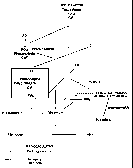
The section of the blood coagulation cascade represented in Figure 1 is of
particular importance for the present invention. A detailed report on the
reaction
cascade leading to blood coagulation can be found in H.R. Roberts, Overview of
the Coagulation Reactions, in: K.A. High and H.R. Roberts (eds.) Molecular
Basis
of Thrombosis and Hemostasis, pp. 35-50, Marcel Dekker: New York, Basel, Hong
Kong (1995).
After injury of the vessel wall, blood comes into contact with tissue cells
that
release on their surface a glycoprotein of 50,000 Dalton which, as the so-
called
thromboplastin or tissue factor (TF), activates the blood clotting system via
the
exogenous pathway. Blood platelets adhering to tissue structures release
phospholipids which activate the intrinsic blood coagulation system. TF forms
with
CA 02288080 1999-10-22
2
factor (F) VII present in plasma a complex that activates the zymogens FX and
FIX
to the serine proteases FXa and FIXa. Via the endogenous pathway, FIXa
together
with its cofactor FVIIIa also forms an efficient activator for FX in the
presence of
calcium ions and phospholipids. FXa forms with FVa, calcium ions and
phospholipids a complex (prothrombinase complex) which converts the zymogen
prothrombin into catalytically active thrombin. The enzyme thrombin converts
fibrinogen by limited proteolysis into fibrin monomer which spontaneously
polymerizes into fibrin.
The factors Va and Vllla are non-enzymatic plasma proteins that strongly
accelerate the activation of FX and prothrombin, respectively, by FIX and FXa,
respectively. The catalytic efficacy of the cofactor, FVa, on the prothrombin
activation is shown in Table 1. It can be seen that the activation of
prothrombin by
FXa, phospholipids and calcium ions is accelerated about 250 times alone by
the
presence of FVa.
The presence of calcium ions and phospholipids is a condition sine qua non for
most reactions of the blood coagulation cascade. Removal of calcium ions by
complexation, precipitation or ion exchange totally inhibits the clotting
capacity of
blood. This property is generally used to obtain blood plasma for analytical
or
therapeutic purposes by mixing freshly collected blood with sodium citrate for
the
complexation of calcium ions, centrifuging and decanting the supernatant,
unclottable blood liquid from the sedimented cells. The coagulability of
citrate
plasma is restaured by addition of a physiological quantity of a calcium salt,
by so-
called recalcification.
Prothrombin activation by the prothrombinase complex
Xa 1
Xa, Ca2+ 1.7
Xa, Ca2+, PL, 8.3 x 10³
Xa, Ca2+, PL, Va 2.0 x 10⁶
Table 1. Catalytic efficacy of the prothrombinase complex
CA 02288080 1999-10-22
3
The anticoagulant protein C system prevents an uncontrolled migration of
activated
blood clotting factors from the site of the vascular injury and hemostasis. As
in the
plasmatic coagulation system, a cell-bound receptor, thrombomodulin (TM), and
a
non-enzymatic cofactor, protein S, as well as the secondary components
phospholipids and calcium ions participate in the activation of the protein C
system.
Thrombin escaping from the site of hemostasis binds to TM, loses thereby its
fibrinogen-coagulant properties and becomes the specific activator for the
zymogen
protein C. Activated protein C (APC) is a serine proteinase which, potentiated
by
protein S, splits off and inactivates the clotting factors FVa and FVIIIa. By
inactivation of these cofactors, the coagulation process is strongly slowed
down:
according to Table 1, a 250-fold delay takes place alone by the inactivation
of FVa.
For a topical description of the protein C system, see K. Suzuki, Protein C:
in: K.A.
High and H.R. Roberts (eds.) Molecular Basis of Thrombosis and Hemostasis, pp.
393-424, Marcel Dekker: New York, Basel, Hong Kong (1995).
The biological significance of the protein C system has been evidenced in 1993
by
B. Dahlback who observed that in patients with thrombosis tendency, unlike in
healthy subjects, the activated partial thromboplastin time (APTT, a
diagnostic
function control for the endogenous coagulation system) is not prolonged after
addition of activated protein C. He defined his observations as õresistance
against
activated protein C" (APC resistance). This resistance is in 97% of the cases
due to
a point mutation in the factor V gene. This genetically inherited defect can
be found
in about 5% of the normal population and in at least 20% of young patients
with
first unexplainable or recurrent thromboembolisms. In the presence of the
mutation,
the activated clotting factor V can no more be split and thus inactivated.
Consequences of the deficiency of this particularly important anticoagulant
component of the blood coagulation system may be coronary heart diseases,
venous thromboses or thromboembolisms. Consequently, heterozygous defect
carriers present a 5-10 times greater thrombosis risk than normal persons and
homozygous defect carriers an even 50 to 100 times higher risk.
Other hereditary or acquired deficiencies or defects in the protein C system
(qualitative or quantitative protein C or protein S deficiency) are also
associated
with higher thrombosis tendency.
CA 02288080 1999-10-22
4
The congenital or acquired APC resistance can be detected by a functional test
or
by the direct detection of the mutation on the DNA level (genotype).
The functional detection can be performed according to Dahlback by an APTT
variant (PCT/SE92/00310; WO 93710261) in which the coagulation of a platelet-
free plasma sample is once triggered off once by calcium chloride without
addition
of activated protein C (APC) and once by calcium chloride with addition of
APC.
The thrombin quantity resulting in the test mixture is determined either by
the
conversion of the natural substrate fibrinogen into a clot (clotting time) or
photometrically by the release of a chromophore from a chromogenic substrate.
The existence of the factor V mutation can be noted by the fact that the
clotting
time is only weakly prolonged by APC, while APC strongly prolongs the APTT of
normal plasma. Dividing the clotting time of the sample with APC by the
clotting
time of the sample without APC gives a ratio of diagnostic significance. A
ratio of
more than 2.0 is found in healthy subjects, a ratio between 1.3 and 2.0 in
heterozygous and a ratio below 1.3 in homozygous defect carriers. However, as
this test system bases on the activation of coagulation in the presence of
calcium
ions, quantitative and qualitative abnormalities on calcium-dependent plasma
clotting factors (Fll, VII, VIII, IX, X) can falsify the result. Deficiency or
dysfunction
of protein S can give erroneously positive values and the presence of
antiphospholipid antibodies (lupus anticogulants) erroneously negative values.
The
presence of platelets in a carelessly prepared plasma sample can influence the
result and finally a therapy with oral anticoagulants or heparin may influence
the
test result of a plasma sample.
Following Dahlback's work, researchers focused on improvements or
modifications
of the original test system. So, for a more specific functional determination
of APC
resistance for example, it is recommended to mix the sample to test with FV-
deficient plasma in the ratio 20:80 before use in the test method
(Behringwerke EP
0711838 A1). The used FV-deficient plasma should contain a normal factor VIII
concentration, as too high or too low FVIII concentrations in the patient
sample
would falsify the results. This method allows to reduce disturbances due to
abnormalities in calcium-dependent plasma clotting factors (Witt I., Kraus M.
APC-
CA 02288080 1999-10-22
Resistenz: Klinik, Pathophysiologie und Diagnostik. H5mostaseologie, 1996;
16:60-
67).
The disturbing influence of heparin can be reduced by addition of a heparin
antagonist, e.g. hexadimethrine bromide (polybrene), whereby the false
clotting
time is however only corrected in the presence of APC. The APTT without APC
addition remains prolonged, so that in this case the APC ratio cannot be used
for
evaluation. Moreover, the presence of lupus anticoagulants, which also causes
an
APTT prolongation, is no more noticeable. In this case, an additional test on
lupus
anticoagulants is thus required. Calculation of the APC ratio must not be
undertaken in the case of an abnormal APTT exceeding 50%.
Exner (PCT/AU95/00474; WO 96/04560) describes modifications of Dahlback's
patent. Here the endogenous factors V and X are activated by the use of snake
venoms, whereby an increased sensitivity should be reached. As this test
principle
also requires the presence of calcium ions, it is neither possible with this
test
variant to identify all APC-resistant plasmas and distinguish them from normal
ones.
For the above mentioned reasons, the prior-art functional tests available,
working in
the recalcified reaction mixture, don't yet allow to diagnose an APC
resistance with
100% certainty. There are always borderline plasma samples, i.e. the
differentiation
between normal plasmas and plasmas from patients with heterozygously acquired
or hereditary FV mutation and the differentiation between heterozygous and
homozygous mutation carriers is often impossible. Consequently, a definite
determination is only possible with the complex, cost- and time-consuming
genome
analysis through PCR (polymerase chain reaction).
It has now been surprisingly found that the FV dependence of prothrombin
activators from defined snake venoms, which are described in the literature as
calcium-, phospholipid- and FV-dependent, is higher in the absence than in the
presence of calcium ions. Moreover, it has been found that the stimulating
cofactor
effect of factor Va is particularly obvious when, instead of calcium ions,
even a
calcium-complexing agent, e.g. the chelating agent ethylenediaminetetraacetic
acid
CA 02288080 1999-10-22
6
(EDTA), is added to the test mixture (Example 1). It has been finally found
that the
said snake venom activator is extraordinarily appropriate for the specific and
diagnostic detection of acquired or hereditary APC resistance in the absence
of
calcium ions in the test mixture.
The method basically comprises incubating a plasma sample with a protein C
activator and/or APC, triggering the coagulation by adding a calcium-
independent,
but FV-dependent prothrombin activator, however without addition of calcium
ions,
measuring the clotting time and comparing the latter to the clotting time of a
reference plasma. The clotting time is prolonged in normal plasma, but not in
plasma with APC resistance, so that comparing the clotting time of the plasma
sample with the clotting time of the reference plasma easily allows to
conclude the
presence or absence of an APC resistance.
The reference plasma without APC resistance can be composed of many normal
plasmas, a mixture of normal and/or heterozygous and/or homozygous plasmas or
a unique plasma (= normal plasma). Comparison of the clotting times of the
plasma
sample with those of the reference plasma can be performed with as well as
without addition of a protein C activator or APC, respectively, to the
reference
plasma.
In addition, part of the plasma sample to investigate may be used as the
reference
plasma.
The devices for measuring the clotting time can be adjusted with a reference
plasma in such a way that the clotting time of the reference plasma amounts to
e.g.
80 - 140 seconds. Thereby, the ratio between mutated and normal factor V in
the
reference plasma can be comprised e.g. between 20:80 and 80:20.
The adjustment of the devices with reference plasmas can be carried out e.g.
once
monthly or in parallel to the measurements of the plasma samples.
However, it is not necessary to adjust the devices for the measurement of the
clotting time. It is generally sufficient to measure the clotting time of a
plasma
CA 02288080 1999-10-22
7
sample and compare it to the pragmatical values of reference plasmas, added
e.g.
to the test kits as tables. From the compared values, it can be finally
concluded on
homozygous, heterozygous or normal blood plasma, what - compared to the prior
art-required measurements - represents a considerable simplification.
Should part of the plasma sample to investigate be simultaneously used as the
reference plasma, it is possible to a) incubate part of a plasma sample with a
protein C activator and/or APC, trigger off the coagulation by addition of a
calcium-
independent prothrombin activator without addition of calcium ions to the test
system, b) trigger off the coagulation in another part of the plasma sample
without
addition of a protein C activator and/or APC by addition of a calcium-
independent
prothrombin activator without addition of calcium ions to the test system and
c)
measure the clotting time and prove the disturbance in the protein C system
from
the comparison of both clotting times or from the value of the quotient of
both
clotting times from parts of the respective sample as previously mentioned
under a)
and b).
The sensitivity of the method is considerably increased by addition of a
calcium-
complexing agent and its specificity for mutated FV can be increased by
dilution of
the plasma sample with FV-free plasma.
Performing one test with and one test without APC addition and calculating a
ratio
is not necessary if the method of the present invention is carried out under
standard conditions referring to reagents, additives and devices. In this
case, the
clotting time or the substrate splitting, respectively, allows to directly
determine the
FV quality in the plasma sample.
The advantage of this method lies in the fact that the Ca2+-dependent,
competitive
or disturbing actions leading to prothrombin activation, increased or reduced
factor
VIII concentration, specific inhibitory antibodies against defined plasma
components, e.g. lupus anticoagulant, presence of platelets, plasmas from
orally
anticoagulated and heparinized patients are repressed by the absence of
calcium
ions.
CA 02288080 1999-10-22
8
In addition, a particular advantage of the method of the present invention
lies in the
specific, functional detection of structural modifications of FV and in
particular of
the frequent FV: Q506 mutation (FV Leiden). This part can be obtained in the
normal
case by determination of the plasma clotting time in the presence of APC
(examples 3 and 5) as well as in the presence of an adequate protein C
activator
(examples 2 and 4) during prothrombin activation in the absence of calcium
ions
and possibly presence of chelating agents such as EDTA and comparison of this
plasma clotting time with that of a reference plasma. As appropriate protein C
activator, e.g. Protac , a product commercially available from the firm
Pentapharm
Ltd. (Kurt F. Stocker and Lars G. Svendsen, EP 0 203 509 B1), can be used. The
specificity of both methods (APC and protein C activator, respectively) is so
high
that homozygous carriers of APC-resistant factor V, heterozygous carriers of
APC-
resistant FV and normal populations can be distinguished in very good
approximation. Moreover, the obtained values also allow to determine whether
heterozygous carriers contain more or less APC-resistant FV.
The method of the present invention can be easily adapted to different
techniques
of blood coagulation analysis. Thus, chromogenic, fluorogenic or amperogenic
substrates or other state-of-the-art methods of determination can also be used
for
determining the values to define. Accordingly, the composition of the test
mixture
can be adapted to the methodical and technical requirements by modifying the
nature or quantity of chelating agents, by specifically adjusting the pH value
or by
adding defined inhibitors of the clotting system (incl. PC system).
Besides the commercial Protac , protein C activators or possibly purified
fractions
from venoms of the snake Agkistrodon contortrix and its subspecies,
Agkistrodon
piscivorus and its subspecies, Agkistrodon bilineatus and its subspecies or
Agkistrodon halys and its subspecies can be basically used as protein C
activators.
As this method basically allows to determine not only factor V but also
protein C or
protein S defects, it can be advantageous to add corresponding deficient
plasmas,
e.g. FV, protein C or protein S-deficient plasma. The test procedure doesn't
react
sensitively to the present quantities of deficient plasma, allowing the
addition of
CA 02288080 1999-10-22
9
small quantities of e.g. 1% up to large quantities of e.g. 99% deficient
plasma
without considerably modifying the determined values.
_ 50% deficient plasmas are advantageously used.
The addition of phospholipids is not required for carrying out the method of
the
present invention. As, however, plasma contains various traces of
phospholipids -
depending on the course of its preparation -, a phospholipid addition could
limit the
range of variation of the test results.
As calcium-independent prothrombin activators, FV-dependent snake venom
enzymes in the form of crude venoms as well as purified venom fractions can be
used in the present invention. Prothrombin activators are calcium-independent
if
they are capable of fully exerting their function according to the present
invention
also without addition of calcium. For their practical use, the prothrombin
activator
preparations may be provided with stabilizing additives known per se.
Appropriate
snake venoms or purified snake venom fractions for the preparation of
prothrombin
activator preparations of the present invention are venoms with a dominant FV-
dependent prothrombin-activating effect, which also develops without calcium
addition. The snake venoms preferentially used in the present invention are
from
the elapid species Notechis, Tropidechis, Cryptophys, Hoplocephalus and
Pseudechis, such as Notechis scutatus scutatus, Notechis ater niger, Notechis
ater
humphreysi, Notechis ater serventyi, Notechis flinders, Notechis occidentalis,
Tropidechis carinatus, Cryptophis nigrescens, Hoplocephalus stephensii and
Pseudechis porphyriacus (Example 6). Besides prothrombin activators from snake
venom, activators produced from microorganisms with a natural or recombinant
genome may basically also be used.
The chelating agent preferred in the present invention due to its wide
distribution is
EDTA, but examination of different Ca2+-chelating agents and calcium-
precipitating
agents has shown that also other substances structurally different from EDTA,
such
as citrate or oxalate, may be applied too (Example 7). Among other chelating
agents, EGTA (ethylenebis-(oxyethylenenitrilo)-tetraacetic acid),
desferoxamine,
tetracycline, BAPTA (1,2-bis-(2-aminophenoxy)-ethane-N,N,N',N' tetraacetic
acid)
CA 02288080 1999-10-22
and their salts and quin-2 (2-[(2-amino-5-methylphenoxy)-methyl]-6-methoxy-8-
aminoquinoline-N,N,N',N'-tetraacetic acid tetrapotassium salt can also be
cited.
Also other methods can be applied to remove the excess of calcium, e.g.
precipitations with sulfate or carbonate, adsorption or processes which exert
the
EDTA effect on the FV-dependent prothrombin activators.
For the detection of the FV-dependent, APC-sensitive prothrombin activation,
the
determination of the plasma clotting time can be carried out manually or
automatically, mechanically, electromagnetically or photometrically. In a test
mixture of the present invention, generated thrombin can also be determined
photometrically, fluorimetrically or amperometrically by using appropriate
synthetic,
chromogenic respectively fluorogenic or amperogenic substrates (Example 8).
For
an overview on synthetic substrates in hemostaseology, see Witt I., Test
systems
with synthetic peptide substrates in haemostaseology, Eur. J. Clin. Chem.
Clin.
Biochem., 1991, 29: 355-374. An adequate chromogenic substrate is, e.g., Tos-
Gly-Pro-Arg-pNA-AcOH (Pefachrome TH) commercially available from the firm
Pentapharm Ltd.
According to the present invention, disturbing influences due to heparinized
plasmas can be avoided with a heparin antagonist, such as polybrene, protamine
salts or heparin-splitting enzymes. With but also without heparin antagonists,
it is -
contrary to prior art - possible to clearly distinguish between homozygous FV
defect, heterozygous FV defect and normal plasmas (Example 9).
The incubation phase in the method of the present invention can be
considerably
shortened by adding a factor V activator to the test mixture. As factor V
activator,
e.g. RVV-V from Vipera russelli or factor V activators from venoms of the
snakes
Bothrops atrox, Bothrops jararaca, Naja n. oxiana, Echis carinatus, Echis
multisquamatus, Vipera ursini, Vipera lebetina, Haemachatus haemachatus, Naja
m. mossambica, Naja nivea, Naja nigricollis, Naja h. haje, Naja n. kaouthia,
Naja
melanoleuca, Pseudechis australis, Pseudonaja t. textilis, Notechis ater,
Oxyuranus
scutellatus or from the caterpillar Lonomia achelous can be used.
CA 02288080 1999-10-22
11
The determination of the APC resistance after collecting and preparation and
stabilization of the sample according to the conditions is preferentially
carried out in
two steps that can nearly not be divided in practice. The two steps may be
designed as incubation phase and prothrombin activation phase. Adequate
samples that for are, e.g., citrate-stabilized plasma samples. However,
stabilizers
such as oxalate or EDTA can also be used. The activation of FV from the
sample,
which can be reached by methods known per se e.g. with RVV-V (from Vipera
russelli) occurs during the incubation phase, preferentially in the presence
of FV-
deficient plasma. This activation reaction with or without APC or a protein C
activator belongs to the incubation phase, the required reagents being added
with
preference at the beginning of the incubation phase. The prothrombin
activation
phase begins with the addition of the FV-dependent snake venom or the
corresponding purified fraction of snake venom, respectively. Adding a
chelating
agent (e.g. EDTA) to the sample may increase the specificity. Such a chelating
agent is typically but not absolutely necessarily added together with the FV-
dependent snake venom enzyme.
It has to be noted that the method of the present invention does neither
exclude
variations with overlapping, step-wise or even continuous addition of
individual or
several of the cited components. The duration of the incubation phase depends
a.o. on the problem and on the nature of the used activation of the APC
system. It
typically ranges from 1 to 40 minutes, preferentially below 30 minutes.
Kits of the present invention which can be used for the detection of defects
in the
protein C system, contain APC or protein C activators, such as Protac , a
prothrombin activator and possibly a factor V activator, such as RVV-V.
Moreover,
phospholipids, factor V-deficient plasma, a Ca complexing agent, such as EDTA,
and - depending on the method of detection applied - one or several reference
plasmas, and a heparin antagonist can be added to these kits.
In the following the invention is explained in more details by means of
examples.
The following abbreviations are used:
APC: activated protein C
RVV-V: FV activator from Vipera russelli
CA 02288080 1999-10-22
12
Protac : Protein C activator from Agkistrodon contortrix contortrix
The way of stabilization of the used plasma samples of the test persons has no
importance as representative results are not only obtained with citrate-
stabilized
samples. The plasma samples were all examined on APC resistance through direct
detection of mutation on the DNA level.
Example 1: FV dependence of the prothrombin activator
from Notechis scutatus scutatus venom
The clotting time was determined by means of a coagulometer KC4 (Amelung,
Lemgo, Germany). 40 l of FV-deficient plasma, 10 l of plasma sample (+ FV)
or
50 l of FV-deficient plasma (- FV) were incubated for 1 minute at 37 C with
50 l
of 50 mM Hepes, pH 7.5. Clotting was triggered off by the addition of 50 l of
5
g/ml prothrombin activator from Notechis scutatus scutatus venom in 25 mM
CaC12, 5 g/ml prothrombin activator without additives or in 10-40 mM EDTA
(Table
2). All the reagents were dissolved in 50 mM Hepes, pH 7.5. The clotting time
is
determined in the presence and absence of FV. A ratio is made from the
clotting
time with FV and without FV.
The results obtained show how the addition of calcium ions impairs the FV
dependence of the test system. The FV dependence of the test system could be
considerably strengthened without addition of calcium ions or with addition of
EDTA.
Table 2
Additives Prothrombin activator Clotting time Ratio
g/ml + FV - FV + FV/-FV
25 mM CaCi2 0.5 51.6 106.0 2.05
without additives 5 46.3 141.3 3.05
mM EDTA 5 39.2 196.3 5.00
mM EDTA 5 48.0 262.6 5.48
mM EDTA 5 59.3 334.5 5.64
1 40 mM EDTA 5 72.0 472.3 6.56
CA 02288080 1999-10-22
13
Example 2: Determination of the APC resistance using Protac
The clotting time was determined with a coagulometer KC4 micro (Amelung,
Lemgo, Germany). 40 l of FV-deficient plasma, 10 l of plasma sample and 50
l
of 2 U/mI Protac , 1 U/mI RVV-V and 0.1 mg/mi of cephalin were incubated for
20
minutes at 37 C. Clotting was triggered off by the addition of 50 l of 5
g/mI
prothrombin activator from Notechis scutatus scutatus venom in 15 mM EDTA. All
the reagents were dissolved in 50 mM Hepes, pH 7.5. The clotting time of a
plasma
sample is compared with the clotting time of a plasma sample from an APC
resistance-free plasma pool or with APC resistance-free normal plasmas,
respectively. A quotient is made from the two clotting times with Protac of
plasma
sample to plasma pool.
The results obtained show that the quotient of the plasma pool over plasmas
with
heterozygous FV defect to plasmas with homozygous FV defect decreases in such
an extent that a distinction is not only possible between normal plasmas and
those
with FV defects, but also within plasmas between heterozygous and homozygous
FV defects.
CA 02288080 1999-10-22
14
Table 3
Plasma sample Clotting time Clotting time
+ Protac plasma sample/
[s] clotting time
plasma pool
1. Plasma pool without APC 97.4 1.00
resistance
2. Homozygous FV defect 44.7 0.46
3. Heterozygous FV defect 59.9 0.62
4. Heterozygous FV defect 66.0 0.68
5. Heterozygous FV defect 61.4 0.63
6. Heterozygous FV defect 60.3 0.62
7. Heteroz ous FV defect 67.2 0.69
8. Heterozygous FV defect 61.9 0.64
9. Heterozygous FV defect 69.7 0.72
10.Normal 96.5 0.99
11.Normal 82.3 0.85
12.Normal 101.4 1.04
13.Normal 93.6 0.96
14.Normal 103.1 1.06
15.Normal 111.1 1.14
16.Normal 97.6 1.00
17.Normal 94.7 0.97
18. Normal 95.3 0.98
19.Normal 98.3 1.01
Example 3: Determination of APC resistance using APC
The clotting time was determined by means of a coagulometer KC4 micro
(Amelung, Lemgo, Germany). 40 l of FV-deficient plasma, 10 l of plasma
sample
and 50 l of 10 g/ml APC, 10 U/mI RVV-V and 0.1 mg/ml of cephalin were
incubated for 8 minutes at 37 C. Clotting was triggered off by the addition of
50 l
of 5 g/ml prothrombin activator from Notechis scutatus scutatus venom in 15
mM
EDTA. All the reagents were dissolved in 50 mM Hepes, pH 7.5. The clotting
time
of a plasma sample is compared with the clotting time of a plasma sample from
an
APC resistance-free plasma pool. A ratio is made from the clotting time of the
plasma sample with APC and the determined ciotting time of the plasmas in the
plasma pool (Table 4).
The results obtained show that the exogenous addition of APC prolongs the
clotting
time of the plasma pool or of the normal plasmas, respectively, while the
clotting
time of the plasmas with heterozygous and, in particular, with homozygous FV
CA 02288080 1999-10-22
defect is significantly shortened in such an extent that it is not only
possible to
distinguish healthy plasmas from those with a FV defect, but also heterozygous
from homozygous FV defects.
Table 4
Plasma sample Clotting time Clotting time
+ APC plasma sample/
[s] clotting time
plasma pool
1. Plasma pool without APC 90.0 1.00
resistance
2. Homozygous FV defect 54.8 0.60
3. Heterozygous FV defect 59.9 0.67
4. Heterozygous FV defect 66.0 0.73
5. Heterozygous FV defect 61.4 0.68
6. Heterozygous FV defect 58.9 0.65
7. Heterozygous FV defect 60.3 0.67
8. Heterozygous FV defect 60.9 0.68
9. Heterozygous FV defect 67.3 0.75
10. Normal 96.5 1.07
11. Normal 77.5 0.86
12. Normal 79.8 0.88
13. Normal 82.3 0.91
14. Normal 101.4 1.13
15. Normal 76.6 0.85
16. Normal 93.6 1.04
17. Normal 103.1 1.15
18. Normal 79.6 0.88
19. Normal 111.1 1.23
Example 4: Determination of APC resistance using Protac
The clotting time was determined with a coagulometer KC4 (Amelung, Lemgo,
Germany). 40 l of FV-deficient plasma, 10 l of plasma sample and 50 l of 2
U/ml Protac , 1 U/mi RVV-V and 0.1 mg/mi of cephalin were incubated for 20
minutes at 37 C. Clotting was triggered off by the addition of 50 I of 5
g/ml
prothrombin activator from Notechis scutatus scutatus venom in 25 mM CaCI2
(Table 5 A), without additives (Table 5 B) or in 15 mM EDTA (Table 5 C). All
the
reagents were dissolved in 50 mM Hepes, pH 7.5. The clotting time is
determined
in the presence and absence of Protac . A ratio is made from the clotting time
with
Protac and that without Protac .
CA 02288080 1999-10-22
16
The results obtained show that the ratio is strongly decreased by the addition
of
calcium ions in normal plasma samples and that it can hardly be distinguished
between normal plasma samples and plasma samples with heterozygous FV
defect. Surprisingly, the addition of EDTA considerably increases the
sensitivity
between normal, heterozygous and homozygous plasma samples (Figure 2).
Table 5
A
Plasma sample Clotting time Clotting time Ratio
+ Protac [s] - Protac [s] +/- Protac
1. Heterozygous FV defect 28.3 27.9 1.01
2. Heterozygous FV defect 31.9 31.5 1.01
3. Heterozygous FV defect 26.0 26.3 0.99
4. Heterozygous FV defect 26.1 25.9 1.01
5. Heterozygous FV defect 43.5 41.7 1.04
6. Normal 31.9 27.4 1.16
7. Normal 34.1 29.7 1.15
8. Normal 37.0 31.2 1.19
9. Normal 37.8 31.4 1.20
10. Normal 37.5 31.4 1.19
11. Normal 46.3 35.8 1.29
12. Normal 50.9 39.9 1.28
B
Plasma sample Clotting time Clotting time Ratio
+ Protac [s] - Protac [s] +/- Protac
1. Heterozygous FV defect 65.5 50.9 1.29
2. Heterozygous FV defect 89.5 63.8 1.40
3. Heterozygous FV defect 55.0 46.6 1.18
4. Heterozygous FV defect 53.5 44.2 1.21
5. Heterozygous FV defect 125.7 82.8 1.52
6. Normal 103.1 46.0 2.24
7. Normal 94.6 42.1 2.25
8. Normal 114.4 47.7 2.40
9. Normal 114.2 47.9 2.38
10.Normal 101.4 47.0 2.16
11. Normal 134.4 54.3 2.47
12. Normal 139.0 62.6 2.22
CA 02288080 1999-10-22
17
C
Plasma sample Clotting time Clotting time Ratio
+ Protac [s] - Protac [s] +/- Protac
1. Homozygous FV defect 44.7 53.6 0.83
2. Homozygous FV defect 57.6 79.9 0.72
3. Heterozygous FV defect 135.6 82.1 1.65
4. Heterozygous FV defect 205.4 119.0 1.73
5. Heterozygous FV defect 101.2 77.9 1.30
6. Heterozygous FV defect 96.9 69.1 1.40
7. Heterozygous FV defect 332.9 186.6 1.78
8. Normal 258.8 76.1 3.40
9. Normal 196.8 62.5 3.15
10.Normal 250.1 77.0 3.25
11. Normal 262.5 81.2 3.23
12.Normal 230.1 73.1 3.15
13. Normal 307.5 88.5 3.47
14. Normal 342.8 109.5 3.13
Example 5: Determination of APC resistance using APC
The clotting time was determined by means of a coagulometer KC4 micro
(Amelung, Lemgo, Germany). 40 l of FV-deficient plasma, 10 l of plasma
sample
and 50 l of 10 g/ml APC, 10 U/mI RVV-V and 0.1 mg/mI of cephalin were
incubated for 8 minutes at 37 C. Clotting was triggered off by the addition of
50 l
of 5 g/ml prothrombin activator from Notechis scutatus scutatus venom in 15
mM
EDTA. All the reagents were dissolved in 50 mM Hepes, pH 7.5. The clotting
time
is determined in the presence and absence of APC. A ratio is made from the
clotting time with APC and that without APC (Table 6).
The results obtained show that the exogenous addition of APC may shorten the
incubation without loss of sensitivity in the test system. The normal plasmas
can in
each case be separated from the heterozygous plasmas. In this example too, no
calcium ions are added.
Table 6
Plasma sample Clotting time Clotting time Ratio
+ APC [s] - APC [s] +/- APC
1. Normal 126.0 43.0 2.93
2. Heterozygous FV defect 64.7 45.0 1.44
3. Heterozygous FV defect 68.8 47.0 1.46
4. Heterozygous FV defect 67.3 45.3 1.49
CA 02288080 1999-10-22
18
Example 6. FV-dependent or independent prothrombin activators from
snake venoms
The clotting time was determined with a coagulometer KC4 micro (Amelung,
Lemgo, Germany). 40 l of FV-deficient plasma, 10 l of plasma sample and 50
l
of 2 U/mi Protac , 1 U/mI RVV-V and 0.1 mg/ml of cephalin were incubated for
20
minutes at 37 C. Clotting was triggered off by the addition of 50 I of 5-50
g/ml
unpurified snake venom from snakes with FV-dependent or FV-independent
prothrombin activators in 15 mM EDTA or 5 g/ml purified prothrombin activator
in
15 mM EDTA. All the reagents were dissolved in 50 mM Hepes, pH 7.5. The
clotting time is determined in the presence and absence of Protac . A ratio is
made
from the clotting time with Protac and without Protac (Table 7).
The results show how purified, FV-dependent prothrombin activators or crude
snake venoms with FV-dependent prothrombin activators may be used for the
determination of APC resistance. FV-dependent prothrombin activators lead to a
ratio with which it is possible to distinguish plasma samples with APC
resistance
from normal plasma.
Table 7
Normal plasma samples:
FV-dependent snake venoms Clotting time Clotting time Ratio
or purified prothrombin activators* + Protac - Protac +/ - Protac
s s
1. Notechis ater occidentalis (25 ~tg/m 166.7 70.8 2.35
2. Notechis ater niger (25 /ml 305.2 105.7 2.89
3. Notechis ater hum hre si (25 /ml 152.6 60.5 2.52
4. Notechis ater serventyi (25 /ml 179.5 59.7 3.01
5. Pseudechis porphyriacus (50 /ml 390.0 144.6 2.70
6. Ho loce halus ste hensii (50 /ml 208.4 78.1 2.67
7. *Tro idechis carinatus 5 /ml 135.6 55.2 2.46
CA 02288080 1999-10-22
19
Heterozygous FV defect:
FV-dependent snake venoms Clotting time Clotting time Ratio
or purified prothrombin activators* + Protac - Protac +/ - Protac
s s
Notechis ater occidentalis (25 /ml 122.8 80.3 1.53
Notechis ater niger (25 /ml 137.3 90.9 1.51
Notechis ater hum hre si (25 /ml 78.5 60.3 1.30
Notechis ater serventyi (25 /ml 79.8 61.3 1.30
Pseudechis porphyriacus (50 /ml 204.8 158.1 1.30
Ho loce halus ste hensii (50 ~Lg/m 114.6 82.4 1.39
*Tropidechis carinatus 5 /ml 94.9 65.4 1.45
Normal plasma samples:
FV-independent snake venoms Clotting time Clotting time Ratio
or purified prothrombin activators* + Protac - Protac +/ - Protac
s s
Akgistrodon rhodostoma (4) (25 /ml 51.5 57.5 0.90
Crotalus adamanteus (12) (25 /ml 70.8 77.2 0.92
Oxyuranus scutellatus (307) (25 /ml 21.9 21.7 1.01
Oxyuranus microlepidotus (337) (25 ltg/ml) 19.0 19.3 0.98
Pseudonaja textilis (25 g/ml) 7.8 7.7 1.01
~Lg/ml) 21.0 21.4 0.98
Bothrops neuwiedi (25 lug/ml) 157.8 160.9 0.98
*Ecarin 5 ~ig/m > 600 > 600 -
*Oxyuranus scutellatus 5 ptg/ml) 21.0 21.0 1.00
*Textarin 5 ltg/ml) > 600 > 600 -
Heterozygous FV defect:
FV-independent snake venoms Clotting time Clotting time Ratio
or purified prothrombin activators* + Protac - Protac +/ - Protac
s s
Akgistrodon rhodostoma (25 [tg/ml) 50.8 53.5 0.95
Crotalus adamanteus (25 /ml 65.2 74.6 0.87
Oxyuranus scutellatus (25 /ml 26.5 25.8 1.03
Oxyuranus microlepidotus (25 /ml 18.2 19.6 0.93
Pseudonaja textilis 5 /ml 21.2 21.6 0.98
Bothrops neuwiedi (25 /ml 155.9 154.5 1.01
*Ecarin 5 ltg/m > 600 > 600 -
*Oxyuranus scutellatus 5 /ml 21.0 20.8 1.01
*Textarin 5 /ml > 600 > 600 -
CA 02288080 1999-10-22
Example 7: Influence of different chelating agents on the test system
The clotting time was determined by means of a coagulometer KC4 (Amelung,
Lemgo, Germany). 40 l of FV-deficient plasma, 10 l of plasma sample and 50
l
of 2 U/mi Protac , 1 U/mI RVV-V and 0.1 mg/mI of cephalin were incubated for
20
minutes at 37 C. Clotting was triggered off by the addition of 50 l of 5
g/ml
prothrombin activator from Notechis scutatus scutatus venom in 15 mM EDTA,
citrate or oxalate. All the reagents were dissolved in 50 mM Hepes, pH 7.5.
The
clotting time is determined in the presence and absence of Protac . A ratio is
made
from the clotting time with Protac and without Protac (Table 8).
The results show that the addition of different chelating agents strengthens
the FV
dependence.
Table 8
Clotting time Clotting time Quotient
+ Protac - Protac +/- Protac
s s
1. 15 mM EDTA 162.1 62.5 2.59
2. 15 mM citrate 115.4 55.8 2.07
3. 15 mM oxalate 92.8 49.1 1.89
4. without additives 76.4 46.3 1.65
Example 8: Determination of APC resistance using a chromogenic substrate
The extinction variation per minute is determined photometrically at 405 nm
(Perkin
Elmer UVNIS LAMBDA BIO 10). 40 l of FV-deficient plasma, 10 l of plasma
sample and 50 l of 10 g/mI APC, 10 U/mI of RVV-V and 0.1 mg/mI of cephalin
were incubated for 8 minutes at 37 C. In a second step 50 l of the above
activated
mixture is added to 750 l of 50 mM Hepes, pH 7.5, 100 l of 4 mM Tos-Gly-Pro-
Arg-pNA-AcOH (Pefachrome TH) and 100 l of 5 g/ml prothrombin activator from
Notechis scutatus scutatus venom after the 8 minutes at 37 C and the
extinction
variation is measured at 405 nm (Table 9). All the reagents were dissolved in
50
mM Hepes, 15 mM EDTA at pH 7.5. Measurements are taken in the presence and
absence of APC. A ratio is again made from the extinction variation per minute
without APC (- APC) and with APC (+ APC).
CA 02288080 1999-10-22
21
The conversion of the chromogenic substrate is more strongly reduced in normal
plasma in the presence of APC, as in this case normal FV is decomposed by APC
and thus less thrombin is formed that splits off the chromogenic substrate.
Consequently, the extinction variation per minute in normal plasma in the
presence
of APC is lower than in the presence of APC resistance.
The obtained results show that the chromogenic determination of APC resistance
is
also possible according to the invention and that in each case normal plasmas
can
be distinguished from heterozygous plasmas. In this example too, the addition
of
calcium ions is unnecessary.
Table 9
Normal plasma Heterozygous FV Leiden
+ APC - APC + APC -APC
0 E/min 0.036 0.115 0.105 0.183
Ratio (- APC/ + APC) 3.18 1.74
Example 9: Influence of heparin on the test system
The clotting time was determined with a coagulometer KC4 micro (Amelung,
Lemgo, Germany). 40 l of FV-deficient plasma (with or without polybrene), 10
i of
plasma sample and 50 l of 2 U/mI Protac , 1 U/mi of RVV-V and 0.1 mg/mI of
cephalin were incubated for 20 minutes at 37 C. Clotting was triggered off by
the
addition of 50 l of 5 g/ml prothrombin activator from Notechis scutatus
scutatus
venom in 15 mM EDTA. All the reagents were dissolved in 50 mM Hepes, pH 7.5.
The clotting time is determined in the presence and absence of Protac . A
ratio is
obtained from the clotting time with Protac and without Protac (Table 10).
The results show that heparin in therapeutic concentrations in the presented
test
has no influence on the ratio. The addition of polybrene in the FV-deficient
plasma
does neither influence the ratio, but the clotting times are prolonged
thereby.
CA 02288080 1999-10-22
22
Table 10
Plasma samples without heparin
Plasma probe FV-deficient plasma FV-deficient plasma
without heparin antagonist with heparin antagonist (Chromogenix )
+ Protac - Protac Ratio + Protac - Protac Ratio
Normal plasma 127.0 56.4 2.25 185.4 80.9 2.29
Heterozygous FV 70.6 65.9 1.07
defect
Heterozygous FV 60.5 59.4 1.02
defect
Heterozygous FV 81.2 69.8 1.16
defect
Heterozygous FV 103.4 78.6 1.32
defect
Heterozygous FV 124.9 94.5 1.32
defect
Heterozygous FV 92.8 69.9 1.65
defect
Heparinized plasma:
(0.5 or 1.0 UI heparin/mI plasma sample).
Plasma sample FV-deficient plasma FV-deficient plasma
without heparin antagonist with heparin antagonist
+ Protac - Protac Ratio + Protac - Protac Ratio
Heterozygous FV defect
0.5 UI heparin 70.3 55.8 1.26 84.9 66.8 1.27
1.0 UI heparin 70.9 56.6 1.25 102.2 73.0 1.40
Heterozygous FV defect
0.5 UI heparin 88.1 62.2 1.42 113.7 83.9 1.36
1.0 UI heparin 86.8 62.2 1.40 119.3 83.0 1.44
Normal
0.5 UI heparin 170.4 61.6 2.77 206.3 84.2 2.45
1.0 UI heparin 179.0 62.4 2.87 206.3 92.1 2.24