Note : Les descriptions sont présentées dans la langue officielle dans laquelle elles ont été soumises.
CA 02293474 1999-12-09
WO 98/56432 PCT/US98/10017
1
TREATING BIOLOGICAL TISSIfE TO MITIGATE CALCIFICATION
Field of the Invention
The present invention pertains generally to methods for preparing
biomedical materials, and more particularly to methods for preparing preserved
biological tissue, such as bovine pericardium, for implantation in a mammalian
body using relative treatment tluid/tissue motion.
Back~~round of the Invention
The prior art has included numerous methods for preserving or fixing
biological tissues, to enable such tissues to be subsequently implanted into
I5 mammalian bodies. Examples of the types of biological tissues which have
heretofore been utilized for surgical implantation include cardiac valves,
vascular
tissue, skin, dura mater, pericardium, ligaments and tendons.
The term "grafting" as used herein is defined as the implanting or
transplanting of any living tissue or organ (See Dorlands Illustrated Medical
Dictionary, 27th Edition, W.B. Saunders Co. 1988). Biological tissues which
are
grafted into the body of a mammal may be xenogeneic (i.e., a xenograft) or
allogeneic (i.e., an allograft).
The term "bioprosthesis" defines many types of biological tissues
chemically pretreated before implantation (Carpentier - See Lonescu (editor),
2 5 Biological Tissue in Heart Valve Replacement, Butterworths, 1972). As
opposed to a graft, the fate of a bioprosthesis is based upon the stability of
the
chemically treated biological material and not upon cell viability or host
cell
ingrowth. Chemical pretreatment includes the "fixing" or tanning of the
biological tissue. Such fixing or tanning of the tissue is accomplished by
CA 02293474 1999-12-09
WO 98/56432 PCT/US98/10017
-,
G
exposing the tissue to one or more chemical compounds capable of cross-linking
collagen molecules within the tissue.
Various chemical compounds have been utilized to fix or cross-link
biological tissues including formaldehyde, glutaraldehyde, dialdehyde starch,
hexamethylene diisocyanate and certain polyepoxy compounds.
In particular, glutaraldehyde has proven to be relatively physiologically
inert and suitable for tixin~ various biological tissues for subsequent
surgical
implantation (Carpentier, A., J. Thorac. Cardiovasc. Surg. 58:467-68 ( 1969)).
In particular, examples of the types of biological tissues which have
heretofore
been subjected to glutaralciehyde fixation include porcine bioprosthetic heart
valves and bovine pericardial tissues.
Clinical experience l3as revealed that glutaraldehyde-fixed bioprosthetic
tissues may tend to become calcified. Such calcification of glutaraldehyde-
fixed
bioprosthetic tissues has been reported to occur most predominantly in
pediatric
patients see, Carpentier et al. and "Continuing Improvements in Valvular
Bioprostheses, J. Thorac Cardiovasc. Surg. 83:27-42, 1982. Such calcification
is undesirable in that it may result in deterioration of the mechanical
properties of
the tissue andlor tissue failure. In view of this, surgeons have opted to
implant
mechanical cardio-vascular valves into pediatric patients, rather than to
utilize
glutaraldehyde-preserved porcine valves. However, pediatric patients who
receive mechanical valve implants require long term treatment with
anticoagulant
medications and such anticoagulation is associated with increased risk of
hemorrhage.
The mechanism by which calcification occurs in glutaraldehyde-fixed
bioprosthetic tissue has not been fully elucidated. However, factors which
have
been thought to influence the rate of calcification include:
a) patient's age
b) existing metabolic disorders (i.e.,
hypercalcemia, diabetes, kidney failure ...)
CA 02293474 1999-12-09
WO 98/56432 PCTIUS98/10017
3
c) dietary factors
d) race
e) infection
f) parenteral calcium administration
g) dehydration
h) distortion/mechanical factors
i) inadequate coagulation therapy during initial period
following surgical implantation; and
j) host tissue chemistry
Methods for treating fixed biological tissue so as to inhibit calcification
thereof following implantation in a mammalian body tend to substantially
increase the usable life of such tissue subsequent to implantation in a
mammalian
body, thereby mitigating the requirement for subsequent tissue replacement. As
those skilled in the art will appreciate, such tissue replacement frequently
causes
substantial trauma to the patient, occasionally resulting in the patient's
death. As
such, it is greatly beneficial to be able to either avoid or postpone the need
for
the replacement of implanted biological tissue.
Various efforts have been undertaken to frnd ways of mitigating
calcification of glutaraldehyde fixed bioprosthetic tissue. Included among
these
calcification mitigation techniques are the methods described in U.S. Patent
No.
4,885,005 (Nashef et al.) SURFACTANT TREATMENT OF IMPLANTABLE
BIOLOGICAL TISSUE TO INHIBIT CALCIFICATION; U.S. Patent No.
4,648,881 (Carpentier et al.) IMPLANTABLE BIOLOGICAL TISSUE AND
PROCESS FOR PREPARATION THEREOF; U.S. Patent No. 4,976,733
(Girardot) PREVENTION OF PROSTHESIS CALCIFICATION; U.S. Patent
No. 4,120,649 (Schechter) TRANSPLANTS; U.S. Pateni No. 5,002,2566
(Carpentier) CALCIFICATION MITIGATION OF BIOPROSTHETIC
IMPLANTS; EP 10 3947A2 (Pollock et al.) METHOD FOR INHIBITING
MINERALIZATION OF NATURAL TISSUE DURING IMPLANTATION;
CA 02293474 1999-12-09
WO 98/56432 PCT/US98/10017
4
Vf084/01879 (Nashef et al ) SURFACTANT TREATMENT OF
IMPLANTABLE BIOLOGICAL TISSUE TO INHIBIT CALCIFICATION;
U.S. PATENT NO. 5,595,571 (Jaffe) BIOLOGICAL MATERIAL PRE-
FIXATION TREATI~gNT; and W095111047 (Levy et. al.) METHOD OF
MAKING CALCIFICATION-RESISTANT BIOPROSTHETIC TISSUE.
Although some researchers believe that glutaraldehyde actually increases
the risk of calcification, it is still the most accepted fixation solution.
For
example, the Levy patent application noted above utilizes an alcohol treatment
for mitigating calcification, in addition to a glutaraldehyde fixation
There is significant research occurring into the extent the mechanisms
mentioned above cause calcification. Many processes are believed to mitigate
calcification, without their proponents knowing exactly why Indeed, the Levy
patent does not offer a mechanism why alcohol is effective in calcification
mitigation, other than it is preferred over aldehydes.
A number of tests are conventionally used to gauge the efficacy of
various calcification mitigation treatments. The most reliable test is actual
implantation into a living organism, preferably a human. Of course, such host
studies are by their nature long-term and the results somewhat skewed by the
variations present in each individual host. Researchers are therefore
constrained
to predict the ultimate calcification mitigation benefits of a particular
treatment
by usin;; laboratow tests on treated tissue, such as calcium uptake studies
Ultimatel~~, there is a substantial amount of extrapolation from the empirical
data
of such laboratory tests, and to date there is no one predominant mechanism
recognized for mitigating calcification.
There remains a need for the development of new methods for inhibiting
or mitigating calcification of chemically-fixed biological tissue.
CA 02293474 1999-12-09
WO 98/56432 PCT/US98/10017
Summar-y of the Invention
These, as well as other advantages of the present invention will be more
apparent from the following description and drawings. It is understood that
changes in the specific structure shown and the described may be made within
5 the scope of the claims without departing from the spirit of the invention
The present invention provides a method for treating at least partially fixed
biological tissue to inhibit calcification of the tissue following
implantation in a
mammalian body, comprising immersing the tissue in a treatment solution,
inducing relative and repeated tissue/solution movement, and heating the
solution during the step of inducing. The step of inducing may comprise
flowing treatment fluid across the tissue and restraining the immersed tissue
from
gross movement, or enclosing the treatment solution in a container and either
shaking the container or stirring the solution within the container, with the
immersed tissue floating free or being restrained from gross movement within
the
container. The step of heating may be applying heat to the outside of the
container to indirectly heat the solution therein, or placing the treatment
container in an enclosure and heating the enclosure. Alternatively, the step
of
heating may comprise applying heat directly to the treatment solution.
The present invention also includes a method for treating at least partially
2 0 fixed biological tissue to inhibit calcification of the tissue following
implantation
in a mammalian body, comprising positioning the tissue in a flow container;
restraining the tissue from gross movement within the container, flowing
treatment solution through the flo~.v container into contact with the tissue,
and
heating the solution during the step of flowing. The step of restraining may
comprise mounting the tissue in a planar configuration substantially parallel
to
the direction of flow of the flowing solution. The tissue may be positioned
within a flow container having a cross-section oriented substantially normal
to
the direction of flow of the flowing solution, the tissue being positioned
downstream of a baffle to create a substantially uniform downstream flow
profile
CA 02293474 1999-12-09
WO 98156432 PCT/US98/10017
6
over the cross-section. In one embodiment, treatment solution is supplied to
an
inlet of the flow container from a reservoir, and fluid is expelled from an
outlet
of the flow container to the reservoir. The treatment solution may be heated
in
the reservoir. Preferably, the treatment fluid flows upward through the flow
container from the inlet to the outlet and into contact with the tissue.
In accordance with the invention, an apparatus for treating at feast
partially fixed biological tissue to inhibit calcification of the tissue
following
implantation in a mammalian body is provided. The apparatus comprises a flow
container, a supply of treatment fluid, a tluid input to the container, a
fluid
output from the container, a tissue mount for positioning the at least
partially
fixed biological tissue within the container between the input and output and
restrain its gross movement therein, and means for heating the fluid. The flow
container is preferably divided into at least two sections in series separated
by
perforated baffles, with at least one tissue mount in each section. The flow
container may be an elongated tube and the baffles circular. The tissue mount
may be configured to mount the tissue in a planar configuration substantially
parallel to the direction of tlow of the solution flowing through the
container.
The apparatus may additionally include at least one baffle positioned in the
flow
container and upstream of the tissue mount, the baffle being configured to
create
a substantially uniform downstream flow profile over a cross-section of the
flow
container.
Brief Description of the Drawings
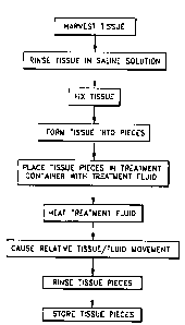
Figure I is a flow diagram illustrating the prior art process for preparing
biological tissue for implantation within a mammalian body comprising fixing
of
the biological tissue with a glutarafdehyde solution;
Figure 2 is a flow chart of the preparation of biological tissue for
implantation in a mammalian body comprising a method for inhibiting
calcification of the biological tissue according to the present invention;
CA 02293474 1999-12-09
WO 98/56432 PCT/US98/10017
7
Figure 3 is a schematic view of an exemplary tissue treatment apparatus
including a closed treatment container and container movement device;
Figure 4 is a schematic view of another exemplary tissue treatment
apparatus including an open treatment container and fluid stirring rod;
Figure 5 is a flow chart of the preparation of biological tissue using the
system of Figures 3 or 4 including the application of heat and motion to a
treatment solution;
Figure 6 is a schematic view of an exemplary tissue treatment apparatus
including a treatment container positioned in a flow stream;
Figure 7 is a flowchart of the preparation of biological tissue using the
system of Figure 5 including the application of heat and flow of treatment
solution past the tissue;
Figure 8 is a perspective view of another preferred tissue treatment
apparatus including an upstanding flow column and a plurality of vertical
sections within which tissues to be treated are mounted;
Figure 9 is an enlarged perspective view of one vertical segment of the
flow column of Fi;;ure 8 illustrating a piece of tissue suspended from a
baffle in a
flow stream;
Figure 10 is a horizontal cross section taken along line 10-10 of Figure 9
through one vertical section ofthe flow column;
Figure 1 1 is a vertical cross section taken along line 1 1-1 1 of Figure 10
and throu;;h a baffle and tissue suspension mount; ;
Figure 12 is a bar graph comparing the measured calcium uptake in
bovine pericardium tissues treated in a conventional manner, solely with heat,
and with heat and motion; and
Figure 13 is a bar graph comparing the measured calcium uptake in
bovine pericardium tissues treated in a conventional manner and with heat and
motion from various sources.
CA 02293474 1999-12-09
WO 98/56432 PCTlI1S98/10017
8
Description of the (referred Embodiments
The detailed description set forth below in connection with the appended
drawings is intended as a description of the presently preferred embodiment of
the invention, and is not intended to represent the only form in which the
present
invention may be constructed or utilized. The description sets forth the
functions
and sequence of steps for constructing and operating the invention in
connection
with the illustrated embodiment. It is to be understood, however, that the
same
or equivalent functions and sequences may be accomplished by different
embodiments that are also intended to be encompassed within the spirit and
scope of the invention.
One method for treating glutaraldehyde fixed biological tissue to inhibit
calcification thereof following implantation in a mammalian body is
illustrated in
Figure 2 which depicts a flow chart of the presently preferred embodiment of
the
invention. Figure 1 depicts a flow chart of the prior art method for preparing
biological tissue for implantation within a mammalian body.
Referring now to Figure l, the prior art process for preparing biological
tissue for implantation within a mammalian body comprises first harvesting the
tissue from an animal or human cadaver 10. As those skilled in the art will
recognize, various different types of tissue are routinely harvested from
different
2 0 animals and/or human cadavers. For example, heart valves are routinely
harvested from pigs, pericardium is routinely harvested from cows or pigs, and
skin is routinely harvested from human cadavers. Those skilled in the art will
further recognize that new tissues are, from time to time, being found to be
implantable within a mammalian body
After harvesting, the biological tissue is rinsed in saline solution,
typically
for a period of 1-6 hours 12.
The tissue is next fixed using a buffered glutaraldehyde solution of
adequate concentration, for example between 0.2% and 0.8%, at room
temperature for at least 3 hours 14. As is well known, ~lutaraldehyde effects
CA 02293474 1999-12-09
WO 98/56432 PCTIUS98/10017
9
cross-linking of the proteins, e.g., collagen, within the tissue. Such cross-
linking
tends to make the tissue more durable and effects preservation thereof. It is
known that cross-linked protein exhibits increased resistance to proteolytic
cleavage and further that one of the major processes by which circulating
blood
may destroy tissue is via enzymatic activity which involves unfolding of the
protein substrate in order to facilitate enzymatic hydrolysis. Cross-linking
of the
protein of a tissue makes the tissue resistant to such unfolding, and
consequently
tends to prevent deterioration thereof due to the enzymatic activity of blood.
The tissue is next sterilized, preferably with an alcohol/formaldehyde
solution for 2 hours at room temperature 16. The preferred solution for
effecting sterilization of the tissue comprises approximately 12 mill of Tween
80;
approximately 2 65 gms/I of l~~gCl? ~ H20; approximately )08 mill of
formaldehyde (37%); approximately 220 mill of ethyl alcohol ( 100%) and
approximately 4.863 gmsll of~ HEPES buffer. The balance of the solution
comprises double filtered H20 The pH of the solution is typically adjusted to
7.4 via the addition of NaOH Those skilled in the art will recognize various
other sterilization solutions are likewise suitable.
Antimineralization treatment 18 is optionally performed so as to inhibit
the accumulation of mineral deposits upon the biological tissue after
implantation
of a mammalian body As those skilled in the art will recognize, various
different
antimineralization treatments are utilized so as to prevent the deposition of
various different minerals upon the biological tissue.
The tissue is trimmed and any non-biological components are then added
thereto 20. For example, it is common to sew a heart valve to a valve holder
which aids in the handling thereof and which may additionally function as a
. mount for the valve when implanted into a mammalian body.
Next, the biological tissue is once again sterilized 22, preferably in an
alcohol/formaidehyde solution as discussed above. Since preparation of the
biological tissue is substantially complete and the biological tissue will
next likely
CA 02293474 1999-12-09
WO 98!56432 PCTIUS98110017
be stored for an extended period of time, a more rigorous sterilization
procedure
from that previously utilized is typically employed. At this stage, the
biological
tissue is typically sterilized for approximately 9 hours at 34-38°C.
After sterilization, the biological tissue is stored in glutaraldehyde at
5 room temperature 24.
Tissue Treatment Usin<, Heat
Referring now to Figure 2, a method for treating glutaraldehyde fixed
biological tissue to inhibit calcification thereof following implantation in a
10 mammalian body comprises the additional step of heating preferably when the
glutaraldehyde is in contact with the biological tissue, to approximately 35-
75°C
for approximately 4-22 weela, and more preferably for a period of a few days
to
22 weeks.
The treatment fluid should be heated to a temperature greater than body
temperature (37°C) but not high enough to damage either the tissue or
the
treatment fluid. Thus, the preferred heat range is between 35-75°C.
However,
the temperature affects the amount of calcification mitigation, and the
process
time, and is preferably between 45°C and 5_5°C, and more
preferably between
50°C ~ 1°C.
Heating of the biological tissue may be performed at any time after
harvesting the tissue from the animal or human cadaver and prior to implanting
the tissue within a mammalian body. However, heating of the biological tissue
is
preferably performed at a point in the process for preparing the biological
tissue
when the biological tissue is already disposed within a bath of glutaraldehyde
2 5 solution, as occurs at various stages of the process according to the
prior art.
Thus, the method for treating glutaraldehyde fixed biological tissues
according to
the present invention is preferably performed either during fixing thereof
with a
glutaraldehyde solution, immediately after fixing thereof with the
glutaraldehyde
CA 02293474 1999-12-09
WO 98156432 PCT/US98/10017
1 7.
solution, or alternatively just prior to or after being stored in a
glutaraldehyde
solution.
As a further alternative, a method for treating glutaraldehyde fixed
biological tissues may be performed during antimineralization treatment by
adding glutaraldehyde to the antimineralization solution and heating the
solution,
preferably to approximately i 5-75°C for approximately 4-22 weeks. More
preferably, the tissue is heat treated at 50°C ~I ° C for a
period of a few days to
22 weeks.
For example, after fixing tissue using a buffered glutaraldehyde solution
of adequate concentration, for example between 0.2% and 0.8%, at room
temperature for at least 3 hours 14, the biological tissue may be heat treated
in
either the same or dit~erent glutaraldehyde solution, preferably at
approximately
35-75°C for a few days to 22 weeks I S
As one of the alternatives discussed above, the biological tissue is fixed
and heat treated simultaneously 13 in the 0.2-0.8% glutaraldehyde solution,
again preferably at approximately 35-75°C for approximately a few days
to 22
weeks. Another alternative is to heat the tissue in saline 17 prior to
fixation 21.
As the other alternative discussed above, the biological tissue may
simultaneously undergo antimineralization treatment and heat treatment 19.
Glutaraldehyde is added to the antimineralization solution so as to effect the
inhibition of calcification of the tissue following implantation in a
mammalian
body.
Tissue Treatment Using Relative Tissue/Fluid Movement
2 5 Figure 3 illustrates one preferred embodiment of a tissue treatment
system 20 of the present invention. One or more pieces of tissue 22 or
leaflets
are immersed in a treatment solution 24 within a closed container 25. The
container 25 rests on a shaker table 26 which reciprocates relative to a base
27 in
one or more directions. One particularly preferred type of shaking device is
an
CA 02293474 1999-12-09
WO 98156432 PCT/US98/10017
12
orbital shaker. In one exemplary embodiment, the orbital shaker 2G is actuated
at a rotational speed of approximately 55 RPM. The container 25 and contents
therein may be subjected to heating, such as with radiant heaters 28 as
illustrated. Of course, any number of means for heating the container 25 are
known, such as resistance heaters, convective flow, and the like.
The solution 24 is preferably a buffered glutaraldehyde, but may be any
chemical solution, such as Denacol » or others, which performs substantially
the
same in this context. The shaking and/or heat may be applied during fixation
or
after. The tissue is preferably at least partially fixed prior to being
subjected to
the calcification mitigation treatment described herein, and more preferably
the
tissue is fully fixed prior to the treatment. The treatment thus can be
designed to
complete the fixation process. In a preferred embodiment, tissue that has been
fixed for a period of between thirty minutes to fourteen days is placed in the
container 25 with a buttered glutaraldehyde solution of adequate
concentration,
for example between 0.2% and 0.8%. The solution is then shaken for thirty
minutes after which the container 25 remains static for fourteen days.
The tissue 22 may be sheets of bovine pericardium tissue, precut leaflets,
or fully formed porcine heart valves. One potential disadvantage of using
precut
leaflets or porcine heart valves is the tissue's nonuniform capacity for
shrinkage
during calcification mitigation treatment. It can be difficult, though not
impossible, to consistently and accurately compensate for this phenomenon. A
detailed map of the fiber orientation, thickness and other properties of each
individual leaflet may be required to predict the final form of the leaflet
after
treatment. Therefore, the preferred procedure is to place sheets or pieces of
tissue in the container and subject it to the shaking and/or heat. Afterwards,
the
leaflets are cut from the treated tissue.
It will be noted that the tissue 22 within the solution 24 may be allowed
to move about freely. In another embodiment, and as will be described below
with respect to the embodiment of Figure 6, the tissue may be restrained from
CA 02293474 1999-12-09
WO 98156432 PCT/US98/10017
13
gross movement but allowed to freely shrink, such as with a device
schematically
shown at 29.
In another variation on the shaking, a treatment system 30 is shown in
Figure 4 wherein a stirring rod 32 is positioned in a container 34 to replace
the
shaking table 28. The stirring rod is preferably actuated magnetically through
the container, but may also comprise a shaft driven apparatus The stirring rod
32 is preferably designed so as not to batter the tissue 36 but instead just
to
cause gentle movement of the fluid 37 relative to the tissue. Therefore, in
the
illustrated embodimern, a piece of filter paper 38, or other such similar
porous
substrate or mesh, is draped over the top rim of the container and the tissue
pieces 36 placed therein In this wa~~, the stirring rod 32 imparts rotational
or
other momentum to the fluid 37 in the container 34, but the tissue 36 remains
above the damaging action of the rotating rod. Also shown in Figure 4 is a
heated enclosure or incubator 39 within which is placed the entire apparatus
30.
In another version of shaking, multiple flasks or containers holding the
treatment fluid and tissue samples are clamped to a rotating ferris-wheel
apparatus. The apparatus includes a wheel rotating about a tilted axis so that
the
flasks follow a tilted circular trajectory. In this manner, the fluid within
the
flasks gently washes over the tissue pieces as the wheel rotates.
2 0 The containers 25 and 34 in Figures 3 and 4 may be open or closed,
primarily dependiny~ on the nature of~ the treatment fluid Glutaraldehyde is a
toxic substance which evaporates to create a dangerous gas. Thus, treatment
with glutaraldehyde is preferably done in a closed container. On the other
hand,
some substances like Denacol0 may be less hazardous and the container may be
2 5 left open under a hood, for example.
Relative movement between the tissue and the treatment fluid is believed
to enhance calcification mitigation A mechanism for this result has not been
fully formulated, although mass transport of the fluid surrounding the tissue
may
be relevant Indeed, one theory is that certain cell material, for example,
CA 02293474 1999-12-09
WO 98/56432 PCT/US98/10017
19
proteins, is extracted or removed from the tissue by the treatment fluid,
which
removal is enhanced relative to static treatment methods by the movement of
the
fluid. In other words, the fluid surrounding any one portion of tissue is
repeatedly replenished by the relative movement of the tissue within the
fluid.
Test results shown in Figures 12 and I 3 for samples of tissue treated in a
variety
of ways in accordance with the present invention indicate that the combination
of
heat and relative tissue/fluid movement decreases the amount of calcium uptake
after implantation in rats, suggesting that such treatment will mitigate
calcification in long or short term implantation in humans.
Figure 5 is a flowchart showing a preferred method for treating tissue
using the system shown in Figures 3 or 4 Many of the specific pre- and post-
treatment steps described v~ith respect to Figures 1 and 2 have been left out
for
clarity, but remain applicable. Initiall~~, the tissue is harvested, rinsed,
fixed and
cut into pieces, preferably squares or rectangles, from which leaflets may be
formed. The pieces of tissue are then immersed in the treatmem fluid within
the
container, and the fluid heated to a predetermined temperature. Relative
movement between the tissue pieces and surrounding treatment medium is
induced and continued for a predetermined time. Inducing relative tissue/fluid
movement may be accomplished by any of the configurations shown herein, such
2 0 as shaking or vibrating a container for the tissue and fluid, or by
flowing
treatment fluid onto the tissue. Finally, the tissue pieces are removed from
the
container, rinsed and stored for later use Of course, rather than storing the
tissue, it may be formed directly into leaflets and assembled into a heart
valve
directly after the treatment process
The solution is heated indirectly through the surrounding air, such as
with the radiant heaters 28 shown in Figure 3, to a temperature of about
50°C
plus or minus 1 °C. The container is shaken or the fluid is stirred to
cause
relative tissue/fluid movemern The treatment time ranges between fourteen
da~~s to two mornhs, but is preferably closer to two months The container 25
is
CA 02293474 1999-12-09
WO 98156432 PCT/US98/10017
preferably a Mass tissue culture flask having a volume of approximately 250
ml.,
and the solution is a buffered glutaraldehyde solution of adequate
concentration,
for example between 0.2°ro and 0.8%. As mentioned above, a number of
pieces
of tissue 22 may be treated at a single time within the container 25. One
5 proposed ratio of tissue to solution is approximately 12 leaflets or leaflet-
sized
pieces of tissue per every 150 ml of solution.
Tissue Treatment Using Relative Tissue/Fluid Flow
Fi~.:ure G illustrates schematically another variation on a treatment system
10 40 which utilizes tlov past tire tissue as opposed to shaking a container
or
stirring the fluid in which the tissue is placed A flow creates the relative
motion
between the treatment solution and the tissue which is believed to result in
the
beneficial calcification mitigation effects.
The system 40 comprises a flow container 42 within which tissue 44 is
15 placed A number of conduits 46 connect one end of the flow container 42 to
a
pump 50 and then to a solution reservoir 48. Conduit 47, shown in dashed line,
may be connected between the other end of the flow container 42 and the
reservoir 48 to complete a closed circulation loop. The pump propels treatment
solution through the system 40 in the direction shown by the arrows 52. The
2 0 tissue 44 is preferably restrained within the flow container 42 using
means
schematically illustrated at 56 Resistance heaters 54 are illustrated
surrounding
the reservoir 48 If immersion heaters are used, they must be able to withstand
the extended exposure to sometimes caustic treatment fluid. Of course, one or
both of the resistance heating elements 54 may be removed from around the
2 5 reservoir, or alternative heating devices may be used. For example,
treatment
system 40, and the system 20 or 30 shown in Figures 3 and 4, for that matter,
may be enclosed in a larger enclosure or room 58 which is heated to the
preferred temperature by internal or external heaters In the illustrated
embodiment, thermocouples S9 are provided to sense the temperature within
CA 02293474 1999-12-09
WO 98/56432 PCT/US98/10017
16
both the flow container 42 and the reservoir 48. The thermocouple 59 in the
reservoir is preferably connected to feedback electronics for controlling the
heaters Sb based on the temperature of the fluid in the reservoir. This is so
that
the temperature does not rise too high to a level which might be detrimental
to
the tissue. The temperature within the flow container is monitored using a
thermocouple both as a safety, and to record the precise temperature profile
of
the treatment fluid.
The basic elements of a method for treating tissue using the system 40
are illustrated in Figure 7. Initially, the tissue is harvested, rinsed, fixed
and cut
into pieces, preferably squares or rectangles, from which leaflets may be
formed.
The tissue (or leaflets in some instances) may be placed within the flow
container 42 and subjected to flow during or after fixation. In a preferred
embodiment, the tissue 44 is at least partially fixed before being subjected
to the
flow within the system 40, and more preferably the tissue is fully fixed prior
to
the treatment. The pieces of tissue are then placed in the treatment
container,
and the solution caused to flow therethrough, initiating relative movement
between the tissue pieces and surrounding treatment medium which is continued
for a predetermined time. The solution is heated directly outside of the
container, or indirectly by heating the container. Finally, the tissue pieces
are
2 0 removed from the container, rinsed and stored for later use. Of course,
rather
than storing the tissue, it may be formed directly into leaflets and assembled
into
a heart valve directly after the treatment process.
With reference to Figure 6, the tissue is first fixed for a period of
between thirty minutes to fourteen days and placed in the flow container 42.
In
2 5 an alternative, the tissue may be first placed within the container 25
shown in
Figure 3 and shaken for a period of thirty minutes. After the fixation (or
after
the shaking, if desired), the tissue is placed in the flow container 42 and
subjected to solution flow of between ten and fifteen gallons per minute (38-
57
lpm) for a period of between fifteen to sixty days. The solution is preferably
CA 02293474 1999-12-09
WO 98/56432 PCT/US98/lOt)17
17
heated directly within the reservoir 48 to a temperature of about 50°C
( 122°F).
The solution is preferably a 0.2-0.8% buffered glutaraldehyde, and the tissue
44
is restrained from movement but allowed to shrink
In an alternative method of treating tissue in the system 40, the treatment
time is between thirty and sixty days. The flow rate is approximately 7.4
gallons
per minute (28 !pm) on average, and is uniform throughout a cross section
normal to the flow within the flow comainer 42. The tissue 44 is preferably a
rectangle of bovine pericardium of about 2 inches by 4 inches in dimension.
This
size of tissue sample may be used to form one or two leaflets after treatment.
Those with skill in the an will recognize that variations to the above
mentioned systems and processes for moving the fluid and/or heating the tissue
are available For example, the flow of solution past the tissue may be
combined
with a vibrational or shaking motion of the flow container 42 to enhance any
calcification mitigation benefits derived from either method. Additionally,
though the system 40 is sho~m as a closed circulation device, fresh solution
may
be pumped to the flow container 42 and discharged after passing through the
container (thus the conduit 47 is shown as optional) Of course, this will
reduire
a significant amount of treatment solution which may be prohibitively
expensive.
Nevertheless, one of the theoretical mechanisms for the beneficial aspects of
the
2 0 present treatment method including,: flow is that the solution is
constantly
replenished in the re~_ion surrounding the tissue so that a maximum mass
transport of chemicals and/or biological material such as protein is realized
from
the tissue to the solution Thus, a system which inputs fresh treatment
solution,
rather than recycling it through a resen~oir, would theoretically be more
effective
in this regard
CA 02293474 1999-12-09
WO 98/56432 PCT/US98/10017
1~3
Flow Column Apparatus
Figure 8 illustrates a perspective view of a flow column 60 which may
represent the flow container 42 illustrated schematically in Figure 6. The
column
60 is preferably a clear acrylic tube 6 I having an inner diameter of
approximately
six inches ( 15.2 cm), a height of about six feet ( 1.8 111), and a capacity
of about
ten gallons (38 1). The top and bottom ends of the cylinder 60 are closed by
caps
62a and 62b, respectively, which are sealed against the inner surface of the
cylinder 60 with O-rims (not shown). A lower inlet fitting 64 centered in the
cap 62b provides a conduit for introducing treatment fluid to the lower end of
the cylinder 60. Likewise, an upper fitting 66 connected to the cap 62a
provides
an outlet for the treatment fluid A length of hose 68 connects the lower
titting
64 to a fluid pump 70, whlCh IS In turn connected by a hose 72 to a fluid
reservoir 74. The circulatory treatment system is completed by a length of
hose
76 connecting the upper fitting 66 to the reservoir 74. Those with skill in
the art
will understand the fluid connections and requirements, which will not be
described further herein.
As mentioned above, the solution within the reservoir 74 is preferably
directly heated to the desired treatment temperature. Although not
illustrated,
the reservoir is desirably provided with one or more immersion resistance
2 0 heaters. A thermocouple 77 senses the temperature of the reservoir and is
preferably connected to feedback electronics for controlling the immersion
heater
so that the solution temperature does not rise too high to a level which might
be
detrimental to the tissue. The temperature within the flow container is
monitored using a thermocouple 78 both as a safety, and to record the precise
temperature profile of the treatment fluid. The treatment solution itself can
be
detrimentally affected by excessive temperatures, and thus the heating must be
done gradually and with a heater having good temperature control.
The vertical flow column or cylinder 60 is segmented into a plurality of
vertical sections 80 (seen enlarged in Figure 9) by a number of regularly
spaced
CA 02293474 1999-12-09
WO 98156432 PCT/US98/10017
19
baffles 82 having perforations 83. The baffles are substantially circular
perforated disks positioned horizontally witi~in the vertical cylinder 60,
normal to
the fluid flow. The outer diameter of each baffle 82 contacts, or comes into
close proximity with, the inner surface of the tube 61. Although the flow
column
60 is illustrated vertically, other arrangements will work. However, the
vertical
flow orientation is preferred to help purge bubbles from the flow column at
start
up. In other words, the bubbles naturally migrate out of the flow column in a
very short time, as opposed to a horizontal flow path, for example. It should
be
also be noted that the perforations are not shown in Figures 8 and 9 for
clarity,
but are shown in Figure 10.
The baffles 82 are commonly mounted on a vertical support rod 84
extending along the axis of the cylinder 60. The support rod 84 contacts the
lower ceiling cap 62b and extends upward into close proximity to the upper cap
62a. As seen at the lower end of Figure 8, the support rod 84 preferably
terminates in a stand member 86 having a pair of bifurcated legs 88 which
contact the top surface of lower cap 62b on either side of an inlet aperture
90.
In this manner, the support rod 84 can be positioned along the axis of the
cylinder 60 while not occluding inlet flow from the pump 70
As mentioned above, the baffles 82 divide the cylinder 60 into a plurality
of vertical sections 80. In this respect, the vertical sections 80 include the
region
between two baffles 82. In the illustrated embodiment, there are eight such
vertical sections 80 having a height of between seven and eight inches (17.8-
20.s
cm). The entire height of the column 60 is approximately 6 feet ( I .8 m), and
thus there is some space left above the top baffle and below the bottom
baffle.
The baffles 82 are slidably mounted on the support rod 84 to enable adjustment
of the spacing therebetween, if desired. Furthermore, the tissue pieces 82 can
be
easily mounted when the baffles 82 are removed from the system, whereupon the
baffles are slid over the support rod which is then positioned within the tube
61.
The tissue pieces to be treated are mounted in a particular manner in a
CA 02293474 1999-12-09...
WO 98/56432 PCT/US98/10017
LO
circumferential array about the support rod 84, as will be apparent from the
description of Figures 9-11.
At the top of the cylinder 60 a vertical space is created between the
upper baffle and the upper cap 62a, in which the central support rod 84
terminates The space is needed to insure that the flow passing through upper
bafl3e 82 is not unduly disturbed so that the flow »ithin the upper vertical
section
80 remains uniform in a horizontal cross section. Indeed, the uniformity of
flow
across any horizontal cross section between the baffles is important in the
present configuration to insure that the flow past any one piece of tissue is
equal
to the flow past other tissues The primary mechanism for insuring such uniform
flow is the baffles 82 themselves Preferably, the perforations 8s are
sufficiently
numerous and have a sufficient diameter so that the cross-sectional area of
the
baffles 82 has less structural material than open flow channels. The baffles
82
are thus designed to maintain a uniform, non-laminar upward flow stream
through each flow section 80.
At the lower end of the cylinder 60, below the lowest baffle 82, a flow
straightener 92 is positioned just above a velocity reducer plate 94, Inlet
flow
through the aperture 90 thus passes upward through the velocity reducer plate
94 and flow strai~htener 92 to impinge on the lowest battle 82 The velocity
reducer plate 94 is a disc like plate having a plurality of apertures 96
formed
therein. The apertures are relatively widely spaced in the plate 94 to create
a
drag on the flow and slow its velocity. The flow straightener 92 resembles a
honeycomb structure with a relatively densely spaced number of individual flow
channels, and has a vertical dimension greater than the velocity reducer plate
94
or baffles 82. Flow enters the column 60 through the aperture 90 and continues
upward through the velocity reducer plate 94 and straightener 92. After flow
passes through the straightener 92, it impinges on the lowest baffle 82. The
treatment solution flows upward through each baffle 82 into each successive
section 80 and out the top of the column 60. The column 60 is initially filled
CA 02293474 1999-12-09
WO 98/56432 PCT/US98/10017
2i
with air which is forced out as the surface of the upwardly advancing
treatment
solution flow passes upward through the column
Now- with reference to Figure 9, a vertical section 80 is enlarged
illustrating a plurality of tissue mounts 100 depending from the upper baffle
82.
The tissue mounts 100 comprise U-shaped members 102, more clearly shown in
Figure 1 1. Figure 10 shows tlje circumferential array of mounts 100
surrounding
the central support rod 84 Each mount 100 has a generally rectangular
configuration and is oriented radially in the baffle 82. That is, free ends of
the U-
shaped members IO? insert within similarly sized downwardly opening apertures
104 in the baffle 8? One of the apertures 104 for each mount i 00 is
positioned
close to the support rod 84, while the other is positioned close to the tube
61
'The apertures ! 0=~ extend approximately halfway through the thickness of the
baffle 82 and a smaller diameter through hole 106 continues upward to the top
surface of the baffle. This hole 106 is needed to push the mounts 100 from the
apertures 104 when treatment is tinished Preferably, the legs of the U-shaped
members 102 are spread outward a sli~_ht amount so that they have to be
scfueezed together to fit into the two apertures 104. This ensures a tight fit
so
the mounts 100 will not fall out of the apertures 104.
Rectangular tissue pieces 108 are attached to the mounts with sutures or
other similar expedient !n the illustrated embodiment, a lower edge I 10 of
each
tissue piece 108 loops around the bride ponion of the U-shaped member 102
and is sewn to the main body of the tissue piece along line i 12. In this way,
the
leading edge of the tissue piece 108 in the upward flow stream is rounded, and
thus protected from friction induced tearing or wear. One or more sutures 1 14
connect the upper comers of the tissue piece 108 to the upper ends of the legs
of
the U-shaped members 1 12 Preferably the tissue piece 108 is only connected at
one or tw-o locations alon;~; its vertical length to prevem gross movement or
flapping of the tissue, while allowing the maximum freedom for the tissue to
shrink An O-ring 116 or other such device placed on each leg of the member
CA 02293474 1999-12-09
WO 98/56432 PCT/US98/10017
22
112 prevents the sutures 1 14 from sliding down the leg. The upward flow I 18
of treatmem solution also assists in maintaining the generally planar
configuration of each tissue piece 108.
Mounting the tissue pieces 108 in a planar configuration substantially
parallel to the direction of flow of the solution ensures that an even amount
of
solution contacts both sides of the tissue. That is, is the tissue pieces were
canted with respect to the flow, the backsides would be exposed to less direct
flow, and eddy currents and the like might be set up, further making the fluid
exposure nonuniform In addition, the preferred parallel orientation minimizes
any stretchin~,= of the tissue during the extensive treatment period, such as
might
occur if the fluid was directed to one face of the tissue or the other.
The radial orientation of the plane of each tissue piece 108 desirably
ensures uniform contact with treatment solution during flow through the column
60. Ideally, the baffles 8? include perforations 120, seen in Figure 10, which
create the uniform, nonlinear flow The same velocity of solution is produced
at
any radial point from the suppon rod 84 outward. Of course, different pieces
of
tissue 108 have been shown to possess widely different properties, even from
the
same pericardial sac. Nevertheless, the present treatment configuration is
designed to maximize the uniformity of conditions seen by each piece of tissue
2 0 108. There may be some variation in treatment conditions between the top
and
bottom reaches of the container due to fluid head differences, but applicants
believe that such variations are minimal for the six foot tall column 60
described
herein.
There are preferably eight vertical sections 80 in which six tissue pieces
2 5 108 are mounted for a total of forty-eight tissue pieces being treated at
once. Of
course, other numbers of sections and tissue pieces per section are possible.
The
present flow column is extremel~~ well-suited for consistently manufacturing
high
duality treated bioprosthetic tissue. The segmented flow column with uniform
flow, and vertical orientation of each tissue piece 108 provides high
uniformity
CA 02293474 1999-12-09
WO 98/56432 PCT/US98/10017
23
of treatment. The modular nature of the column with the entire support rod 84
having all of the baffles 82 attached thereto is a significant advantage in
manufacturing. One batch of tissues may be treated, and then removed so that
after flushing the system a new batch can be ready for installation and
treatment.
Furthermore, the flow column lends itself to a high degree of control over the
system parameters such as the relative tissue/fluid velocity and the
temperature. -
Significantly, there are no large stagnant zones of flow within the column,
and
especially not within each vertical segment 80.
Rat Subcutaneous Studies
Figures 12 and 13 are results of calcium uptake measurements from
tissue treated in a variety of ways, implanted subcutaneously in rats for
several
months, and then removed. These graphs indicate that heat alone reduces
calcium uptake in comparison with a control, and that heat and motion reduces
the calcium uptake even further. A number of shaking, stirring or movement
apparatuses were used at two different temperatures, with the same general
results.
Figure 12 shows the results from three groups of samples of untreated
and treated bovine pericardium tissue. The first group (GLUT CONTROL)
exhibited an average measurement of about 16% calcium from 12 tissue samples
which were subjected to a post-fixation treatment of unheated and static
glutaraldehyde. The second group (HEAT) exhibited an average measurement
of about 7% calcium from 8 tissue samples which were subjected to a post-
fixation treatment of static glutaraldehyde heated to a temperature of
50°C.
Finally, the third group (HEAT AND SHAKING) exhibited an average
measurement of about 4% calcium from 7 tissue samples which were subjected
to a post-fixation treatment of static glutaraldehyde heated to a temperature
of
50°C. The treatment solution for all three groups was identical - 0.6%
HEPES-
glutaraldehyde at a pH of 7.4 - and the treatment period was equal - 2 months.
CA 02293474 1999-12-09
WO 98156432 PCT/US9811,0017
24
The third group was shaken in a bottle or container using a reciprocal orbital
shaker actuated at 80 RPM. The rats were all approximately 12 days old, and
the tissue samples were left implanted for eight weeks.
Figure 1 s shows the results from a number of groups of samples of
untreated and treated bovine pericardium tissue. The calcium uptake results
for
the groups are indicated by bars with different shading depending on the
overall
treatment regimen. Thus, the black bars for group 1 are the control (no heat
or
shaking), the middle shaded bars are for samples subjected to shaking and heat
treated to 50°C, and the ri~hthand white bars are for samples subjected
to
shaking and heat treated to 42°C.
Group 1 on the left is a control and shows results for two subgroups of
7 and 4 samples each. The control samples were treated for 2 months in 0.6%
HEPES-glutaraldehyde at a pH of 7.4 with no heat or movement. Each sample
was implanted in 16 day old rats, and left implanted for a period of between 3
and 4 months before being removed to test for calcium.
Groups 4-6 in the middle were all heat treated at 50°C in the same
treatment solution as group t for the same period. The dif~'erences between
the
treatment regimen for groups 2-6 are the methods used to induce relative
tissue/fluid movement. The methods are shown graphically below each group.
Group 2 7 includes two subgroups of 7 and 8 samples each subjected to
reciprocal orbital shaking. Group ~ 7 includes two subgroups of 2 and 11
samples each placed in a flash with a magnetic stirring bar in the bottom.
Group
4 is the same method as group 3 but with two subgroups of 8 samples each
placed on a filter instead of being allowed to float around the flask. Group 5
2 5 included two subgroups of 12 samples each placed in a first container and
subjected to a rolling motion, using a tilted ferris wheel arrangement. Group
6
included two subgroups of 20 and 12 samples each placed in a second container
and also subjected to a rolling motion.
CA 02293474 1999-12-09
WO 98/56432 PCT/US98/10017
Groups 7-9 on the right were all heat treated at 42°C in the same
treatment solution as groups 1-8 and for the same period. Again, the
differences
between the treatment regimen for groups 7-9 are the methods used to induce
relative tissue/fluid movement, shown graphically below each group. Group 7
5 includes two subgroups of 8 and 4 samples each subjected to reciprocal
orbital
shaking. Group 8 includes two subgroups of 8 and 1 I samples each placed in a
flask with a magnetic stirring bar in the bottom. Group 9 is the same method
as
group 8 but with two subgroups of 8 samples each placed on a filter instead of
being allov~ed to float around the flask.
10 It is apparent ti~om these tests that the shaking and heat treatment
reduced calcium intake over the control group, as well as over the heat
treatment
alone. Also, treatment at 50°C was substantially more effective than
treatment
at 42°C. Comparisons of the different shaking? stirring methods
indicates that
stirring with a magnetic rod within the flask produced the least amount of
15 calcium uptake, regardless of temperature, although perhaps not by a
significant
margin at 50°C.
It is understood that the exemplary methods and apparatuses for treating
glutaraldehyde fixed biological tissue described herein and shown in the
drawings
represent only presently preferred embodiments of the present invention.
2 0 Indeed, various modifications and additions may be made to such
embodiments
without departing from the spirit and scope of the invention. For example,
various fixing agents, such as Denacol rt or aldehydes other than
glutaraldehyde,
may exhibit properties similar to those of glutaraldehyde so as to make them
suitable for use in the present invention and, thus, may likewise be utilized.
25 Accordingly, these and other modifications and additions may be obvious to
those skilled in the art and may be implemented to adapt the present invention
for use in a variety of ditferent applications. Furthermore, the scope of the
invention should be determined with reference to the appended claims.