Note : Les descriptions sont présentées dans la langue officielle dans laquelle elles ont été soumises.
CA 02295973 2006-10-25
METHOD OF AND SYSTEM FOR
CONE-BEAM TOMOGRAPHY RECONSTRUCTION
BACKGROUND OF THE INVENTION
The present invention is directed to a method of and system for computed
tomography
(CT) density image reconstruction. More particularly, the present invention is
directed to the
three-dimensional reconstruction from two-dimensional projections acquired
with x-ray cone-
beam CT and single photon emission computed tomography (SPECT) scanners. Even
more
particularly, the present invention is directed to a method of and system for
intravenous
volume tomographic digital angiography imaging.
For about the past twenty years, computerized tomography has revolutionized
diagnostic imaging systems as well as non-destructive test imaging techniques.
Conventional
CT scanners use a fan-shaped x-ray beam and one-dimensional detector in order
to
reconstruct a single slice with a single scan of an object. However, current
CT technology is
limited by a trade-off between high longitudinal resolution and fast volume
scanning. One
method which has been utilized to address the shortcomings of CT scanner
technology is the
use of cone-beam tomography. A cone-beam volume CT scanner uses a cone-beam x-
ray
source and a two-dimensional detector to reconstruct the whole volume of an
object with a
single scan of that object. The data obtained from the scan of the object is
processed in order
to construct an image that presents a two-dimensional slice taken through the
object. The
current technique for reconstructing an image from 2-D is referred to in the
art as the filtered
back projection technique. That process converts the attenuation measurements
from a scan
into integers called "CT numbers" or "Hounsfield units" which are then used to
control the
brightness of a corresponding pixel on a cathode ray display.
In a 3-D scan technique, a cone-shaped x-ray beam is used which diverges to
form a
cone-beam that passes through the object and impinges on a two-dimensional
array of
detector elements. In that manner, the volume scanning time of a 3-D object
can be at least
CA 02295973 2006-10-25
times shorter than a standard CT on a spiral CT. In contrast to existing CT
with an
intraslice plane resolution of 1.0 1 p/mm, the reconstructions of cone beam CT
will have isotropic spatial resolution along all three axes (0.5-2.0 lp/mm).
Each view is thus a 2-D
array of x-ray attenuation measurements and the complete scan produces a 3-D
array of
attenuation measurements.
At present, either of two methods are commonly used to reconstruct a set of
images
from the acquired 2-D attenuation measurements. The first technique is that
developed by
Feldkamp, Davis & Kress, which is described in "Practical Cone-Beam
Algorithm", J. Opt.
Soc. Am., Vol. 1, pp. 612-619 (1984). The Feldkamp, et al. technique, which
uses an
algorithm which was derived using approximations of a tilted fan beam fonnula,
is a
convolution-back projection method which operates directly on the line
integrals of the actual
attenuation measurements. That method can be easily implemented with currently
available
hardware and is a good reconstruction for images at the center or "mid-plane"
of the cone-
beam. While the algorithm of Feldkamp, et al. provides excellent computational
efficiency
and minimal mechanical complexity in data acquisition, its major shortcoming
is that it is
based on single circle cone-beam geoc_netr~!.. Single eircle eone-bearn
geometry, in which the
source always lies on a circle, cannot provide a complete set of data to
exactly reconstruct the
object. For that reason, Feldkamp, et al.'s algorithm causes some unavoidable
distortion in
the non-central transverse planes, as well as resolution degradation in the
longitudinal
direction.
In order to address the problems of Feldkamp's algorithm, several other
approaches
have been proposed using different cone-beam geometries including dual
orthogonal circles,
helical orbit, orthogonal circle-and-line, and Smith's curve. Such geometries
can-achieve
exact reconstructions when using the approach of Tuy, Smith, or Gangreat.
In addition to the Feldkamp, et al. approach for analytic cone-beam
reconstruction, a
second commonly used method is that disclosed by Pierre Grangeat in,
"Mathematical
Framework of Cone-Beam 3-D Reconstruction Via the First Derivative of the
Radon Transform", Mathematical Methods in Tomography, Herman, Lewis, Natterer
(eds.) Lecture
Notes in Mathematics, No. 1497, pp. 66-97, Spring Verlag (1991). That
algorithm provides
an accurate solution to the image reconstruction task based on a fundamental
relationship
2
CA 02295973 1999-12-30
WO 99/01066 PCT/US98/12468
between the derivative of the cone-beam plane integral through the derivative
of the parallel
beam plane integral. While the Grangeat method is theoretically accurate, it
requires
mathematical operations that can be solved only using finite numerical
calculations that are
approximations. Thus, errors can be introduced by the implementation of the
Gangreat
method that can be greater than those produced using the Feldkamp, et al.
method and such
errors are not correlated with the cone-beam angle.
A third method has been disclosed by H.K. Tuy in "An Inversion Formula for a
Cone-
Beam Reconstruction", SAIMJ. Appl. Math. 43, pp. 546-552 (1983). Using Tuy's
approach,
in order to generate a complete or sufficient set of data, every plane which
passes through the
imaging field of view must also cut through the orbit of the focal point at
least once. The
single plane or orbit of Feldkamp, et al. does not satisfy this condition.
Still yet another approach that has been proposed is the inversion of the cone-
beam
data sets if the assumption is made that for any line that contains a vertex
point and a
reconstruction point, there is an integer M which remains constant for the
line such that
almost every plane that contains this line intersects the geometry exactly M
times.
Mathematical improvement to the reconstruction algorithms was described in an
article by
B.D. Smith entitled "Cone-Beam Tomography: Recent Advances and a Tutorial
Review,"
Opt. Eng., Vol. 29 (5) pp. 524-534 (1990). However, such an integer
requirement condition
is too restrictive for practical application since the only known source point
geometry which
meets that condition is a straight line.
Two somewhat recent patents were issued in the United States directed to the
cone-
beam reconstruction problem. The first, U.S. Patent No. 5,170,439 to Zeng, et
al., was
issued on December 8, 1992 and utilizes the above-described cone-beam
reconstruction
method using combined circle and line orbits. However, that technique requires
the removal
of redundant and unnecessary data, which necessarily requires more computing
time and
complexity than the method and system of the present invention.
Another approach to the reconstruction of images from cone-beam data is
disclosed in
U.S. Patent No. 5,400,255, which issued to Hu on March 21, 1995. The
methodology
disclosed in the Hu patent represents a minimal improvement from Feldkamp's
algorithm and
it is still an approximate method based on a single circle cone beam geometry.
It cannot
3
CA 02295973 1999-12-30
WO 99/01066 PCT/US98/12468
result in exact reconstruction and it is not acceptable in many clinical
applications when the
cone angle is large.
In contrast to the prior art approaches, the present invention discloses an
exact cone-
beam reconstruction system and method using a circle-plus-arc data acquisition
geometry in
which the locus of a source and a detector is a circle plus an orthogonal arc.
In that manner,
the best image quality of a cone-beam volume CT is achieved without
introducing any
additional mechanical complexity compared to a regular CT gantry. If the locus
of an x-ray
source and a detector is a single circle during cone-beam scanning (single
circle cone-beam
geometry), an incomplete set of projection data will be acquired. The
incompleteness of the
projection data results in some unavoidable blurring in the planes away from
the central z
plane and a resolution loss in the z direction (i.e., Feldkamp, et al.'s
algorithm). The
reconstruction error due to the incompleteness of the projection data could be
up to 50% of
the signal when using Feldkamp, et al.'s algorithm with a 22 cone angle.
However, using the
data acquisition geometry of the present invention, the locus of an x-ray
source and a detector
is a circle plus an arc perpendicular to the circle. That corresponds to
rotating the x-ray tube
and detector on the gantry, and then acquiring the arc projections on a
perpendicular arc while
tilting the gantry at a relatively small angle ( 15 to 30 ). Such geometry
results in a
complete set of data for an object with a 25-40 cm length in the z direction,
which
corresponds to a 37-60 cm field size at the detector in the z direction with a
magnification of
1.5. Using the system and method of the present invention, the 3-D
reconstruction is exact
and no image blurring or resolution loss occurs.
The method and system of the present invention is based upon the three-
dimensional
Radon transform. The algorithm used with the present invention first
transforms the cone-
beam projections acquired from a circle-arc orbit into the first derivative of
the 3-D Radon
transform of an object using Grangeat's formula. Then, the object function is
reconstructed
using the inverse Radon transform. In order to reduce the interpolation errors
in the re-
binning process required by Grangeat's formula, new re-binning equations have
been derived
exactly, therefore transforming 3-D interpolations into one-dimensional
interpolations. The
inventive cone-beam acquisition method and system disclosed herein provides a
complete set
of projection data such that the cone-beam image reconstruction algorithm
achieves exact
4
CA 02295973 1999-12-30
WO 99/01066 PCT/US98/12468
reconstructions. The result is a 3-D cone-beam reconstruction which introduces
no obvious
artifacts and only a practical acceptable reduction of reconstruction
accuracy.
The 3-D volume tomographic imaging and system described above can also be used
to
achieve a 3-D or volume tomographic digital angiography imaging method and
system which
= is capable of providing clinically useful 3-D vascular images for enhancing
diagnostic and
therapeutic decisions. In particular, the volume tomographic digital
angiography imaging
method and system disclosed herein is particularly useful for intravenous (IV)
volume
tomographic digital angiography (IV-VTDA). Such IV-VTDA is superior to
conventional
angiography because it provides a direct, unambiguous and accurate 3-D
measurement of
stenosis and other irregularities and malfunctions, including the caliber,
geometry and spatial
orientation in the structure. Moreover, the IV-VTDA method and system
disclosed herein
requires only a single IV injection of contrast media and uses fast volume
scanning, thus
reducing the invasiveness of the procedure as well as the procedure time,
while also providing
a substantial reduction in the total x-ray exposure to the patient.
A need for the accurate and detailed assessment of atherosclerotic disease has
been
reemphasized by the growth of new therapeutic techniques, such as
thrombolysis,
endarterectomy, atherectomy, angioplasty, embolization and the placement of
vascular stents,
as well as the need to facilitate and improve the success rate of such
therapeutic procedures.
Cerebrovascular disease is the third leading cause of death in the United
States and claims
approximately 500,000 new victims each year. New surgical and endovascular
techniques
greatly improve patient survival and their quality of life. Thus, there has
been an increase in
the therapeutic procedures enumerated above in the last several years. As the
result of such
procedures, patient survival has increased and the quality of the patient's
life has improved.
The identification of patients who can benefit from a specific therapeutic
procedure
requires both accurate and detailed information about the severity of the
stenosis, their
geometry and their spatial orientation. However, most therapeutic decisions
are based on
information obtained through standard projectional angiographic techniques.
Projection
images using such standard projectional techniques do not provide sufficient
information with
which to detect and completely characterize all vascular lesions. That lack of
complete data
impairs the ability of the physician to determine the optimal therapeutic
procedure.
Obviously, an inappropriate choice of intervention based on improper knowledge
of the
CA 02295973 1999-12-30
WO 99/01066 PCTIUS98/12468
patient's anatomy can lead to unnecessary interventions, a sub-optimal
outcome, injury or
death.
As discussed above, all of the standard projectional angiographic techniques
contain
major shortcomings with respect to providing a complete characterization of
vascular lesions.
For example, intraarterial (IA) digital subtraction angiography (IA-DSA),
which is currently
used for examining most patients for vascular disease, has two principle
limitations. First,
IA-DSA provides only a 2-D projection of 3-D anatomical structures. Second, IA-
DSA
images are of reduced usefulness due to vessel overlap, particularly when non-
selective
injections are used. Obviously, the knowledge of the geometry of the stenosis
and the spatial
orientation of the arteries is a major step in the performance of successful
surgical or
transvascular interventional procedures. Thus, using IA-DSA techniques,
multiple views are
utilized to attempt to detect all lesions as well as to evaluate the geometry
of the stenosis and
to integrate the 2-D views into correct spatial relationships. That, in turn,
requires the use of
multiple contrast injections as well as a multiple series of x-ray exposures.
However, even with multiple views, the number of views is limited, which often
results in non-detected lesions because of the failure to achieve orthogonal
projection and
overlap. Consequently, the angiographic procedure can become prolonged,
increasing patient
morbidity from lengthened catherterization time, increasing contrast as well
as the radiation
dose, while also increasing procedure costs. There is also an added risk of
complications
related to percutaneous cannulation of an artery and the manipulation of the
IA catheters and
wires in critical vessels which are often affected by vascular disease. The
risk of procedure
related vascular injury and stroke is also present, with major morbidity.
Moreover, such
angiography technique is frequently repeated as the vascular disease
progresses, thus
multiplying costs and risks.
In the past, in order to avoid the shortcomings and risks of IA-DSA, attempts
have
been made to utilize intravenous digital subtraction angiography (IV-DSA).
However, in
addition to the shortcomings inherent with all types of DSA, there are two
additional
important technical deficiencies which are specific to IV-DSA. First, image
misregistration
often occurs due to patient motion. Such misregistration too often masks the
vascular
anatomy to be imaged. Second, there is an inability to attain a sufficiently
high concentration
of contrast media through intravenous injection to overcome the quantum noise
inherent in
6
CA 02295973 1999-12-30
WO 99/01066 PCTIUS98/12468
the DSA technique. Due to those deficiencies, the resulting image is generally
of poor quality
and, thus, IV-DSA has become an infrequently used clinical technique.
In the past fifteen years, many attempts have been made to improve the image
quality
of IV-DSA. Such techniques have been only partially successful in reducing the
severity of
motion artifacts and in improving problems with vessel overlaps. Thus, even
with such
improved IV-DSA techniques, there still exists a significant amount of missing
3-D
information which would be very useful to obtain.
An improvement over IA-DSA can be obtained by incorporating the volume
tomographic imaging principles discussed herein with digital angiography. As
is disclosed in
more detail herein, a cone-beam volume CT scanner using an image intensifier
coupled to a
CCD camera as a 2-D detector can be used to obtain CT-like 3-D reconstructions
of blood
vessels from a single IA contrast media injection and a single fast volume
scan. In contrast to
the DSA technique, such an image intensifier-based volume based tomographic
imaging
method and system provides the ability to tomographically isolate an object of
interest, such
as a blood vessel, from the structures in an adjacent plane, such as other
blood vessels or
bone. The 3-D reconstructions eliminate vessel overlap and provide a complete,
true 3-D
description of the vascular anatomy. Such reconstructions have isotropic
spatial resolution
along all three axes. Others have reported similar results on selective intra-
arterial volume
tomographic angiography reconstructions, thus demonstrating the advantages of
IA-VTDA
over IA-DSA. See, for example, an article entitled "3 D computed x-ray
angiography: first in
vivo results," by D. St-Felix, R. Campagnalo and Y. Rolland, et al. in
Radiology 1992,
185:304, a paper presented by R. Fahrig, A.J. Fox and D.W. Haldsworth,
entitled "Three-
Dimensional CT Angiography from a C Arm Mounted XRII," which was presented at
RSNA
82nd Scientific Assembly, December 1, 1996, and a paper presented by K.
Sekihara, H.
Kawai, K. Yamamoto and T. Kumazaki, entitled "Cone Beam CT Angiography," at
Proc. of
JAMIT Frontier '95, pp. 23-28, 1995.
One of the drawbacks of IA-VTDA is that it is based upon IA injections, which
are
generally much more invasive than IV injections. Although when compared to
DSA, IA-
VTDA represents a significant advance, IV-VTDA represents an even greater
advance
compared to IA-DSA because it has all of the advantages IA-VTDA has over IA-
DSA and at
the same time makes the angiographic procedure much safer. IV-VTDA also
provides a
7
CA 02295973 1999-12-30
WO 99/01066 PCT/US98/12468
significant reduction in the cost of the angiographic procedure because it
eliminates the need
for arterial puncture and catherterization.
One of the difficulties of using IV-VTDA in place of IA-VTDA is that IV
injections
result in a much lower signal compared to IA injections. Whereas a selective
IA injection
results in almost no dilution of injected iodine concentration and a non-
selective IA injection
results in a factor of 3-4 dilution, the dilution of central and peripheral IV
injections depends
on cardiac output, transit time, venous capacitance, the injection rate and
the length of the
injection. Dilution factors on the order of 20:1 - 30:1 are common. The result
is that an IV-
VTDA system must compensate for a significantly lower signal compared to IA-
VTDA,
which in turn requires that the IV-VTDA system have a much better low contrast
resolution
than an IA-VTDA system.
There are two IV injection protocols. One is the central IV injection which is
performed at the vena cava near the right atrium. The other is the peripheral
IV injection,
which is performed through the antecubital fossa or other peripheral veins. If
necessary, veins
in both antecubital fossae can be injected simultaneously to achieve even
higher rates of
contrast administration intravenously. The injection can be performed using an
injector, and
contrast solution can be iodinated contrast materials.
Other modalities could potentially also be used for 3-D angiographic imaging,
such as
helical CT, magnetic resonance angiography (MRA) and ultrasound (US). However,
IV-
VTDA is clearly preferable to all three of these modalities.
Spiral CT angiography (CTA), while having proven useful for the evaluation of
cerebrovascular and aorto iliac disease, has some major disadvantages when
compared to IV-
VTDA. First, the long volume scan time of CTA limits the rate of contrast
injection and at
least a 30 second breathhold is required by the patient. Therefore, CTA is
more sensitive to
patient motion than IV-VTDA techniques. Also, due to tube loading limitations,
the
resolution in the section direction of CTA is practically limited, and small-
vessel resolution
may be limited by partial volume effects. IV-VTDA, on the other hand, requires
a much
shorter volume scanning time, which allows a higher contrast media injection
rate so that a
much higher IV injected iodine signal can be achieved, which produces a better
image quality,
requires less contrast media and a much smaller tube loading. Thus, compared
to CTA
8
CA 02295973 1999-12-30
WO 99/01066 PCT/US98/12468
techniques, IV-VTDA can cover a much larger segment of the body in the
direction
orthogonal to the slices than conventional CTA, with a single injection.
It is contemplated that the use of IV-VTDA for imaging a patient's body will
produce
significant benefits over the images produced by the present CTA techniques.
IV-VTDA is
also superior for cross-sectional pulmonary angiography because of its shorter
breathhold
requirements as well as the isotropic resolution it attains. Furthermore, IV-
VTDA can also be
used for lower extremity angiography where spiral CTA cannot be used because
of the limited
tube capacity and total amount of contrast media which can be safely
administered to a
patient.
MRA has already proven useful for the evaluation of vascular disease. However,
current MRA procedures have some deficiencies, including limited spatial
resolution,
overestimation of stenosis and other artifacts, particularly at regions of
flow disturbances,
tradeoffs between maximizing the field of view (FOV), signal to noise ratio
and spatial
resolution and relatively long scanning times, which make it sensitive to
patient motion.
While many attempts have been made to solve these problems, even if they are
finally solved,
the IV-VTDA system and method of the present invention will be less expensive.
IV-VTDA
can also be used for patients with contraindications to MR scanning, such as
claustrophobia,
pacemakers, cerebral aneurysm clips, implanted defibrillators, previous
surgery with metal
implants, or prior trauma with residual metal fragments, and will also allow
visualization
inside metallic endovascular stents.
Transcutaneous duplex ultrasound (US) has the advantages of real-time non-
invasive
imaging that provides spectral information in a relatively inexpensive package
while also
providing extraluminal information. However, compared to IV-VTDA, a principle
limitation
of ultrasound is the need for an appropriate acoustic window. Intervening air
or bone prevent
the acquisition of diagnostic information at a substantial number of potential
vascular sites.
For that reason, ultrasound has a primary diagnostic role in the carotid and
lower extremity
arteries, but is not useful in adult (closed fontanelle) skull and central
chest areas and is
limited in use in the deep abdomen. Furthermore, calcific plaque obscures
visualization,
often right at the stenosis. Other disadvantages of ultrasound include a
limited FOV,
dependence on Doppler angle, dependence on operator skill, and the inability
to distinguish
9
CA 02295973 1999-12-30
WO 99/01066 PCT/US98/12468
total occlusion from severe stenosis as well as a poor 3-D depiction of the
anatomy for
surgical planning.
SUMMARY AND OBJECTS OF THE INVENTION
In view of the foregoing, it should be apparent that there still exists a need
in the art
for a method of and apparatus for producing a 3-D image from two dimensional
projections
acquired with x-ray cone-beam volume CT and SPECT scanners such that exact
reconstructions with no image blurring or distortion are produced. It is,
therefore, a primary
object of this invention to provide a method of and apparatus for obtaining an
exact 3-D
reconstruction from 2-D projections acquired with cone-beam volume CT and
SPECT
scanners which is characterized by lack of image blurring and distortion.
More particularly, it is an object of this invention to provide a new method
for cone-
beam reconstruction using a circle-plus-arc data acquisition geometry to
provide a complete
set of data such that an exact 3-D reconstruction can be obtained using a cone-
beam x-ray
source and a 2-D detector with a conventional CT scanner gantry.
Still more particularly, it is an object of this invention to provide for a
circle-plus-arc
data acquisition geometry for use with volume CT scanner using a cone-beam x-
ray source
and a 2-D detector in which a standard CT gantry is utilized without
introducing mechanical
complexity to achieve exact 3-D reconstructions of an object.
Briefly described, these and other objects of the invention are accomplished
by the use
of a new analytic cone-beam reconstruction algorithm which uses a circle-plus-
arc data
acquisition geometry to provide a complete set of data so that an exact 3-D
reconstruction is
obtained even in cases where Feldkamp's algorithm fails severely. The novel
data acquisition
scheme disclosed herein is applied to a volume CT scanner which uses a cone-
beam x-ray
source and a 2-D detector, such as a selenium or silicon thin-film flat-panel
x-ray imager.
The circle-plus-arc data acquisition scheme is implemented by acquiring one
set of cone-
beam projections while rotating an x-ray tube and a detector on a standard CT
gantry and,
then, acquiring another set of projections while tilting the gantry by a small
angle of
approximately 15 to approximately 30 with the x-ray tube and the
detector fixed on the
gantry. That scanning method is accomplished on a standard CT gantry without
introducing
CA 02295973 1999-12-30
WO 99/01066 PCT/US98/12468
mechanical complexity and achieves exact 3-D reconstructions of an object with
a 25-40 cm
diameter.
In practice, the arc length and arc sampling rate can be reduced (for example,
by 50%)
without the introduction of any obvious artifacts and with only a practically
acceptable
reduction of reconstruction accuracy. Thus, data acquisition time on the arc
is significantly
reduced by decreasing the arc length or arc sampling rate with the result that
the desired 3-D
image reconstruction may be computed in less time.
In its method aspects, the present invention is carried out by first obtaining
the cone-
beam projection data from a volume CT or SPECT scanner. Then, that projection
data is
preweighted and the partial derivatives of the preweighted projection data are
calculated.
Next, the calculated partial derivatives are rebinned to the first derivative
of the Radon
transform, for both the circular orbit data and the arc orbit data. The second
partial derivative
of the Radon transform is then calculated. Finally, the reconstructed 3-D
images are obtained
by backprojecting using the inverse Radon transform.
In view of the foregoing discussion concerning the need for an accurate
assessment of
atherosclerotic disease and the limitations of the current techniques for
examining most
patients for vascular disease, it should be apparent that there still also
exists a need in the art
for a method of and apparatus for producing a 3-D image of vascular anatomy
utilizing IV
injections and a volume tomographic digital angiography imaging system which
uses an x-ray
cone-beam volume CT scanner. It is, therefore, a primary object of this
invention to provide a
method of and apparatus for obtaining an exact 3-D vascular image for enabling
diagnostic
and therapeutic decisions using intravenous volume tomographic digital
angiography.
More particularly, it is an additional object of this invention to provide a
method of
and system for using cone-beam volume CT techniques to produce direct,
unambiguous and
accurate 3-D measurement of stenosis and other irregularities and
malformations associated
with vascular disease which requires only a single IV injection of contrast
media, thus
reducing the invasiveness of the procedure.
Still more particularly, it is an object of this invention to provide a method
of and
system for intravenous volume tomographic digital angiography in which a
direct,
unambiguous and accurate 3-D measurement of vascular disease is obtained using
an x-ray
11
CA 02295973 2006-10-25
cone-beam volume CT scanner, thus reducing the procedure time while at the
same time
providing a substantial reduction in the total x-ray exposure to the patient.
Briefly described, these and other objects of the invention are accomplished
by the
use of an x-ray cone-beam volume CT scanner which utilizes a new analytic cone-
beam
reconstruction algorithm with a circle-plus-arc data acquisition geometry in
order to provide
a complete set of data so that an exact 3-D image of the vascular diseased
anatomy is ob-
tained with the use of only a single IV injection of contrast medium. The
novel IV volume
tomographic digital angiography method and apparatus disclosed herein is
accomplished
using a volume CT scanner which uses a cone-beam x-ray source and a 2-D
detector, such as
a selenium silicon thin-film flat panel x-ray imager which achieves high
resolution, high
frame rate and a high dynamic range while at the same time having only a small
image lag
and excellent linearity. In an alternate embodiment, computer-controlled table
movement is
utilized and synchronized with the x -ray exposures such that a circle-plus-a
straight line
cone-beam geometry is utilized in order to optimize computational efficiency.
In its method aspects, the present invention is carried out by first injecting
into the
vein of patient a contrast fluid using an injector. Then, the portion of
interest of the patient's
body is scanned using the volume tomographic digital angiography system
described herein.
The projection data thus obtained is then pre-weighted and the partial
derivatives of the pre-
weighted projection data are calculated. The remaining steps described in
conjunction with
constructing an image from a volume CT or SPECT scanner discussed above are
then utiliz-
ed in order to generate the 3-D vascular images of interest for diagnostic and
therapeutic
uses.
The invention thus provides according to an aspect, for an apparatus for
generating a
three-dimensional image representative of an interior portion of an object.
The apparatus
comprises: a radiation cone-beam scanner which generates cone-beam projection
signals, the
radiation cone-beam scanner generating cone-beam circular projection signals
representative
of a circular orbit around the object and cone-beam arc projection signals;
means for pre-
weighting the cone-beam circular and arc projection signals with pre-weighting
factors based
on a distance along which each of the circular and arc projection signals is
taken to produce
pre-weighted cone-beam circular and arc projection signals; means for
calculating partial
derivatives of the pre-weighted cone-beam circular and arc projection signals
with respect to
spatial coordinates in a detector-based coordinate system; means for rebinning
the pre-
weighted cone-beam circular and arc projection signals from the detector-based
coordinate
12
CA 02295973 2006-10-25
system to Radon space to produce Radon transform signals; means for inverse
Radon trans-
forming the Radon transform signals; backprojection means for reconstructing
the Radon
transform signals into a three-dimensional image representation; and an image
memory
means for storing the three-dimensional image representation. The apparatus is
characteriz-
ed in that the radiation cone-beam scanner comprises a gantry frame with a
first arc orbit for
mounting a radiation source and a second arc orbit for mounting a radiation
detector so that
the radiation source and the radiation detector are movable to obtain both arc
projections and
circular projections in a circle-plus-arc geometry without tilting the gantry
frame.
With these and other objects, advantages and features of the invention that
may
become hereinafter apparent, the nature of the invention may be more clearly
understood by
reference to the following detailed description of the invention, the appended
claims and to
the several drawings attached herein.
BRIEF DESCRIPTION OF THE DRAWINGS
Figure 1 is a drawing showing the geometry of the circle-plus-arc orbit
utilized by the
present invention;
12a
, ,. .
CA 02295973 1999-12-30 + = .
. .
) ) . .
Figure 2 is a drawing of the cone-beam geometry as used in connection with the
present invention;
a =
Figure 3 is a drawing of the Radon transform geometry as used in connection
with the
present invention;
Figure 4 is a drawing the cone-beam geometry showing the intersection of a
Radon
plane;
Figure 5 is a diagram showing projection data taken from the circular orbit;
Figure 6 is a diagram showing projection data taken from the arc orbits;
Figure 7 is a drawing of the geometry of a Defrise Phantom and circle-plus-arc
orbit;
Figure 8 is a diagram of a flow chart showing the steps performed in
converting the
projection data from a volume CT scanning apparatus to the cone-beam
reconstruction matrix
and a desired 3-D tomography display; and
Figure 9 is a schematic block diagram showing the use of the inventive
intravenous
volume tomographic angiography imaging system.
DETAILED DESCRIPTION OF TIIE PREFERRED EMBODIMENT
The following description of the theoretical underpinnings of the present
invention is
provided for background purposes.
As described above, this invention is directed to a method of and an apparatus
for
cone-beam tomography, which allows the processing of projection data which
will be
described herein to provide an artifacts-free reconstruction of a 3-D image.
In cone-beam tomography, the data sufficient condition must be fulfilled in
order to
obtain exact 3-D reconstructions. Tuy showed that the sufficient data
condition requires that
each plane passing through an object intersect the orbit of the focal point.
In fact, dual
orthogonal circles, orthogonal circle-and-line, and a helical orbit all
satisfy Tuy's data
sufficient condition for exact 3-D reconstructions. However, a single circular
orbit does not,
because planes parallel to the dircular orbit do not contain any focal points
on the orbit. In the
present invention, a combination of a circular orbit and a small arc orbit is
used. As shown in
Figure 1, the plane of the arc orbit is perpendicular to the circular orbit,
and the two orbits
intersect at the center of the arc. It is assumed that the two orbits are
concentric at point 0,
and therefore have the same radius D (being concentric is assumed for the
simplicity of
13
AMENDED SHEET
CA 02295973 1999-12-30
WO 99/01066 PCT/US98/12468
mathematical derivation). The introduction of the arc orbit provides focal
points for the
planes which will not intersect the circular orbit. It is also assumed that
the object function
f(3e) has a finite boundary.
One extreme situation to the circle-plus-arc orbit is that the arc extends to
a whole
circle, therefore constructing two orthogonal circles. In that case, the
radius R of the sphere
that constrains the object function f(9) has to satisfy the inequality:
DZ V/2-R. (1).
As shown in Figure 1, the cone-beam originating from point S should fully
cover the object,
i.e.,
Ymin ? 2 sin-1( R ) D(2)
where D is the radius of the circular orbit and y m,,, is the minimum required
cone-angle.
In order to satisfy Tuy's data sufficient condition theorem, the arc orbit
should supply
focal points to planes which will not intersect the circular orbit. The outer-
most one of those
planes is tangential to both the circular orbit and the sphere of radius R
which constrains the
object and is perpendicular to the arc orbit plane. If the minimum arc
spanning angle is
represented as Smin, then from the geometry shown in Figure 1, it follows that
the inequality
below should be satisfied:
smin 2! 2Ymin_ 4 sin-1 ( R). (3)
()
The inequalities 2 and 3 guarantee that any plane that intersects the object
will also
intersect either the circular orbit or the arc orbit, therefore providing the
data sufficient
condition. Thus, the minimum spanning angle of the arc orbit should be no less
than two
times the minimum cone-angle.
14
CA 02295973 1999-12-30
WO 99/01066 PCTIUS98/12468
The cone-beam projections and the 3-D Radon transform of an object will now be
expressed in terms of the coordinate systems defined in this application. The
cone-beam
geometry is shown and defined in Figure 2.
In the 3-D spatial space shown in Figure 2, the point 0 is the origin of the
coordinate
system and OS is the position vector of the cone-beam focal point S. For
purposes of
the discussion herein, it will simplify mathematical derivation if the
detector plane ~ is
defined in such a way that l; is perpendicular to the vector OS and always
contains the point
0. That convention will be used throughout this specification. Also, point A
is any point in
the detector plane and ~ is the unit directional vector of SA .
In Figure 2, a local detector Cartesian coordinate system uvw - 0 is also
defined.
The u-axis is coincident with the vector OS and the v-axis and w-axis are in
the detector
plane ~((D) . Those local coordinates are discussed later herein in connection
with the
formula developed by Grangeat.
Cone-beam projections are generally defined as line integrals. If the object
is
characterized by some function f( z), z F R 3, the cone-beam projection g of
that object can
be expressed as:
(4)
g(~, ~) = f((D +tO)dt,
- -~
where ~ is also called the directional vector along the ray of the line
integral.
The Radon transform of a 3-D object is defined as plane integrals. Thus, the
radon
transform are integrals of the object function f(3e) in the planes C (~, p),
where b is the
normal vector of the plane C and p is the distance from the plane C to the
origin of the
coordinates, point O. In the 3-D Cartesian space as shown in Figure 3, any
plane ~ can be
uniquely defined by a unit vector b and a scalar p. Thus,
~=(sin6cossp, sin8sinsp, cosO), (5)
is the normal vector to the plane C (~,p) and p is the distance from that
plane to the origin 0
CA 02295973 1999-12-30
WO 99/01066 PCT/US98/12468
of the coordinate system, 0 E[0, n), ~p E[0, 7E) and p E(-~, The 3-D Radon
transform R
of an object f(2) is defined as plane integrals:
(6)
R P) _ ,f( z) a(x =p)dz
where the d function constrains the 3-D integration in the plane C (t, p) .
The object function
f(,Y) can be exactly reconstructed by using the inverse 3-D Radon transform:
TE
Z
f(Y)=- 12 dcp dO sinO 2 R(b,p) I p = x=~, (7)
4n fo fo ap
if R(b, p) is known for every (b, p) on set M.-
M _ {(b,p) 10 E[0, n), tp E[0, n), p E(-~, +~)}.
Thus, in cone-beam tomography, the 3-D reconstruction of the object function
f(
from its cone-beam projection data can be accomplished if the relationship is
established
between those projections and the object's 3-D Radon transform R.
P. Grangeat, in his work entitled "Mathematical framework of cone-beam 3-D
reconstruction via the first derivative of the radon transform," Mathematical
Methods in
Tomography, G.T. Herman, A.K. Lous, F. Natterer, Eds., Lecture Notes in
mathematics,
Springer Verlag, 1990, developed an exact formula in establishing the
relationship between
the cone-beam projections g( (b, ~) of the object function f( z) and the first
derivative of its
3-D Radon transform R(b,p). That formula is introduced here based on the
coordinate
systems defined in this specification.
Referring now to Figure 4, the detector plane g is defined in such a way that
& is
H
perpendicular to the vector OS= (0) and always contains the point 0, which is
the origin of
16
CA 02295973 1999-12-30
WO 99/01066 PCT/US98/12468
the coordinate system shown in Figures 2 and 4. Therefore, the plane ~ is
uniquely defined by
the vector 4) , i.e., ~ = ~ ( 4)).
The orientations of the v-axis and the w-axis of the local detector coordinate
system
uvw - 0 are arbitrary but normally they take the physical orientations of the
detector arrays.
The Radon plane C (b , p) , where the plane integral takes place, goes through
the focal point S
and intersects the detector plane ~((1) at line D1DZ.
As shown in Figure 4, another local Cartesian coordinate system upq - 0 is
defined
with the rotation of the v-axis and the w-axis about the u-axis by an angle a,
where a E
[- 2,+ 2). The p-axis should be perpendicular to the line D1DZ and their
intersection point is
C. Point A can be located anywhere on the line D,DZ and is assigned the
coordinate (0, p, q)
in the upq - 0 coordinate system. Therefore, the projection of the object
function f( x)
along the line SA can be expressed in the local upq - 0 coordinates as:
g.p+-o (~,p,q) _ +~ f + SA t)dt. (8)
- ISAI
Having defined the cone-beam geometry and 3-D Radon plane, Grangeat's formula
can be
expressed as:
p R( ~ P) _ /SC' / a +~ g (9)
ISC~ / f/SO/upqo(p&dq
~ ISfT l
Both 4i and p in Equation 9 are functions of band p, and the rebinning process
is necessary
to transform ib and p to the 3-D Radon space.
Rebinning to the Radon Domain
(1) Preweighting of the Cone-beam Projections
According to Equation 9, a preweighting of the cone-beam projections should be
performed prior to the rebinning process. The direct calculation of the
preweighting can be
17
CA 02295973 1999-12-30
achieved by utilizing the local uvw - 0 coordinate system, which is detector
array oriented.
(2) Integration and Partial Derivative
As shown in Appendix A, the relationship between the first derivative and the
preweighted cone-beam projections is given by:
+00
a R(P) cosa a G._-0 ( (f, v, w) + sina a Gu.~.o (T, v, w) dq. (10)
ap 1SOF - av aw
Since the partial derivatives a Gw--o (iD, v,w) and a G~-o (4), v,w) on the
right-hand side
av aw
of Equation 10 need to be calculated only once, the computational complexity
is significantly
reduced. In implementing the present invention, these partial derivatives are
calculated by
convoluting (using FFT) a 1-D ramp filter with Gu_-o ( di; v,w) for a fixed w)
and a fixed
(ib, v) , respectively. To get the best results, the ramp filter is first
implemented in the
spatial domain to avoid any dc-shift and then multiplied with a Hamming window
in the
frequency domain to reduce the reconstruction noise. A line integral algorithm
based on a
linear interpolation between pixels is applied to Equation 10 for the
integration calculations,
as shown in the article by Y. Weng, et al. entitled "A Reconstruction
Algorithm for Helical
Cone-Beam SPECT," IEEE Transactions in Nuclear Science, Vol. 40, No. 4, pp.
1092-1101,
August 1993.
(3) The Rebinning Process
The rebinning process maps the results on:the right-hand side of Equation 10
to the
Radon space, i.e., from uvw - 0 coordinates to (cb, p) coordinates. A unit
vector ~ can be
expressed by two scalar parameters 6 and cp, as in Equation 5; thus the Radon
space can be
represented by the three scalars 6, cp and p. In this specification, 0, (p and
p are all linearly
quantized into 2561evels in the domain
M {(b, p) I 6 E[0, n), (P E[0, n), p E(- R , +R)} .
Each point (6, (p, p) in the Radon domain is then mapped back to the
projection domain
v,w) and interpolation is accomplished in the projection domain. For that
purpose, a new
18
AMENDED SHEET
CA 02295973 1999-12-30
WO 99/01066 PCT/US98/12468
set of rebinning equations have been derived for the circle and arc orbit
separately. In the
above-cited article, Weng et al. have suggested one method in which the
parameters p, a and
(3 are discrete and the interpolation is accomplished in the 3-D Radon space.
While such a
process is appropriate to a helical orbit and can reduce the computational
load, it is not
suitable to the circle-plus-arc orbit geometry of the present invention
because the finite
quantization levels ofp, a and P will introduce large discontinuities in the
Radon domain and
therefore severe artifacts will be shown in the reconstructed images.
(a) Rebinning from the Circular Orbit
As shown in Figure 5, any Radon plane that intersects the circular orbit has
two intersection points, except when the Radon plane is tangential to the
circular orbit. Either
intersection point represents a corresponding focal point position. In order
to improve the
quality of the reconstructed images, both projections from the two focal
points are used.
First, the two intersection points are named B, and BZ , respectively, and the
position
arrangement for B, , B2 , 0 is counter-clockwise. Second, the angle between
0.91
and the x-axis is P , and that between OA2 and the x-axis is 0Z. Then, for a
given point (6, cp,
p) in the Radon space, P , and (3Z can be calculated directly from the
coordinates of point B1
and point B2, respectively. As derived in Appendix B, p and a can be solved
exactly for a
given 6, cp and p: for 0 , :
p = Dipi (lla)
D2 - pl
sin -' ( Dcos B) forp z 0,
D 2 - p2
a _ (lib)
sin Dcos 6) forp < 0,
Da - pz
19
CA 02295973 1999-12-30
WO 99/01066 PCT/US98/12468
and for (32:
p = - Dlp)
D2 p2 (12a)
-sin-I ( DcosO ) for p z 0,
D2_p2
a = (12b)
sin ~ DcosO
for p< 0,
( D2-p2)
Consequently, if 0, cp, p and (3 are discrete parameters, for a given (0, (p,
p) in the Radon
space, only a 1-D interpolation relative to (3 needs to be calculated for the
rebinning process,
which greatly reduces the interpolation errors. From the above solutions, to
find the region
where the projection data from the circular orbit can contribute to the Radon
space:
Icos61 = Dsina D =VD2-p2
for a E r- ~TE),
I D 2 2 2 2 D 22 +p D + p
i.e.,
2-
D Dp2 (13)
6c B s ~ - 6c, where B, = cos -1 J
CA 02295973 1999-12-30
, , = . , , , , , ,
which is the mathematical proof why a single circular orbit does not satisfy
Tuy's data
sufficient condition.
(b) Rebinning from the Arc Orbit
From Equation 13, it can be seen that the region of the Radon space that the
projection data can contribute to the arc orbit is:
( V D Z - p7
6s 6< 6 or 0 < B< ~, where 6= cos -1 /
c D ) (14)
Referring to Figure 6, it is seen that the arc orbit comes with the rotation
of the focal
point S about the y-axis by an angle P and OS is defined as the u-axis. As
derived in
Appendix C, p and a can be solved exactly for a given (8, cp, p):
Dp
P _ D 1 - pZ
(15a)
D sin 6cos V - p cos,8
D1 _pz D2 _pz (15b)
a = sin -~
sin,(3
Once again, only the 1-D interpolation with regard to P needs to be calculated
for the discrete
values of the parameters 6, (p,'p and P.
(4) Reconstruction of the Object Function
After the first derivative of the Radon transform PR( p) is obtained from the
rebinning process, the calculation of the second derivative can be
accomplished by
convoluting 9 R( p) with a 1-D ramp filter. In order to obtain the best
results, the ramp
ap
21
CA 02295973 1999-12-30
WO 99/01066 PCT/US98/12468
filter is first implemented in the spatial domain to avoid a dc-shift and then
multiplied with a
Hamming window in the frequency domain in order to reduce the reconstruction
noise. The
object function can then be reconstructed by using backprojection as indicated
in Equation 7.
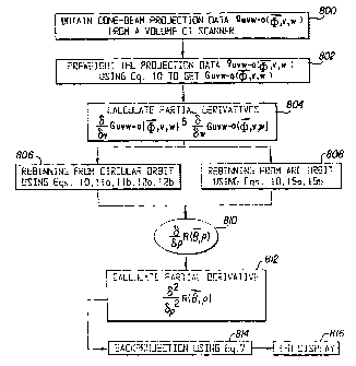
Referring now to Figure 8, which is a diagram of a flow chart showing the
steps
performed in converting the projection data from a CT scanning apparatus to
the desired 3-D
display, in the first step 800, the cone-beam projection data is obtained from
a volume CT
scanner. Then, the projection data g.-o (0, v, w) is preweighted using
Equation 10 in order
to obtain the preweighted projection data G.o ((D, v, w) in Step 802. Then, at
Step 804, the
partial derivatives C? G..o (T), v, w) and 13 GY.,.-o (0, v, w) are
calculated. In Step 806, the
results from the partial derivatives obtained in Step 804 are used to rebin
the data from the
circular orbit, using Equations 10, 11 a, l lb, 12a and 12b. At Step 808, the
partial derivatives
calculated in Step 804 are used to rebin data from the arc orbit, using
Equations 10, 15a and
15b.
In Step 810, the results from the rebinning from the circular and arc orbits
are utilized
to obtain the partial derivatives ~R( p). Next, the partial second derivative
~ R( b, p) is
calculated at Step 812. Then, at Step 814, the back projection data is
calculated,~ using
Equation 7. Finally, at Step 816, the 3-D image is displayed.
In a standard CT, a 3-D reconstruction is obtained by stacking a series of
slices. In a
volume CT, a direct reconstruction of an object can be obtained. Referring now
to Figure 9, it
is shown how the cone-beam tomography system 900 of the present invention can
be used to
obtain a direct 3-D reconstruction of an object. It should be understood that
the volume CT
scanning apparatus 900 is illustrated in a simplified block diagram form. The
invention may
preferably be employed in conjunction with such a volume CT scanning apparatus
to generate
a 3-D reconstruction matrix of the object. Based on the 3-D reconstruction
matrix, the
desired three dimensional display can be obtained.
A volume CT scanning apparatus examines a body P which rests on a motorized
table
906 using a cone shaped radiation beam 904 which traverses a set of paths
across the body.
As shown in Figure 9, an x-ray source 910 and a 2-D detector 911 are mounted
on a gantry
frame 902 that rotates around the body P being examined. The operating voltage
for the x-ray
source is obtained from a conventional high-voltage generator 908 in such a
manner that the
x-ray source 910 produces the desired cone-shaped beam of radiation when the
high-voltage
22
CA 02295973 1999-12-30
WO 99/01066 PCT/US98/12468
is applied to it. The high-voltage generator 908 is energized by means of a
power source 918,
through a switch 916.
A first motor 912 is also powered by the power source 918 such that it drives
the
gantry frame 902 in its orbit about the body, for example, in a clockwise
direction as shown
by the arrows adjacent to the frame. The power source 918 is turned on by
means of the
switch 920 or other conventional control devices, in order to initiate a
measurement sequence.
A speed control circuit 914 is used to control the speed of rotation of the
gantry frame 902
and to provide an output control signal which indicates when the speed of the
motor 912 is at
the desired level for taking measurements. The output from the rotational
contro1914 may
also be utilized to operate the switch 916 such that the high-voltage
generator 908 is only
turned on when the gantry frame 902 is driven at the desired speed for making
measurements.
In order to obtain the arc measurements as previously discussed, a tilt
contro1915 is
utilized to cause the gantry frame 902 to tilt by a relatively small angle of
t15 to 30 , by
means of the gantry frame tilt motor 913. That tilting allows the acquisition
of arc projection
data on the perpendicular arc. Such geometry results in a complete set of data
for an object
with a 25-40 cm diameter corresponding to a 3 7-60 cm field at the detectors
911 with a
magnification of 1.5. Although the tilting of the gantry 902 is generally
available in a
standard CT gantry, to acquire arc projections, the minimal modification of a
standard CT
gantry has to be made such that the tilting of the gantry, x-ray exposure
timing and the
projection acquisition are synchronized by the system control computer 924 as
shown in
Figure 9. The system control computer 924 also functions to control the
movement of the
motorized examination table 906 in relation to the gantry frame 902, for
utilizing a circle-
plus-line geometry as described later herein.
In addition to the method described above to acquire circle and arc
projections,
alternatively, the circle-plus-arc geometry can be implemented in one of the
following two ways.
In the first and preferred of the three methods, the gantry 902 is tilted to a
small angle (+15 to
+ 30 ) and then the x-ray tube 910 and the 2-D detector 911 are rotated while
the gantry 902 is
tilted. A half set of arc projections will be acquired only when the x-ray
tube 910 and the 2-D
detector 911 are at the rotation angle of 0 . When the tilted angle becomes
zero, the circle
projections will be acquired at the preset rotation angle positions. When the
circle projection
acquisition is completed, the gantry 902 will be tilted toward -15 to -30 .
Another half set of arc
23
CA 02295973 1999-12-30,
projections will be acquired only when the x-ray tube 910 and the 2-D detector
911 are at the
rotation angle of 0 .
. ,
The second alternative method is to mechanically modify a standard CT gantry
such that
two short arc orbits are added to the gantry, and the x-ray tube 910 and the 2-
D detector 911 can
be moved on the arc to acquire the arc projections and on the circle to
acquire the circle
projections. One arc constitutes the orbit of the x-ray tube 910 and the other
arc is the orbit of
the 2-D detector 911. The two arc orbits are mounted 180 apart from each
other, as shown in
Fig. 6. The x-ray tube 910 and the 2-D detector 911 are synchronously moved on
the arc orbits
to acquire arc projections. Then, the x-ray tube 910 and the 2-D detector 911
are rotated on the
gantry to acquire circle projections.
Mounted on the gantry frame 902 opposite the x-ray source 910 is a 2-D
detector 911
which has a dynamic range equal to or greater than 1000:1 and an image lag of
less than 10%,
for example a selenium thin film transistor (STFT) array or a silicon STFT
array, in order to
provide 2-D projections that correspond to an x-ray attenuation signal
pattern. The x-ray source
910 and the 2-D detector 911 are mounted on the gantry ~frame 902 in such a
manner that they
both move synchronously.
The cone-shaped beam of radiation 904 generated by the x-ray source 910 is
projected
through the body or object under test. The 2-D detector cone measures the
radiation transmitted
along the set of beam paths across the cone.
Alternatively, a continuous series of two-dimensional detectors (not shown)
can be
fixedly mounted proximate to the gantry frame 902 and the x-ray source 910 is
mounted to the
gantry frame such that, upon rotation of the gantry frame, the cone-shaped
radiation beam 904
is projected through the body P under test and sequentially received by each
of the series of
detectors.
A 2-D projection acquisition control and A/D conversion unit 926, under
control of
the scanning pulses sequentially obtained from the system control computer
924, which
includes the clock 922, receives a sequence of outputs corresponding to
different lines of the
2-D detector 911. Each line of the 2-D detector consists of many detection
cells (at least >
100). The output of each detector cell represents a line integral of
attenuation values
measurable along one of the respective beam paths. The cone-shaped beam 904
subtends a
cone angle sufficient to include the entire region of interest of the body.
Thus, a complete
24
AMENDED SIt~,
CA 02295973 1999-12-30
WO 99/01066 PCT/US98/12468
scan of the object can be made by merely orbiting the gantry frame 902
supporting the x-ray
source 910 and the 2-D detector 911 around the body to acquire the 2-D
projection signals at
different angular positions.
The analog-to-digital conversion unit 926 serves to digitize the projection
signals and
to save them in the 3-D image reconstruction array processor 928 and storage
device 930.
The method employed by the 3-D image reconstruction array processor 928 is the
invented
algorithm described in this application. The 3-D image reconstruction array
processor 928
serves to transform the digitized projection signals into x-ray attenuation
data vectors. The x-
ray attenuation data matrix corresponds to x-ray attenuation at spaced grid
locations within
the body trunk being examined. Each data element of the matrix represents an x-
ray
attenuation value and the location of the element corresponds to a respective
3-D grid
location within the body.
In accordance with the principles of the invention discussed previously, a
display
processor 932 obtains the data stored as 3-D x-ray attenuation signal patterns
in the memory
storage 930, processes that data as previously described, for example, in
connection with
Figure 8, and then the desired 3-D images are displayed on a 3-D display
device 934.
The 3-D image reconstruction array processor 932 may, for example, be
comprised of
an ULTRA SPARC-1 model workstation, available from Sun Microsystems, Inc. of
Mountain
View, California 94043.
The volume CT scanner system described above and shown in Figure 9 can also be
used to obtain clinically useful 3-D vascular images for enabling diagnostic
and therapeutic
decisions when used as an IV-VTDA system.
The IV-VTDA system of Figure 9, when operated to perform IV-VTDA 3-D imaging,
preferably uses a 2-D detector 911 such as a selenium or Silicon thin film
transistor (TFT)
plat panel detector, available from Sterling Diagnostic Imaging, Inc. of
Newark, Delaware
19714 or Varian Associates, Inc., of Palo Alto, California 94304 (for example,
model VIP-
540X/ARM TFT). Preferably, such TFT detector is capable of acquiring
projections at a rate
of 30 or more frames/second, with each frame containing 512 x 512 x 12 or
higher bits of
data. Thus, using such a detector, a single volume scan of a selected object
of interest can be
completed within about 5.0 - 8.0 seconds. Therefore, IV-VTDA is relatively
insensitive to
motion artifacts.
CA 02295973 1999-12-30
WO 99/01066 PCT/US98/12468
Alternatively, the 2-D detector 911 may be formed by an image intensifier (II)
coupled
to a charge coupled device (CCD), such as a CCD camera. However, a flat panel
detector as
described above provides better contrast and spatial resolution, as well as
better geometric
accuracy, than an image intensifier based detector. In addition, if an II
based detector, such as
an II-CCD detector is utilized as the detector 911, then pincushion and "S"
distortion
algorithms need to be utilized to correct for the curvature of the input
surface of the II
(pincushion distortion) and for the earth's magnetic field (S distortion).
Such algorithms are
known in the art. For example, see X. Wang and R. Ning, "Accurate and
Efficient Image
Intensifier Distortion Correction Algorithm for Volume Tomographic
Angiography," Proc.
SPIE 1997; 3032:427-440.
Three different data acquisition geometries can alternatively be utilized with
the IV-
VDTA system of the present invention, depending on the cone beam angle. If the
cone beam
angle is less than 5 degrees, then a single circle cone beam data acquisition
geometry using
Feldkamp's reconstruction algorithm for 3-D image reconstructions of vascular
structure can
be used. If the cone beam angle is greater than 5 degrees, then the IV-VTDA
system of the
present invention should use either a circle-plus-arc or circle-plus-line cone
beam data
acquisition geometry in order to obtain a complete set of projection data for
the exact 3-D
reconstruction of the imaged vascular structures.
In order to obtain an exact 3-D reconstruction when using a circle-plus-arc
cone beam
data acquisition geometry, the algorithm discussed above in connection with
volume CT
scanning should be utilized. The circle-plus-line cone beam data acquisition
geometry is
another method which can be used to solve the problem of the incompleteness of
projection
data from the single circle cone beam geometry. That geometry corresponds to
rotating the x-
ray tube 910 and the 2-D detector 911 on the gantry 902 and then acquiring the
line
projections by moving the table 906. An appropriate algorithm for the circle-
plus-line cone
beam data acquisition geometry was developed by G.S. Zheng and G.T. Gullberg
and is
described in "A cone-beam tomography algorithm for orthogonal circle-and-line
orbit," Phys.
Med. Biol. 1992; 37:563-577, and has been applied by H. Hu, "A new cone beam
reconstruction algorithm for the circle-and-line orbit," Proceeding of 1995
Int'1 Meeting on
Fully 3D Image Reconstruction in Radiology and Nuclear Medicine, pp. 303-310.
26
CA 02295973 1999-12-30
WO 99/01066 PCT/US98/12468
As will be obvious to those of ordinary skill in the art, the IV-VTDA system
of the
present invention can be used for performing whole body vascular imaging
procedures. The
IV-VTDA system of the present invention uses an intrinsically high object
contrast associated
with CT imaging. For example, even a 70:1 dilution of 350 mgI/ml contrast
material within
an artery provides more than 100 HUs of image contrast at 60 keV, the
effective energy at
which a standard CT scanner functions when the voltage of the x-ray tube is
100 kV.
Thus, the IV-VTDA system of the present invention is able to image a contrast
having
a concentration about three times more dilute than that usually achieved
during a
conventional IV-DSA study, while still obtaining acceptable image quality.
Thus, using the
IV-VTDA system of the present invention with a flat-panel detector, sufficient
low contrast
resolution is obtained to isolate the injected iodinated contrast signal in a
2 mm artery and
spatial resolution to detect a 25% stenosis of a 2 mm artery in the human body
is achieved.
Since TFT detectors are capable of acquiring projections at a rate of 30 or
more
frames/seconds, each of which contains 512 x 512 x 12 or higher bits of data,
a single volume
scan can be completed within 5.0 - 12.0 seconds. Thus, IV-VTDA is relatively
insensitive to
motion artifacts and is superior to IA angiography. By using the IV-VTDA
system of the
present invention in place of IA, the arterial puncture and IA catheter risks
and expense are
eliminated.
The imaging protocols used with the IV-VTDA system of the present invention
significantly improved the delineation, localization and visualization of non-
cardiac vascular
anatomy and disease. The IV-VTDA system of the present invention has five
important
advantages over current conventional digital angiography systems. First, the
IV-VTDA
system of the present invention provides true 3-D reconstructions that can be
viewed at any
angle or plane and that can be rotated around any axis. As is known to those
of ordinary skill
in the art, multiple views of the same vascular anatomy often convey more
information than
do only one or a few views. In addition, changing the viewing angle can make
the difference
between detecting and missing a significant lesion.
Second, the IV-VTDA system of the present invention allows for the direct
measurement of the area of lumina stenosis which allows a more objective
decision regarding
whether or not the patient should undergo an invasive procedure or should be
treated with
medication. Presently, radiologists and surgeons can only estimate the area of
stenosis based
27
CA 02295973 1999-12-30
WO 99/01066 PCT/US98/12468
on one or several 2-D projection images. The improved accuracy of disease
measurement
obtainable with the IV-VTDA system of the present invention will also allow
for a finer
analysis in outcomes research for the evaluation of medical or surgical
therapy. A 3-D data
set is also more amenable to automated 3-D computerized lesion quantification
techniques.
A third advantage of the IV-VTDA system of the present invention over
conventional
DSA systems is that the IV-VTDA system decreases the risk of embolization of
atherosclerotic plaque by eliminating the catherterization which is necessary
for IA
angiography.
A fourth improvement is that the IV-VTDA system of the present invention
provides a
more objective evaluation of the results of a particular interventional
procedure because
VTDA allows the identical angle view to be computed at different times such as
before and
after intervention. That greatly facilitates the evaluation of the progression
or correction of a
narrowing. Using conventional angiography, however, a radiologist may not see
a particular
vessel post-procedure from the same angle as it was seen pre-procedure.
The fifth advantage of the IV-VTDA system of the present invention over
conventional digital angiography is that the IV-VTDA system eliminates the
need for
angiographers to perform trial runs to identify the correct angle from which
to view the
lesion. Since only a single volume scanning and a single fast contrast
injection are necessary
for data acquisition using the IV-VTDA system of the present invention, the
total x-ray
exposure necessary as well as the procedure time is reduced compared to those
necessary in
conventional DSA. For example, for a typical VTDA scan, the total patient
entrance
exposure for a typical 288 exposures is 836 mR, which represents more than a
factor of 50
reduction in total patient entrance exposures used in a routine DSA procedure.
A typical
DSA procedure uses about 100 exposures at an average of 400 mR per exposure.
The IV-VTDA system of the present invention operates as follows to obtain a 3-
D
reconstruction image of a vascular structure of interest. First, the patient
P, who has already
been placed on the table 906, is moved into place within the gantry 902. Then,
a single
peripheral or central IV contrast injection is made into the venous structure
of interest. The
injector 940 will then inject iodinated contrast solutions, for example,
OMNIPAQUE 300,
available from Winthrop Pharmaceuticals, New York, New York 10016, preferably
into one
28
CA 02295973 1999-12-30
WO 99/01066 PCT/US98/12468
or both antecubital fossa (for peripheral injection) or superior vena cava
(for central
injection).
The volume CT scanning apparatus shown in Figure 9 is then placed in its
operational
state and the patient is instructed to hold his or her breath. Then, using a
cone beam x-ray
source 910 and a 2-D detector 911, such as the Selenium or Silicon TFT flat
panel detector
discussed above, fast volume scanning of the vascular anatomy of interest is
performed when
the injected contrast solution, flowing from the injection site mentioned
above, arrives at the
image site, i.e., the vascular anatomy of interest (after more than about a 4
second time delay,
counted from the beginning of the injection). Assuming that the circle-plus-
arc acquisition
geometry is utilized, a 3-D reconstruction matrix of the vascular structure
under study is
obtained as discussed above in connection with the description of the use of
the volume CT
scanner of Figure 9. The arc measurements are obtained, as previously
discussed, using the
tilt control 915. The projection data obtained from the CT scanning apparatus
is converted to
the desired 3-D display using the steps 800-816 shown in Figure 8. The
injector 940 is
available from E-Z-EM, Inc., Westbury, New York 11590.
In the event that a circle-plus-line data acquisition geometry is utilized,
then the table
906 is moved relative to the gantry 902 in a known manner in order to generate
the straight
line projection data. Then, utilizing the steps 800 through 816, in which
certain of the
equations would be modified in accordance with the circle-plus-straight line
data acquisition
geometry, the cone-beam projection data can be converted to the desired 3-D
display.
3-D images can be reconstructed from both subtraction projections and
nonsubtraction
projections. Subtraction projections will be formed in the following way: two
sets of
projection images, each set equally spaced over 360 degrees, will be acquired.
One set of
projections will be acquired without injected iodine contrast solution as a
mask projection and
another set of projections will be acquired with injected iodine contrast
solution. Then, the
subtraction will be performed on the angularly paired and logarithmically
transformed images
to form a set of subtraction projections. Preferably, the mask projections
will be acquired
first, and then the projections after injecting iodine contrast solution will
be acquired.
Subtraction and nonsubtraction projections for vascular reconstruction have
different
advantages. Subtraction increases the efficiency of the reconstruction by
reducing the number
of required projections and improves image quality by reducing the impact of
scatter and
29
CA 02295973 1999-12-30 = õ .,
beam hardening problems. Nonsubtraction allows for a simpler and possibly
faster data
acquisition protocol with the reconstruction protraying both arteries and
reference structures,
4 ,
making them useful as anatomical landmarks.
Using the IV-VTDA system of the present invention, efficient contrast and
spatial
resolution is provided in order to visualize all vascular vessels presently
amenable to
interventional techniques. In addition, the IV-VTDA system of the present
invention covers a
much larger segment of the human body in the direction orthogonal to the
slices within a
single scan (approximately 3-4 times larger) than a spiral CT can cover, and
without
sacrificing image quality.
.,R.
.. õ
CA 02295973 1999-12-30
APPENDIX A
With the relationship between the upq - 0 and the uvw - 0 coordinate systems
(See
Figure 4),
1 0 0
u u
v = 0 cosa sina p (16)
w 0 sina cosa q
and variable substituting,
~p G.~_o p, q) = p ~ G~,~_o ( v, w) + ~ ~ G.~._o v, w) (17)
=cosa ~GL~~-o(~,v,w) f sina ~G.~~_ov,w)
As is known in the prior art, great computational efficiency and accuracy can
be obtained by
swapping the integral with the derivative in Equation 9. With Equation 10 in
mind, by
putting Equation 17 into Equation 9 and swapping the integral with the
derivative,
a R( p) _ ~ SC f a f+~ ISO I
g~o (~,P, q) dq
ap JsOf ap ISAI
=ISCp a f + ~
G ,,o(4Z,p, 4) dq
I SO F ap
+~
S'C f a G-~.o (~,p, q) dq (18)
I SO ~' -
_ I S C F 5 1c0sa'o(,v,w) + sina a Gu-_o ((f, v, w) dq'
ISOR av aw
31
AMENDED SF#~T
CA 02295973 1999-12-30
WO 99/01066 PCTIUS98/12468
Since the partial derivatives a G..-o (4), v,w) and a G...o (4), v,w) on the
right-hand side
av aw
of Equation 18 need to be calculated only once, the computational complexity
is significantly
reduced.
32
CA 02295973 1999-12-30
WO 99/01066 PCT/US98/12468
APPENDIX B
To fill the Radon cube (0, (p, p) with the projection data from the circular
orbit, the
transformation function should be found between the local uvw - 0 coordinates
and the
absolute xyz - 0 coordinates (see Figure 5). The circular orbit lies in the xy
plane and the
focal point S will be restrained on this orbit. The angle between the x-axis
and the vector IfS
is defined as (j and the angle between the p-axis and the v-axis is a. If the
point C is
represented by (0, p, 0) in the local upq - 0 coordinate system, the Radon
plane which
contains the line D,DZ and the point S can be described in the uvw - 0
coordinate system as:
up + vDcosa + wDsina - Dp=O. (19)
Referring to Figure 5, the transformation between the local uvw - 0
coordinates and absolute
xyz - 0 coordinates can be expressed as:
u cos(3 sinp 0 x
v = -sin(3 cosP 0 y . (20)
w 0 0 I Z
Therefore, the Radon plane represented by Equation 19 can be rewritten in the
absolute xyz -
0 coordinates in terms of the parameters a, 0 and p as:
x( Dcosasin,(3 f pcos,6) fy(Dcosacos,Q f psin,8) +zDsina -Dp =0. (21)
Comparing Equation 21 with the other representation of this Radon plane in
terms of 0 and p:
xsin0coscp + ysin8sincp + zcosO - p = 0, (22)
it can be shown that:
sin0coscp - -Dcosasin(3 +pcosp (23a)
~
D2 + p2
33
CA 02295973 1999-12-30
WO 99/01066 PCT/US98/12468
The symbol "-" is used instead of "=" in Equations 23a-23d because there may
be a factor +1
or -1 involved. Any Radon plane (Equation 21) that intersects the circular
orbit (x2 +.)~ = D2)
has two intersection points except when the Radon plane is tangential to the
circular orbit.
sin6sinT - Dcosacosp + psin(3
D2 +p2 ' (23b)
cos6 - Dsina
(23c)
D2 +p2
Dp
p D2+p2 (23d)
Either point represents its corresponding focal point position. In order to
improve the quality
of the reconstructed images, both projections from the two focal points are
used. First, the
two intersection points are named B, and B2 respectively, and the position
arrangement for B,
- B2 -= 0 is counter-clockwise. Second, the angle between 0,91 and the x-axis
is P, and that
between 0A2 and the x-axis is (3Z. Then, for a given point (0, (p, p) in the
Radon space, (3,
and P2 can be calculated directly from the coordinates of point B, and B2,
respectively. The
solutions to Equations 23a-23d can also be expressed as:
for (3, :
p = D1 PI
D 2 - ~ (24a)
34
CA 02295973 1999-12-30 ' ' ' '=
_ , , = , ~ , ,
sin Dcos 0~ f'orp Z 0,
Dz _ p2
sin Dcos B> f.orp < 0. (24b)
D2 -p2
and for (3z:
Dipl
p (24c)
D2_p2
4
-sin-i DcosB = for p z 0,
( DZ - p,)
sin-t ( DcosO ) f.or p< 0 (24d)
Dz_pz
Therefore, if 0, , p and jQ are discrete parameters, for a given (0, , p) in
the Radon
space, only a 1-D interpolation relative to P need be calculated, which
greatly reduces the
interpolation errors.
{J~ ~ ... }'.,~
CA 02295973 1999-12-30
WO 99/01066 PCT/US98/12468
APPENDIX C
Referriag to Figuce 7, the arc orbit comes with the rotation of the focal
point S about
they-axis by an angle (3 and ON is defimed as the u-axi,s. The transformation
between the
local uvw - 0 coordinate system and the absolute xyz - 0 coordinate system can
be expressed
as:
u coso 0 -sznR z
v= 0 1 0 y (25)
'y sino 0 cosp z
Again, the R.a.don plane represented by Equation 19 can be rewritten in the
absolute xyz - 0
coordinates as:
x (Dsinaasin)6 + pcos,8) +yDcosa +y(DsinxcosO -psin,6) -Dp = 0. (26)
Comparison with Equation 22, wbich is the representation of the Radon plane in
terms of
parameters and p, yields the following resuJ.ts:
sin (~os q~ _ Dsin asinp * pcos,8 (27a)
bZ ;p 2
sin6 sin(p = Dcosa
VD p ' (2'7b)
cos0 - DsinarosQ -psr*np
(27c)
(27c)
Dz +p2
D
O - -- (27d)
D' fpz
36
RECTIFIED SHEET (RULE 91)
ISA/EP
. .,
CA 02295973 1999-12-30
, , . ~ . . .
Therefore, the solution to the above equations for a given (6, (p, p) are:
DP
p D2 -~' (28a)
D sin6 cosV - p cos'6
a = sin-' D2 -pl Dz -PZ (28b)
sin,(3
Once again, only a 1-D interpolation relative to needs to be calculated for
the discrete
values of parameters 8, cp, p and ~3.
,~.
37
AMENDED SHEE~
~....