Note : Les descriptions sont présentées dans la langue officielle dans laquelle elles ont été soumises.
CA 02306476 2000-04-13
WO 99/18996 PCT/US98/21561
ENTEROCOCCUS ANTIGENS AND VACCINES
BACKGROUND OF THE INVENTION
The present invention relates to antigens from
Enterococcus that are useful as vaccines, and to methods --
for obtaining and using such antigens.
S The prevalence of Enterococcus infection is increasing
steadily. Strains of Enterococcus now are responsible for
12~s of all the nosocomial infections among hospitalized
patients and they are the second most common organism
isolated from patients with nosocomial infections. This
increased prevalence of Eaterococcus is due at least in
part to the appearance of strains of enterococci that are
resistant to antimicrobial agents and therefore difficult
to treat with currently available antibiotics. The
increase in antibiotic resistance among Eaterococcus has
increased the importance of alternative prophylactic and
therapeutic approaches against enterocpccal infections.
Various groups have disclosed polysaccharides isolated
from Enterococcus. For example, lipoteichoic acids which
contain a 1,3-linked polyglycerophosphate backbone have
been isolated from "S. faecalis," which according to
current classification is E. faecalis. Position 2 is
glycosylated with disaccharides or trisaccharides of
glycosyl residues which may be esterified with alanyl
residues, and is denoted intracellular teichoic acid
because of its predominance between the cell wall and the
protoplast membrane. Wicken et al., J. Gen. Microhiol. 33:
231-39 (1963) .
Pazur et al., J. Biol. Chem. 246: 1793-98 (1971), have
isolated two other polysaccharides from the cell wall of E.
faecalis strain N. One of these polysaccharides is
characterized as a diheteroglycan consisting of glucose and
D-galactose, while the other polysaccharide is said to be
a tetraheteroglycan of 2-acetamide-2-deoxy-galactose,
galactose, rhamnose, and glucose in molar ratio of 1:1:2:4.
CA 02306476 2000-04-13
WO 99/18996 PCT/IJS98I21561
_ - 2 - _
Bleiweis et al., J. Bacteriol. 94: 1381-87 (1967),
have isolated a third polysaccharide from strain D76 of
group D Streptococci. The sugar composition of this
material includes glucose, glucosamine, galactosamine,
rhamnose, ribitol, and phosphorus; structural information
is not provided, however. It is postulated that this
material may be ribitol phosphate teichoic acid with
attached sugar substituents. It also has been hypothesized
that glucose and N-acetyl glucosamine are the possible
components of the antigenic site.
Enterocvccus antigens) capable of eliciting
protective antibodies would provide an effective means of
preventing and/or treating Enterococcus infection. While
the art discloses a variety of Enterocvccus antigens, not
every antigen is effective as a vaccine. Indeed, none of
the material reported in the literature has been shown to
be effective in protecting against infection by
Eaterococcus. In this regard, even a disclosure that an
antigen is immunogenic, i.e., that it causes the production
of antibodies, provides an insufficient basis for a
conclusion that the antibodies are protective and that the
antigen therefore is useful in a vaccine.
Finally, the art suggests that Enterococcus
serologically is a very diverse genus. This serologic
diversity suggested that a vaccine comprised of a practical
number of active components was not feasible. Maekawa et
al., Microbiol. Immunol. 36: 671-681 (1992).
SUI~iARY OF THE INVENTION
It is therefore an object of the present invention to
provide Enterocvccus antigens, particularly antigens from
E. faecalis and E. faecium, that are capable of eliciting
the production of protective antibodies.
It is a further object to provide a vaccine that
contains Enterococcus antigens, more particularly a vaccine
CA 02306476 2000-04-13
WO 99/18996 PC'fIUS98/21561
_ - 3 - _
that contains antigens from both E. faecalis and E.
faeci um.
It is another object to provide a hyperimmune globulin
composition that contains antibodies directed against
Enterococcus antigens, particularly antigens from E.
faecalis and E. faecium.
In accordance with these and other objects according
to the invention, there is provided an isolated
Enterococcus antigen that reacts with antibodies to cells
from one of ATCC 202013, ATCC 202014, ATCC 202015, ATCC
202016, and ATCC 202017. More particularly, an isolated
Frsterococcus antigen is selected from the group consisting
of an E. faecalis antigen comprising 2-acetamido-2-deoxy-
glucose and rhamnose in an approximate 1:2 molar ratio, an
E. faecalis antigen comprising a trisaccharide repeat which
comprises a 6-deoxy sugar, and an E. faecium antigen
comprising 2-acetamido-2-deoxy-galactose and galactose.
The antigen can be used in diagnostic assays or in
immunotherapy methods. A conjugate in which the antigen is
covalently bonded to an immunocarrier, preferably a
recombinantly-produced, non-toxic mutant strain of
Pseudomonas aeruginosa exotoxin A or diphtheria toxoid, is
provided. The antigen-carrier conjugates are useful in a
vaccine, particularly a multivalent vaccine, for active
immunotherapy. The antigen or vaccine also can be used to
produce immune globulin for passive immunotherapy, or in
the production of monoclonal antibodies for diagnostic or
therapeutic use.
Other objects, features and advantages of the present
invention will become apparent from the following detailed
description. It should be understood, however, that the
detailed description and the specific examples, while
indicating preferred embodiments of the invention, are
given by way of illustration only, since various changes
and modifications within the spirit and scope of the
invention will become apparent to those skilled in the art
from this detailed description.
CA 02306476 2000-04-13
WO 99/18996 PCT/US98/21561
_ _ 4 _ _
BRIEF DESCRIPTION OF THE DRAWINGS
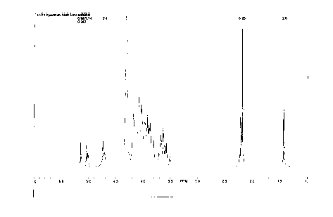
Figure 1, 2 and 3 are NMR spectra for Enterococcus
antigens according to the invention.
DESCRIPTION OF PREFERRED EI~ODIMENTS
It surprisingly has been discovered that the majority
of E. faecalis clinical isolates fall into two groups, and
that the majority of E. faecium human clinical isolates
fall into three groups. The discovery that the majority of
clinical isolates are characterized by only a few common
antigens is unheralded in the art, and permits development
of multivalent vaccines that comprise a minimal number of
active components yet are protective against the majority
of clinical isolates.
Antigens characteristic of each of the two groups of
I5 E. faecalis and three groups of E. faecium can be
extracted, purified and identified. In this regard, an
antigen is characteristic of a group or strain of bacteria
if it is expressed by the bacteria in a quantity sufficient
to cause a significant immune response when a whole cell
vaccine of the group or strain is injected into an animal,
i.e., an animal produces protective antibodies when so
injected.
The E. faecalis characteristic antigens are denoted
herein as EFS1 and EFS2, and the E. faecium characteristic
antigens as EFM3, EFM4 and EFM5. These antigens are
referred to collectively herein as "Enterococcus antigens."
A strain of bacteria is called an EFS1 strain if a whole
cell vaccine of the strain produces a significant immune
response primarily toward EFS1 when injected into a
subject, and only a minor response to EFS2. Similarly, a
strain of bacteria is called an EFS2 strain if a whole cell
vaccine of the strain produces a significant immune
response primarily toward EFS2 when injected into a
subject, and so forth.
CA 02306476 2000-04-13
WO 99/18996 PCT/US98/21561
.. _ 5 _ _
While each of the major clinical groups of E, faecalis
and E. faecium expresses a different characteristic antigen
that may be readily extracted and purified in recoverable
amount, the groups also may express antigen characteristic
of the other groups) in minor amounts. However, when
immunized with whole cells from one of the groups, rabbits
mount a significant immune response only toward the
characteristic antigen of that group, and not at all or
only poorly to the minor amounts of the antigen most
characteristic of the other group(s), as shown by the
absence of a precipitin band between antibodies from the
immunized rabbit and purified antigen characteristic of the
other group.
The degree to which a non-characteristic antigen is
I5 expressed by cells varies. For example, antisera generated
against a whole cell vaccine of an EFS1 strain contains
antibodies to EFS2 in amounts, detectable both by slide
agglutination and by opsonophagocytosis assay (infra).
Antisera generated against a whole cell vaccine of an EFS2
strain, on the other hand, does not contain antibodies that
precipitate with EFS1.
The Enterococcus antigens are readily obtained from
strains of E. faecalis and E. faecium, pursuant to
protocols provided herein, and are capable of eliciting
production of protective antibodies when conjugated to
immunocarriers. They therefore can be used to prepare
vaccines that provide protection of humans and other
mammals, e.g., horses, cattle, swine, dogs, and cats,
against infection by clinically significant isolates of
Enterococci. in this regard, a ~~clinically significant~~
isolate is one that is pathogenic in humans or other
mammals.
E. faecalis and E. faecium clinical isolates can be
grouped by slide agglutination experiments, using an
appropriate antibody preparation for agglutination of
bacteria. Slide agglutination experiments with E. faecalis
show that the majority of clinical isolates fall into two
groups, EFS1 and EFS2. Antisera generated against an EFS1
CA 02306476 2000-04-13
WO 99118996 PCT/US98/21561
_ _ 6 _ _
strain of E. faecalis agglutinates both EFS1 and EFS2
strains of E. faeca3is. The reactivity of antisera
generated against an EFS1 strain of E. faecalis can be
absorbed out with cells from the EFS1 strain. The absorbed
sera may then continue to agglutinate only an EFS2 strain.
Antisera generated against an EFS2 strain of E.
faeca3is agglutinates only EFS2 strains, and this
reactivity cannot be absorbed out with EFS1 bacteria. As
expected, absorption with cells from an EFS2 strain removes
ZO the reactivity of this antisera with cells from an EFS2
strain. While not wishing to be bound by theory, it is
hypothesized that EFS1 and EFS2 strains of E. faeca3is
contain EFS2 antigen, but that this antigen is covered or
otherwise masked by EFS1 antigens on EFS1 cells.
Slide agglutination experiments with E. faecium show
that the majority of clinical isolates fall into three
groups. Antisera raised against two of the groups give
results similar to that obtained with E. faecalis. That
is, antisera generated against a EFM3 strain of E. faecium
agglutinates both EFM3 and EFMS bacteria, and the
reactivity of this antisera with an EFM3 strain can be
absorbed out with cells from an EFM3 strain. The absorbed
sera then agglutinates only EFMS strains of bacteria. This
absorption also causes a reduction in reactivity with cells
from EFMS strains, indicating that small amounts of EFM5
antigen is exposed on the surface of EFM3 cells.
Antisera generated against a EFM5 strain of E. faecium
agglutinates only isolates in that group, and this
reactivity cannot readily be absorbed out with cells of an
EFM3 strain. As expected, absorption with cells from an
EFM5 strain reduces the reactivity of this antisera with
cells. Similarly EFM3 and EFM5 strains of E. faecium both
contain EFM5 antigen. Again, this antigen is hypothesized
to be covered or otherwise masked by EFM3 antigen on EFM3
cells.
Antisera raised against an EFM4 strain of E. faecium
is specific only to cells of EFM4 strains in slide
CA 02306476 2000-04-13
WO 99/18996 PCT/US98/21561
_ _ 7 _ _
agglutination experiments. This antisera demonstrates no
cross reactivity with EFM3 and EFM5 bacteria.
Antibodies generated against the whole cell vaccine
generally are not directed toward proteins on the cell
surface, as shown by treatment of formalin-killed cells
with pronase E. When killed cells are incubated for 3
hours at 37~C with 500 ~,g/ml pronase E, and then tested in
slide agglutination against whole cell sera, there is no
difference in the agglutination pattern from that observed
with untreated E. faecium or E. faecalis, i.e., the pronase
treatment does not remove the surface antigen against which
the antibodies are directed.
Representatives of each of the two E. faecalis and
three E. faecium strains have been deposited under the
Budapest Treaty with the American Type Culture Collection,
and have been given Accession Nos. 202013 (E. faecalis
EFS1), 202014 (E. faecalis EFS2), 202015 (E. faecium EFM3),
202016 (E. faecium EFM4), and 202017 (E. faecium EFMS)
respectively. Antigen according to the invention can be
isolated from the deposited strains, or the deposited
strains can be used to identify other strains which express
antigen according to the invention, from which antigen may
be extracted and purified in accordance with protocols
described herein.
Enterococcus antigens according to the invention can
be obtained in recoverable amount, and in substantially
pure form, from their respective E. faeca.Iis and E. faecium
isolates cultured pursuant to the protocols described
herein. A ~~recoverable~~ amount in this regard means that
- the isolated amount of the antigen is detectable by a
methodology less sensitive than radiolabeling, such as
immunoassay, and can be subjected to further manipulations
involving transfer of the antigen per se into solution.
In an illustrative approach to obtaining antigen
according to the present invention, a strain of E. faecalis
or E. faecium first is grown on a blood agar plate and then
transferred to a 2~ NaCl/Columbia starter flask. An
80-liter fermentor that contains the same medium with added
CA 02306476 2000-04-13
WO 99/18996 PCT/US98/21561
_ _ g _
4% glucose is inoculated with the starter flask. Cells are
fermented for 16-24 hours. The cells were centrifuged to
separate the cells from the supernatant. Each of the five
antigens can be extracted from either cell paste or
supernatant .
When cell paste is used, antigen is extracted by -'
stirring the paste with cold 10% trichloroacetic acid
(TCA), and then precipitated from the TCA solution by one
or more sequential precipitations with cold ethanol/CaCl2.
When supernatant is used, the supernatant is subjected
directly to precipitation with cold ethanol/CaCl2. This
produces a crude antigen extract.
The crude extract is redissolved in water, dialyzed
and lyophilized. The lyophilized material is dissolved in
buffer and purified by ion exchange chromatography.
Fractions containing antigen can be pooled, dialyzed,
concentrated, and lyophilized, and size exclusion
chromatography is used to purify the antigen further by
size on a suitable column. Antigen-containing fractions
are pooled, concentrated, dialyzed and lyophilized.
Purified antigen is analyzed by 'H-NMR spectroscopy.
A composition of the Enterococcus antigen according to
the present invention "consists essentially of" the
antigens) or a conjugate of the antigen(s), which means
that the composition does not contain any material that
interferes with elicitation of an immune response to the
antigens) when the composition is used in a therapeutic
context, or with the antigen-antibody coupling
characteristic of a diagnostic assay. In a preferred
embodiment, the composition contains both E. faecalis and
E. faec.ium antigens.
The antigens according to the invention are useful in
the production of diagnostic assays for detecting the
presence of Enterococcus antigen and/or anti-Enterococcus
antibody in a sample. Either the Enterococcus antigen or
antibody specific to the Enterococcus antigen is mixed with
a sample suspected of containing Enterococcus antibody or
antigen and monitored for antigen-antibody binding. The
CA 02306476 2000-04-13
W0 99/18996 PCT/US98/21561
_ _ _ g _ _
antigen or antibody is labelled with a radioactive or
enzyme label. In a preferred embodiment, the antigen or
antibody is immobilized on a solid matrix such that the
antigen or antibody is accessible to complementary antibody
or antigen contacting a surface of the matrix. The sample
then is brought into contact with the surface of the
matrix, and the surface is monitored for antigen-antibody
binding.
For example, the antigen or antibody can be used in an
enzyme-linked immunosorbent assay (ELISA) , in which antigen
or antibody is bound to a solid phase and an enzyme
antibody or enzyme-antigen conjugate is used to detect
and/or quantify antibody or antigen present in a sample.
Alternatively, a western blot assay can be used in which
solubilized and separated antigens are bound to
nitrocellulose paper. The antibody then is detected by an
enzyme or label-conjugated anti-immunoglobulin (Ig), such
as horseradish peroxidase-Ig conjugate by incubating the
filter paper in the presence of a precipitable or
detectable substrate. Western blot assays have the
advantage of not requiring purity greater than 50% for the
desired antigen. Descriptions of ELISA and western blot
techniques are found in Chapters 10 and 11 of Ausubel, et
al. (eds.), CURRENT PROTOCOLS IN MOLECULAR BIOLOGY, John
Wiley and Sons (1988), the contents of which are hereby
incorporated by reference.
In a vaccine context, it is preferable to conjugate
the antigens) to an immunocarrier, usually a polypeptide
or protein, to improve the interaction between T and B
cells for the induction of an immune response against the
antigen. This is particularly important for vaccines
intended for use in patients with reduced resistance. An
immunocarrier enhances immunogenicity both for active
immunization and for preparing high-titered antisera in
volunteers for passive immunization. Suitable
immunocarriers according to the present invention include
tetanus toxoid, diphtheria toxoid, Pseudorrronas aeruginosa
Exotoxin A or its derivatives, recombinantly-produced non-
CA 02306476 2000-04-13
WO 99/18996 PCT/US98/21561
- - 10 - -
toxic mutant strains of exotoxin A, as described, for
example, in Fattom et al., Inf. and Imrn. 61: 1023-32
(1993), as well as other proteins commonly used as
immunocarriers.
In order to conjugate the antigen to a carrier, the
antigen is first derivatized. Various methods can be used -'
to derivatize antigen and covalently link it to an
immunocarrier. In a preferred method, hydroxyl groups on
the antigen are activated using 1-cyano-4-dimethylamino
pyridinium tetrafluoroborate, and the antigen is then
derivatized with a six carbon bifunctional spacer adipic
acid dihydrazide (ADH), according to techniques known in
the art, according to the method of Kohn et al. FEES Lett.
154: 209:210 (1993). This material is then linked to
diphtheria toxoid (DT), recombinant exoprotein A from
Pseudomonas aeruginosa (rEPA), tetanus toxoid (TT) or
another suitable carrier protein by 1-ethyl-3-(3
dimethylaminopropyl) carbodiimide (EDAC). The resulting
conjugates can be separated from unreacted antigen by size
exclusion chromatography.
Preferably the antigen conjugate is administered with
an adjuvant which promotes the protective IgG subtype 2
antibodies. Typical adjuvants include complete Freund~s
adjuvant (CFA), incomplete Freund~s adjuvant (IFA), alum
and other adjuvants suitable for human and animal use.
Dextran sulfate has been shown to be a potent stimulator of
IgG2 antibody against staphylococcal cell surface antigens,
and also is suitable as an adjuvant.
Induction of bacteremia in some mammals, e.g.,
laboratory animals, requires extremely high numbers of
organisms or some previous maneuver to lower the host
resistance. In vitro phagocytosis, however, can be studied
as a correlate of protective immunity in vivo for humans
and other mammals. In this model, the ability of antigen
specific monoclonal and polyclonal antibodies to opsonize
Enterococcus strains in vitro is measured by phagocytosis,
according to the method described in Kojima et al., Infect.
Dis. Immun. 58: 2367-74 (1990). In vitro
CA 02306476 2000-04-13
WO 99/18996 PCT/US98/21561
. - - 11 -
opsonophagocytosis assays are recognized in the field as
being predictive of efficacy as a vaccine. For example,
Fischer et al. discloses a correlation between functional
antibody determined with an in vitro opsonic assay and in
vivo activity. J. Inf. Dis. 169: 324-9 (1994).
Antibodies induced by the Enterococcus antigens
according to the invention are opsonic and facilitate type-
specific phagocytosis. Rabbit antibodies raised against
the Enterococcus antigens are able specifically to mediate
the opsonophagocytosis of the strains carrying the antigens
by human polymorphonuclear leukocytes (PMN) cells in the
presence of human complement. The in vitro phagocytosis
assays thus indicate that antibodies to the Enterococcus
antigens are protective against infection by E. faecalis
and E. faecium. A vaccine based on a combination of E.
faecalis and E. faecium antigens can be used to protect
against infection from the majority of clinical
Enterococcus strains.
In vivo results are consistent with results of in
vitro opsonophagocytosis assays. Antibodies to EFS1
conjugate lowered bacteremia in mice challenged with E.
faecalis.
The present invention also relates to the use of the
Enterococcus antigens) to produce polyclonal~antibodies or
monoclonal antibodies (mouse or human) that bind to or
neutralize Enterococcus. Illustrative protocols for
producing these antibodies are described in Chapter 11 of
MOLECULAR CLONING: A LABORATORY MANUAL, Cold Spring Harbor
Laboratory (Cold Spring Harbor, NY); in METHODS OF
HYBRIDOMA FORMATION 257-271, Humana Press (Clifton, NJ); in
Vitetta et al., Immunol. Rev. 62: 159-83 (1982); and in
Raso, Imntunol. Rev. 62: 93-117 (1982).
Inoculum for polyclonal antibody production typically
is prepared by dispersing the antigen-immunocarrier in a
physiologically-tolerable diluent such as saline, to form
an aqueous composition. An immunostimulatory amount of
inoculum, with or without adjuvant, is administered to a
mammal, and the inoculated mammal then is maintained for a
CA 02306476 2000-04-13
WO 99/18996 PCT/US98r11561
- - 12 - -
time period sufficient for the antigen to induce protecting
anti-Enterococcus antigen antibodies. Boosting doses of
the antigen-immunocarrier may be used in individuals that
are not already primed to respond to the antigen.
Antibodies can include antibody preparations from a
variety of commonly used animals, such goats, primates,
donkeys, swine, rabbits, horses, hens, guinea pigs, rats,
and mice, and even human antibodies after appropriate
selection, fractionation and purification. Animal antisera
may also be raised by inoculating the animals with
formalin-killed strains of E. faecalis and/or E. faecium by
conventional methods, bleeding the animals and recovering
serum or plasma for further processing.
The antibodies induced in this fashion can be
harvested and isolated to the extent desired by well known
techniques, such as by alcohol fractionation and column
chromatography, or by immunoaffinity chromatography; that
is, by binding antigen to a chromatographic column packing
like Sephadex~'", passing the antiserum through the column,
thereby retaining specific antibodies and separating out
other immunoglobulins (IgGs) and contaminants, and then
recovering purified antibodies by elution with a chaotropic
agent, optionally followed by further purification, for
example, by passage through a column of bound blood group
antigens or other non-pathogen species. This procedure may
be preferred when isolating the desired antibodies from the
sera or plasma of humans that have developed an antibody
titer against the pathogen in question, thus assuring the
retention of antibodies that are capable of binding to the
antigen. They can then be used in preparations for passive
immunization against strains of E. faeca~is and E. faecium.
A monoclonal antibody composition contains, within
detectable limits, only one species of antibody combining
site capable of effectively binding to the Enterococcus
antigen. Suitable antibodies in monoclonal form can be
prepared using conventional hybridoma technology.
To form hybridomas from which a monoclonal antibody
composition of the present invention is produced, a myeloma
CA 02306476 2000-04-13
WO 99118996 PCTIUS98/21561
_ - - 13 - -
or other self-perpetuating cell line is fused with
lymphocytes obtained from peripheral blood, lymph nodes or
the spleen of a mammal hyperimmunized with the Enterococcus
antigen. It is preferred that the myeloma cell line be
from the same species as the lymphocytes. Spienocytes are
typically fused with myeloma cells using polyethylene
glycol 1500. Fused hybrids are selected by their
sensitivity to HAT. Hybridomas secreting the antibody
molecules of this invention can be identified using an
ELISA.
A Balb/C mouse spleen, human peripheral blood, lymph
nodes or splenocytes are the preferred materials for use in
preparing murine or human hybridomas. Suitable mouse
myelomas for use in the present invention include the
hypoxanthine-aminopterin-thymidine-sensitive (HAT) cell
lines, a preferred myeloma being P3X63-Ag8.653. The
preferred fusion partner for human monoclonal antibody
production is SHM-D33, a heteromyeloma available from ATCC,
Rockville, Md. under the designation CRL 1668.
A monoclonal antibody composition of the present
invention can be produced by initiating a monoclonal
hybridoma culture comprising a nutrient medium containing
a hybridoma that secretes antibody molecules of the
appropriate specificity. The culture is maintained under
conditions and for a time period sufficient for the
hybridoma to secrete the antibody molecules into the
medium. The antibody-containing medium is then collected.
The antibody molecules then can be isolated further by well
known techniques.
Media useful for the preparation of these compositions
are both well known in the art and commercially available,
and include synthetic culture media, inbred mice and the
like. An exemplary synthetic medium is Dulbecco's Minimal
essential medium supplemented with 20~ fetal calf serum.
An exemplary inbred mouse strain is the Balb/c.
Other methods of preparing monoclonal antibody
compositions are also contemplated, such as interspecies
fusions, since it is primarily the antigen specificity of
CA 02306476 2000-04-13
WO 99/18996 ~ PCT/US98/21561
. - - 14 -
the antibodies that affects their utility in the present
invention. Human lymphocytes obtained from infected
individuals can be fused with a human myeloma cell line to
produce hybridomas which can be screened for the production
of antibodies that recognize the Enterococcus antigen.
More preferable in this regard, however, is a process that
does not entail the use of a biological sample from an
infected human subject. For example, a subject immunized
with a vaccine as described herein can serve as a source
for antibodies suitably used in an antibody composition
within the present invention. Purified monoclonal
antibodies can be characterized by bacterial agglutination
assays using a collection of clinical isolates, or by ELISA
using plates coated with purified antigen.
The monoclonal and polyclonal antibody compositions
produced according to the present description can be used
by passive immunization to induce an immune response for
the prevention or treatment of infection by strains of E.
faecalis and E. faecium. In this regard, the antibody
preparation can be a polyclonal composition. Such a
polyclonal composition includes antibodies that bind to the
Erlterococcus antigen (s) . The polyclonal antibody component
can be a polyclonal antiserum, preferably affinity
purified, from an animal which has been challenged with the
Enterococcus antigen(s). Alternatively, an "engineered
oligoclonal" mixture may be used, which is a mixture of
monoclonal antibodies to the Enterococcus antigens from
both E. faecalis and E. faecium.
In both types of mixtures, it can be advantageous to
link antibodies together chemically to form a single
polyspecific molecule capable of binding to both E.
faecalis and E. faecium antigens. One way of effecting
such a linkage is to make bivalent F(ab')Z hybrid fragments
by mixing two different F (ab' ) 2 fragments produced, e. g. , by
pepsin digestion of two different antibodies, reductive
cleavage to form a mixture of Fab' fragments, followed by
oxidative reformation of the disulfide linkages to produce
a mixture of F(ab')2 fragments including hybrid fragments
CA 02306476 2000-04-13
WO 99/18996 ~ PCT/US98/Z1561
. - - 15 -
containing a Fab' portion specific to each of the original
antigens. Methods of preparing such hybrid antibody
fragments are disclosed in Feteanu, LABELED ANTIBODIES IN
BIOLOGY AND MEDICINE 321-23, McGraw-Hill Int'1 Book Co.
(1978); Nisonoff et al., Arch Biochem. Biophys. 93: 470
(1961); and Hammerling et al., J. Exp. Med. 128: 1461
(1968); and in U.S. patent No. 4,331,647.
Other methods are known in the art to make bivalent
fragments that are entirely heterospecific, for example,
use of bifunctional linkers to join cleaved fragments.
Recombinant molecules are known that incorporate the light
and heavy chains of an antibody. See, for instance, the
products of a methodology described by Boss et a.I., U.S.
patent No. 4,816,397. Analogous methods of producing
recombinant or synthetic binding molecules having the
characteristics of antibodies are included in the present
invention. More than two different monospecific antibodies
or antibody fragments can be linked using various linkers
known in the art.
An antibody component produced in accordance with the
present invention can include whole antibodies, antibody
fragments, or subfragments. Antibodies can be whole
immunoglobulin of any class, e.g., IgG, IgM, IgA, IgD, IgE,
chimeric antibodies or hybrid antibodies with dual or
multiple antigen or epitope specificities, or fragments,
e.g., F(ab')2, Fab', Fab and the like, including hybrid
fragments, and additionally includes any immunoglobulin or
any natural, synthetic or genetically engineered protein
that acts like an antibody by binding to a specific antigen
to form a complex. In particular, Fab molecules can be
expressed and assembled in a genetically transformed host
like E. coli. A lambda vector system is available thus to
express a population of Fab's with a potential diversity
equal to or exceeding that of subject generating the
predecessor antibody. See Huse, W.D., et al., Science 246:
1275-81 (1989) .
Antigen conjugates) according to the present
invention can be the active ingredient in a composition,
CA 02306476 2000-04-13
WO 99/18996 PCT/US98/21561
. - - 16 -
further comprising a pharmaceutically acceptable carrier
for the active ingredient, which can be used as a vaccine
to induce a cellular immune response and/or production in
vivo of antibodies which combat Enterococcus infection. In
this regard, a pharmaceutically acceptable carrier is a
material that can be used as a vehicle for administering a
medicament because the material is inert or otherwise
medically acceptable, as well as compatible with the active
agent, in the context of vaccine administration. In
addition to a suitable excipient, a pharmaceutically
acceptable carrier can contain conventional vaccine
additives like diluents, adjuvants, antioxidants,
preservatives and solubilizing agents.
Pursuant to the present invention, such a vaccine can
be administered to a subject not already infected with E.
faecalis or E. faecium, thereby to induce a Enterococcus
protective immune response (humoral or cellular) in that
subject. Alternatively, a vaccine within the present
invention can be administered to a subject in which E.
faecalis and/or E. faeciurr~ infection already has occurred
but is at a sufficiently early stage that the immune
response produced to the vaccine effectively inhibits
further spread of infection.
By another approach, a vaccine of the present
invention can be administered to a subject who then acts as
a source for immune globulin, produced in response to
challenge from the specific vaccine that contains
antibodies directed against Enterococcus. A subject thus
treated would donate plasma from which immune globulin
would then be obtained, via conventional plasma-
fractionation methodology, and administered to another
subject in order to impart resistance against or to treat
Enterococcus infection. Immune globulins according to the
invention are particularly useful for immune-compromised
individuals, for individuals undergoing invasive procedures
or where time does not permit the individual to produce his
own antibodies in response to vaccination.
CA 02306476 2000-04-13
WO 99/18996 PCT/US98/21561
. ' - 17 - _
Similarly, monoclonal or polyclonal anti-Eaterococcus
antibodies produced according to the present invention can
be conjugated to an immunotoxin, and administered to a
subject in whom Enterococcus infection has already occurred
but has not become widely spread. To this end, antibody
material produced pursuant to the present description would
be administered in a pharmaceutically acceptable carrier,
as defined herein.
The present invention is further described by
reference to the following, illustrative examples.
Example 1: Fermentation of E. faeca.Iis and E. faecium
E. faeca3is and E. faecium were cultivated in Columbia
broth supplemented with 2% NaCl and 4% glucose in an
80-liter fermentor containing 60 liters of broth medium at
37°C. The fermentation was started with one liter of a 16
hour old seed culture . The cells were grown with agitation
at 200 rpm for 16-24 hours.
Cells to be used as a vaccine to prepare whole cell
antiserum were formalin fixed overnight at room
temperature. Cells for purification were harvested by
centrifugation at 14,500 x g and stored at -70°C until use.
Approximately 500 g, 180 g, and 350 g of cell paste (net
weight) were obtained from an 80-liter fermentor for EFS1,
EFS2 and EFM3, respectively.
Examx~le 2: Preparation of whole cell antiserum
Killed and formalin-fixed cells from two strains of E.
faecalis and three strains of E. faecium cultivated as in
Example 1 were adjusted at ODD=1 and were injected
intravenously into rabbits. No adjuvant was used. The
rabbits received 10 injections and were bled at weekly
intervals after the last injection and positive whole cell
serum was collected and pooled. IgG was purified from
whole cell serum by a protein G affinity column.
CA 02306476 2000-04-13
WO 99/18996 PCT/US98/21561
_ _ 18 _ _
Example 3: Aactlutination studies with E. faecalis and
E. faecium
Immune rabbit sera obtained from rabbits immunized
with the two killed and formalin-fixed strains of E.
faecalis and three killed and formalin-fixed strains of E.
faecium were used to type isolates of E. faecalis and E. --
faecium by slide agglutination. The antisera were used to
type 67 clinical isolates of E. faecalis and 85 clinical
isolates of E. faeciurri. Sixty of the 67 isolates of E.
faecalis (89.5%) reacted with antisera obtained by
immunization of rabbits with cells of ATCC 202013. Forty-
one of the 85 clinical isolates of E. faecium reacted with
antisera obtained by immunization of rabbits with cells of
ATCC 202015.
Example 4: Antigen purification
Based on the results reported in Example 3, antigens
were isolated from ATCC 202013, ATCC 202014, and
ATCC 202015, respectively. Antigens were extracted from
cell paste or from supernatant obtained according to
Example 1.
Purification of EFSl antigen
This antigen was isolated from the cell paste of ATCC
202013. The antigen was extracted from the cell surface by
stirring the cell paste (434 g) with cold 10% TCA (1735 ml)
at 4°C for a period of 48 hours. A clear supernatant was
obtained by centrifugation. This supernatant was
concentrated to its original 1/5th volume by evaporation
under reduced pressure below 40°C. An equal volume of 95%
ethanol was added to this solution and the solution was
incubated at 4°C overnight. A small amount of precipitates
was separated from the supernatant by centrifugation.
Another four volumes of ethanol were added to the clear
supernatant and a sufficient amount of 1 M CaCl2 was added
to make a final l0 mM CaCl2 concentration in the solution.
The mixture was incubated again at 4°C overnight. The
precipitates were recovered by centrifugation.
CA 02306476 2000-04-13
WO 99118996 PCT/US98/21561
_ _ lg _ _
The precipitates were redissolved in a minimum amount
of cold 10% TCA and the above 50% and 80% ethanol
precipitation steps were repeated to remove more
impurities. Final precipitates recovered after 80% ethanol
precipitation step were dissolved in water, dialyzed _
against cold distilled water and lyophilized. This
material was dissolved in 0.41 M Tris-HC1 buffer, pH 7.0,
and loaded on to Q-Sepharose anion exchange column. The
column was eluted sequentially with 0.01 M Tris-HC1 buffer
containing 0.1 and 0.2 M NaCl. The 0.2 M NaCl fraction was
dialyzed against cold distilled water and lyophilized. The
lyophilized material was further purified on Sephacryl
S-300 column and eluted with phosphate buffered saline
(PBS) to obtain 258 mg of the final purified antigen.
Purificatioa of EFS2 antigea
Antigen was purified from the supernatant obtained
from the fermentation of ATCC 202014. Crude material was
obtained from the supernatant by 25-75% ethanol
precipitation containing 10 mM CaCl2. The fraction obtained
from the 75% ethanol precipitation was partially purified
by ion exchange chromatography on a Q-Sepharose column.
The column was eluted sequentially with 0.01 M Tris-HCl
buffer containing 0.2 and 0.5 M NaCl. The 0.5 M NaCl
fraction was treated with protease overnight to remove
contaminating proteins and subsequently further purified by
size exclusion chromatography on a Sephacryl S-300 column.
The fractions reacting with whole cell antisera to ATCC
202014 were pooled and further purified by a second ion
exchange step on a Q-Sepharose column. The material was
eluted with a linear 0.2-0.5 M sodium chloride gradient in
Tris-HC1 buffer at pH 7. The same material also was
isolated from the cells following similar steps after the
release of this material from the cell surface by chemical
or enzymatic treatment.
Purificatioa of EFM3 antigen
Antigen was extracted from ATCC 202015 by stirring the
cell paste with cold 10% TCA at 4°C for 48 hours, as
described for EFS1. A clear supernatant was obtained by
CA 02306476 2000-04-13
WO 99/18996 PCT/US98/21561
- - 20 - -
centrifugation. This supernatant was concentrated to its
original 1/5th volume by evaporation under reduced pressure
below 40°C. An equal volume of 95% ethanol was added to
this solution and the solution was incubated at 4°C
overnight. A small amount of precipitates was separated
from the supernatant by centrifugation. Another four
volumes of ethanol were added to the clear supernatant, and
a sufficient amount of 1 M CaClz was added to make a final
mM CaCl2 concentration in the solution. The mixture was
10 incubated again at 4°C overnight. The precipitates were
recovered by centrifugation.
The precipitates were redissolved in a minimum amount
of cold 10% TCA and the above 50% and 80% ethanol
precipitation steps were repeated to remove more
impurities. Final precipitates recovered after 80% ethanol
precipitation step were dissolved in 0.01 M Tris-HC1
buffer, pH 7.0, and loaded on to Q-sepharose anion exchange
column. The column was eluted by the above buffer
containing 0.1 M NaCl. The fraction was dialyzed against
cold distilled water and lyophilized. The lyophilized
material was further purified on a Sephacryl S-300 column
and eluted with PBS. Antigen-containing fractions were
pooled, dialyzed against cold distilled water and
lyophilized.
Example 5: Antigen characterization
The antigens isolated in Example 4 were analyzed to
determine their composition. EFS1 comprises major amounts
of four sugars: 2-acetamido-2-deoxy-glucose, rhamnose,
glucose and 2-acetamido-2-deoxy-galactose in an approximate
calculated molar ratio of 1:2:2:2. A complete biochemical
analysis of the antigen is given in Table 1.
CA 02306476 2000-04-13
WO 99/18996 PCT/US98/21561
- 21 -
Table 1. EFS1
Assay performed Result
Phenol-sulfuric acid 25-41%
Phosphorus 1.5-2.5%
Residual nucleic acid <1%
Residual protein <1%
Uronic acid undetectable
O-acetyl undetectable
The material also was analyzed by H'-NMR spectroscopy. The
maj or down f field peaks observed were at b 5 .14 ( s ) , 5 . 03
(s) , 5.01 (d, Jt.2=7.8 Hz) , 4.78-4.67 (complex) . In the high
field region the spectrum showed resonances at 2.21 and
2.18 due to N-acetyl groups, and at 1.43 (d, J5.6 = 6Hz) due
to the 6-methyl group of the 6-deoxy sugar. A complete
spectrum of the material is shown in Figure 1.
EFS2 antigen comprises a trisaccharide repeat, as
determined by 'H-NMR (Figure 2). One of the sugars is a
6-deoxy sugar. The constituent sugars do not contain N- or
O-acetyl substituents. The antigen gave a positive color
in phenol-sulfuric acid assay, indicating the presence of
neutral sugar residues. The antigen was eluted from anion
exchange column with buffer containing >0.20 M NaCI and it
moved towards the anode in rocket immunoelectrophoresis,
which means that it contains acidic groups.
Sugar analysis of EFM3 antigen revealed the presence
of 2-acetamido-2-deoxy-galactose and galactose as the two
major sugars. A complete biochemical analysis of the
antigen is given in Table 2.
CA 02306476 2000-04-13
WO 99/18996 PCT/US98/21561
- - 22 - -
Table 2. EFM3
Assay performed Result
Phenol-sulfuric acid 23-39% _
Phosphorus 1.12-3.6%
Residual nucleic acid <1%
Residual protein <2%
Uronic acid undetectable
O-acetyl undetectable
EFM3 antigen also was analyzed by H'-NMR spectroscopy
and the full spectrum is shown in Figure 3. The
characteristic resonances observed in the downfield region
were at b. 5.01 (s), 4.73 (d, J=7.8 Hz), 4.6-4.55,
(complex) and 4.52 (d, J=7.8 Hz). Protons from N-acetyl
groups resonated in the high field region at b. 2.14, 2.20
and 2.21.
EFS1 and EFS2 each reacted specifically in capillary
with antisera obtained from rabbits immunized with a whole
cell vaccine of ATCC 202013 and ATCC 202014, respectively.
Additionally, EFS2 reacted with whole cell antisera to ATCC
202013 in capillary, due to the expression of minor amounts
of EFS2 by EFS1 strains. EFSl did not react with whole
cell antisera to ATCC 202014 in capillary, and more
sensitive techniques such as dot blot are required to
detect the presence of EFS1 specific antibodies in EFS2
immunized rabbit sera. EFM3 antigen reacted specifically
with sera from rabbits immunized with cells of ATCC 202015.
EFM3 antigen did not cross react with specific antisera
obtained from rabbits immunized with either ATCC 202013 or
ATCC 202014.
In an in vitro assay, rabbit antisera against ATCC
202013 specifically deposited C3b component of human
complement on plates coated with EFS1 antigen, and rabbit
CA 02306476 2000-04-13
WO 99/18996 PCT/US98/21561
. - - 23 -
antisera against ATCC 202015 specifically deposited C3b
component of human complement on plates coated with EFM3
antigen. No cross deposition of C3b occurred.
Example 6: Preparation of anti4en-immunocarrier
cony uctates -.
A solution of antigen in water (10 mg/ml) was cooled
in an ice-water bath. A cold aqueous solution (100 mg/ml)
of 1-cyano-4-dimethylaminopyridine tetrafluoroborate (CDAP)
was added to this solution, in an amount 1.2 times the
volume of above antigen solution. A volume of 0.2 M
aqueous triethyl amine solution equal to the volume of CDAP
solution added earlier then was added dropwise. After
stirring the mixture for a total of 3 minutes at 4°C, an
equal volume of 0.5 M ADH solution prepared in 0.5 M sodium
hydrogen carbonate was added. The above solution was
stirred at 4°C overnight, dialyzed against cold distilled
water and lyophilized to obtain derivatized final product.
The amount of ADH incorporated into antigen was determined
colorimetrically by trinitrobenzene sulfonic acid (TNBS)
assay.
Equal amounts of ADH-derivatized polysaccharide and DT
were dissolved in water to obtain final concentration of 5-
10 mg/ml of each component. This solution was adjusted to
pH 5.6 using 0.1 M hydrochloric acid. To this solution was
added a freshly prepared solution of 1-ethyl-3-(3-
dimethylaminopropyl) carbodiimide (EDAC) in minimum amount
of water, in an amount four times by weight of the antigen.
The solution was stirred vigorously at room temperature and
pH of the solution was maintained at 5.6 using 0.1 M HCL.
The reaction was stopped after 1 hour by bringing the pH to
7.0 with 0.1 M NaOH. Pure conjugate was obtained by size
exclusion chromatography on Sephacryl S-100 column eluted
with PBS. The amount of antigen and protein in the
conjugates was determined by phenol sulfuric acid assay and
BCA assay using the corresponding antigen or BSA as
standards, respectively.
CA 02306476 2000-04-13
WO 99118996 PCT/US98/21561
- - 24 - -
Example 7: Preuaration of antisera to Enterococcus
antigen-immunocarrier coniugates
White female New Zealand rabbits were immunized by
subcutaneous injection with 50 ~.g of antigen-immunocarrier
conjugate prepared according to Example 6 on days 0, 14 and
28. The first injection was given with an equal volume of
complete Freund's adjuvant (CFA) and subsequent injections
were given with incomplete Freund's adjuvant (IFA). Test
bleeds taken from rabbits were monitored for the presence
of precipitating rabbit antibodies specific to the antigen
with which they were immunized. Further injections were
given as needed to boost the titer.
Rabbits were bled to obtain high titered rabbit
antisera that contained antibodies specific to the antigen
with which they were immunized. The antisera were used to
evaluate the ability of the specific antibodies to mediate
opsonophagocytosis of corresponding Enterococcus bacteria
by HL-60 cells in in vitro assays.
Sera obtained from rabbits immunized with E. faecalis
EFS1-DT conjugate were high titered and gave precipitates
with EFS1 in capillary. The antibodies were able to
mediate killing of cells carrying EFS1 by HL 60 in the
presence of complement. Rabbits immunized with E. faecium
antigen-DT conjugate were also able to elicit antigen
specific antibodies. These antibodies gave precipitates
with E. faecium antigen in capillary.
Example 8: In vitro opsonophacrocvtosis assays
Bacteria were transferred from stock beads to a new
Todd Hewitt thioglycolate agar plate. The plate was
incubated for 18-2o hours at 37°C in 5~ C02. The bacteria
were scraped from the plate and suspended in two
milliliters of sterile saline. The tube was centrifuged at
2000 rpm for 10 minutes at 25-35°C, and the supernatant was
removed. The pelleted bacteria was resuspended in two
milliliters of sterile saline, and used to prepare a
suspension of bacteria of an optical density of 0.1 at
540 nm.
CA 02306476 2000-04-13
WO 99/18996 PCTIUS98I21561
- - 25 - -
A 1:100 diluted sample prepared from the above-
described bacterial suspension in RP-5 medium was used as
working stock of bacteria solution. This bacterial
preparation was tested against corresponding antisera for
positive slide agglutination. The bacterial working stock
was loaded into microtiter plate wells with the appropriate
dilution of RP-5 medium.
PI~Ts were obtained from HL-60 cells adjusted to a
concentration of 1.0 x 10' cells per ml in RP-5 medium. The
PMI~1 cells were centrifuged at 1000 rpm for 10 minutes at
30-35°C. The pelleted cells were resuspended in five
milliliters of RP-5 medium and centrifuged at 1000 rpm for
10 minutes. The pelleted cells were resuspended in one
milliliter of RP-5 medium to yield a working concentration
of 1x10'/ml.
A human complement prepared from human serum was
diluted to 1:40 in RP-5 medium. The reaction mixture in
the microtiter plate wells contained 50 ~,1 of bacteria
[106-10' cells/ml] , 50 ~1 of diluted sera, 50 ~C1 PN~T [1x10'
cells/ml] and 50 ~.1 of complement [1:40] , to give a total
volume of 200 ~.1. At time zero, a 20 ~,1 sample from the
reaction plate was serially diluted 10-', 10-2, 10-3 and 10'x.
A 10 ~,1 sample from each dilution was plated onto a tryptic
soy agar (TSA? plate. The TSA plates were incubated
overnight 37°C, 5% C02. After the time zero dilution, the
reaction plate was incubated at 37°C for 90 minutes. The
samples were remixed. A 20 ~.1 sample from the reaction
plate was serially diluted 10-1, 10-2, 10'3 and 10'x. A 10 ~,1
sample from each dilution was plated onto a TSA plates,
which then were incubated overnight 37°C, 5% CO2.
CA 02306476 2000-04-13
WO 99/18996 ~ PCT/US98/21561
_ - 2s - _
The bacterial colonies were counted for each
dilution/sample/plate, and percentage kill of bacteria was
calculated by the formula:
X kill = No. of colonies at 10 - no. of colonies at T~ x 100
..
ncanber of colonies at To
Both whole cell antiserum from rabbits immunized with
ATCC 202013 and rabbit antibodies raised against EFSl-DT
conjugates mediated the opsonophagocytosis of E. faecalis
by HL-60 in the presence of human complement. Opsonic
activity of anti-EFS1-DT conjugate rabbit antibodies was
absorbed out completely by EFS1-DT conjugate. Opsonic
activity of whole cell antisera was only partially absorbed
with EFS1-DT conjugate, indicating that part of the opsvnic
activity of the whole cell antisera arises from antibodies
directed towards an antigen other than EFS1. Opsonic
activity of both anti-EFS1-DT conjugate and whole cell
antibodies were completely absorbed out by ATCC 202013.
Whole cell antibodies raised against ATCC 202014 did not
react with EFSI in agglutination assays, clearly indicating
that EFS1 and EFS2 are distinct antigens.
Whole cell antiserum from rabbits immunized with
ATCC 202014 mediated the opsonophagocytosis of E. faecalis
by HL-60 in the presence of human complement. The whole
cell rabbit antibodies also were able to mediate
opsonophagocytosis of multiple E. faecalis isolates,
including EFS1 isolates, by HL-60 in the presence of human
complement. This opsonic activity could be absorbed out by
the EFS2. EFS1-DT conjugate failed to absorb out the
opsonic activity of whole cell antiserum from rabbits
immunized with ATCC 202014. This observation suggests that
the immune response elicited by EFS2 isolates in rabbits is
against EFS2 antigen and that the antibodies against EFS2
antigen can mediate opsonophagocytosis of multiple E.
faecalis isolates by HL-60 in the presence of human
complement.
CA 02306476 2000-04-13
WO 99/18996 PCT/US98/21561
_ _ 27 _ _
Example 9: In vivo protection of mice from E. faeca.~is
challencre by EFS1-DT coniuQate antibodies
A total of 42 ICR mice were divided into three groups
with 15 mice in each of the first two groups and 12 mice in
the third group. The mice in the first two groups were
immunized with an intraperitoneal injection of 0.75 mg of --
protein G column-purified rabbit IgG obtained either from
conjugate immunized rabbits (I-IgG) or normal rabbits
(N-IgG). The third group was immunized with PBS. Twenty
four hours later, all animals were challenged with 5x10' CFU
of an EFS1 strain other than ATCC 202013, mixed with 5% hog
mucin. Blood samples were taken from all the mice through
their eyes at 6, 24, 48, 72 and 158 hours. These samples
were plated on TSA plates and levels of bacteremia in the
mice was quantitated by bacterial counts in the blood.
Results are shown in Table 3.
After 48 hours, only 17% of mice were bacteremic in
the I-IgG group, while in the NOIgG and PBS immunized
groups the corresponding number were 60% and 79%,
respectively. After 7 days, all the animals were
sacrificed and their livers and kidneys were isolated and
these organs were sampled for bacterial colonization.
Fewer animals in the I-IgG group (4/30) had detectable
bacterial colonization in the kidneys compared to the N-IgG
group (9/30) or the PBS group (13/24). These observations
clearly demonstrate antibodies specific to EFS1 antigen are
able to protect mice from E. faecalis bacterial challenge.
CA 02306476 2000-04-13
-WO 99118996 _ 28 _ - PCT/US98/21561
~~ _~
:<:~<::.>.:.::
?~f~<;~ ~:~ ;:
,;.r::>>r.'
..y
i.''~si:' ~f:>.
of jChf:
:.-'C#$. _y,a. '.~~.
i ~~ . ~i
W~:%:y~ M M N N M M
:.~,f,C.,. . r ~ ~':'3
;,,~~~'.~,'.~~'.~r$r:.j;. O O O O O O O
:x~~~f ~ i:e,~,:#: t N t N t N t C~~1 t N t N + N
:#?' ... ~'.:E: M \ 10 \ ~ \ 'W-ii \ eW-1 \ ~ \ p
:./ hf~d~ N 01 l0 ri M
' N
~~.t~,. MN ~.N ~,~ ~~ ~rl ~r-i NN
'ri N M 01 M rl O1
ri
r';~:~.;Y
:x. sc x:
F~sV.. ::
h.
. ii~. .:i:;.
:ii: i.i...:::i:
' .f
O y.1 ~ <~:~#,:. ?! r;'#
V -~-~ ~ :~3: ::: ~'' ::::
.~r:~,f .., ..
O w ~3:.::~ ~::
. ...{~n~. rs
':C.u,T.rK/ ~f.lv
U
Q ~ =' ~'~''~ . ~' M N N M N N M
r.
< :#~s: ..
y~t~r: ~ ;'~~ '~, ~~ tf, O p 0 0 0 0 0 0 0 0 0 0 O O
u... ~« ;~,, W ~''~ W ~''~ W ~'''~ W ~"~ W M W M W M
::;riF Y'%S' or,:; \
N ~ .: k.~;;~.~s.~..: \ \ \ \
n ;.Y.; . ~ .: 00 O ~-'I ~ 00 p ~--1 ~ M \ N \ Qv p
~r1 ~ .t%~. x;~..,..'~!i.;.r_~
O 'E.~a%~; ~ M ~ N W -i '~ ~ l0 01 d~ Q1 Lf) N
:::?ir, r . .
Q ~ ~ '''.~:f;''':'' ::01 l~ Q1 ,-1 Ci' M M
v'f'~r~'~r;~'~';:
::: .sr...
O :.., .. ..~::>.::::
:....s:>~><rY f..:.
C~ ~ >:.:Ws:
. ~ ~ ify r ~~~
::. r:':.':: ;~<;r
M ~syr'.~ ,fx'.f. .:
:ø ~':: ' ° .:
~ :.f:4,,:yJ..~.,.,~,~.Y,.~.~1~.,, .y.
tj~ii:;:?. >,, ~ it
.~..','%.,.~.':.'..'~':~~ia.'',%
.' : ~:v:3:rKc> '
ax~i,..~.
H Q iJy v . ' 3 .:
k:hi>~aY.~
.F
v c3~ ;y
a~,.y,s ~s.
s;~:. . . ;s-::
,~,' ~ :::'~ . :ra,: M N N N N N M
1~ ~ ~ y;.,s. ' ~ O O O O O O O
-~ ,.~,.:~;..~h,F. t O t O t Oi i' O t O t O t O
~ v ,i~" ~;r W ~ W ~ W M W M W M W M W M
O y _~~.,~~~~~; ~ ~, m N N \ oo \ ~ \ oo \ O
l3~ .,..~ . :c: .,.:..: ,: ~ l~ N l~ lIl t0 d~ 00 N d~ d' 01
i~' . N . r"~
O -rl ':'..:~TW.,~.~~.,,<; .~'~ N N M CO M I'~ rl
uS.:..:k
O . ::r~~c~:.
:.y~~f~,<./:'.''°i~y,'~.. ~ '~,.
:~,~~.::~ .. :.
:.
.La ~' ::Jiy;~.>.f. .
'~ ..~ .~::x.. ".~.'~.:.:r. ~:
O
~~.:,.~'. ::...' '-ir
U %' s~~~ :~:~,>' ..~.;
-rl :J~:~ ~ ~~. n:
-1 ~o'ni,'
1.1 :~ ~ ~ '~: a1 O U7 dl d! U1
v ' 'Q~.ax$ . ~~~.I ~.I ~ ~r 'r
'Y~a::. ~~~~~
O 'U1 O U1 O UI O U1 T3 U1 b d b d1
v . :>v .~ v .~ v .~ v v ~ v
C7 f~~ ' . : :~~ ~ ~ ~ G
.' / ':;r f~l S~ R~ .r-~ i1 O il, v p, v E v p,
t:.:
~. ~r..:~~': ~n ra o rt rn ~a ~ rt v rt v m v ~a
~;(C, . :, .'i..'~
U) 4-1 fn -~ fl! I U~ U~ U! U1 Ul UI
.'d.~S' ~ r~'yY
:~ ~.: ;r~.?:.,,:~r:r O 'Cy ?~ 'L~ t 'L3 J~ 'Zf ~1 'L3 f-1 v f-1
w~wf~w <:x o ~ o ~ o ~ 0 0 0 0 ~ o v
3 : ":<:
kv.r:. o ~ o ~ o v o o ~cs
~~-~, w 3 w o w v m -~ w -~ x ..~ a
'!X :;J' l: (n ~ ~,.~ ~r ~ ~' ~ " ~ v yr y..