Note : Les descriptions sont présentées dans la langue officielle dans laquelle elles ont été soumises.
CA 02312743 2000-06-02
wo oo~m pcrrtrs9snosso
~ia~ ar T~ =avaa~~
This invention relates to systems that
identify tissue type in a patient by the use of
combinations of optical and electrical measure-
manta on tissue surfaces. The measurements
are compared with data gathered from prior
patient studies, and the patient's tissue is
then categorized.
~cacixvu~w~ vs Txa iwwrri~
The identification of tissue type based
upon responses to incident light and/or elec-
trical stimulation is well known. This has
led to diagnostic techniques and apparatus for
identifying tissue types such as cancerous or
pre-cancerous. Existing techniques for identi-
fying cancers run the gamut from microscopic
examination of tissue smears by trained cell
pathologists, to the study of the fluorescence,
electrical and other physical properties of
tissues. Much research has been devoted to the
identification and comparison of optical and
electrical characteristics of healthy and
damaged tissue in the hope that it could lead
to new diagnostic techniques. The research is
driven by the fact that none of the present
methods for the detection of cervical cancers
are sufficiently accurate, and the risks of
incorrect diagnosis are severe. Many cancerous
conditions, especially cervical cancers, are
treatable by removal of the involved area if
caught in time, but become deadly if not.
The Papanicolaou ("Pap") smear has been
the method of choice for cervical screening for
over 50 years. The sensitivity limitations of
8U8SZTfUTE SHEET (RULE 26)
CA 02312743 2000-06-02
WO 00/19886 PCT/US98/20850
the Pap smear have been wall documented, and
include an overall false negative rate vari-
ously reported as between 20 - 40%, and between
6 - 55%. False negative rates for pre-can-
cerous lesions have been assessed as 28%, and
between 20-50%. In addition, the estimated
specificity for the test has been profoundly
affected by the widespread introduction in the
USA of the Bethesda-cytology classification
system. The system, introduced in 1989 and
revised in 1991, introduced a new cytologic
category, Atypical Squamous Cells of Undeter-
mined significance (ASCUS). It has been noted
that "ASCUS is not a morphologic entity, but
rather an 'I don't know' category": "ASCCP
Practice Guideline: Management guidelines for
follow-up of Atypical Squamous Cells of
Undetermined Significance (ASCUS)",~
So~,y~osco~'ist 1996: XXVII(1), 1-12. Other
equivalent cytological categories including
morphologic changes bordering on mild dyskary-
osis, atypical cells,: minor atypic and minimal
atypia also represent a high false positive
rate if all women with screening results in
these categories are referred for diagnostic
examination.
Most research has focused on isolated
techniques, either optical (reflecting or
scattering light or infra-red radiation from
tissue), or electrical .(studying the conduc-
tivity of tissue at different depths below the
surface), or otherwise responding to such
things as magnetic fields or pressure. Fricke
and Morse, in 1926, conducted a study involving
the electrical measurement of breast tumors;
Fricke H and Morse S, "The electric capacity of
tumors of the breast". ~ Cancer Res 1926: 10,
2
SU85T1TUTE SHEET (RULE 26)
CA 02312743 2000-06-02
wo oon9ss6 rcrrus9snosso
340-76. This was followed in 1949 with a study
of electrical parameters derived from measure-
ments of cervical tissue by Langman and Burr
who found "significant differences in cancerous
and non-cancerous tissue". Langman L7 and Burr
IiS, "A technique to aid in the detection of
malignancy of the female genital tract",
Obstet Gvnecol 1949: 57, 274-81. Researchers
have measured various physical properties of
tissue samples for many years, many having con-
centrated on bulk properties of tissue rather
than concentrating on the epithelial layers.
Very few groups have been successful in a tran-
sition to ~vivo studies. Some examples of
work which specifically focused on examination
of epithelial tissue are as follows:
The impedance of single layers of cultured
cells grown across electrodes has been used to
assess their growth and physiological activity
under various circumstances. Iiyun, C.~i. , et
al., "Morphological factors Influencing
Transepithelial Conductance in a Rabbit Model
of Ileitis," G~tstroentsroloav, 1995; 109:13-23.
Epithelium has been removed from the body, pre-
pared and placed in experimental apparatus for
detailed measurement of its electrical proper-
ties. Kottra, G. at al., "Rapid Determination
of Intraepithelial Resistance Barriers by
Alternating Current Spectroscopy," Pfluaers
3p Archiv: European Journal of Physioloav, 1984:
402:409-420. Electrical impedance tomography
has been used to develop a technique for imag-
ing deeper structures in the body by mapping
impedance measurements across the surface of
the skin. This technique tries to deliberately
eliminate the effect of the surface epithelium.
Webster, J.G., Electrical Im e~dance Tomoarayhv,
3
SUBSTITUTE SHEET (RULE 28)
CA 02312743 2000-06-02
WO 00/14886 PGT/US98~20850
Bristol & New York: IOC Publishing, 1990. The
use of the scattering of light to characterize
tissue is known. Bigio, I.J. et al., "Optical
Diagnostics Based on Elastic Scattering: An
Update of Clinical Demonstrations with the
Optical Biopsy System", , 2324:46-54, 1994.
Representative patents are US 4,407,300,
"potentiometric diagnosis of cancer in viva":
US 5,353,802, "Devise for measurement of elec-
trical impedance of organic and biological
materials": U.S. 5,439,000, "Method of diagnos-
ing tissue with guide-wire": and US 5,560,357,
"D. C. epidermal biopotential sensing electrode
assembly and apparatus for use therewith".
Representative publications are: Avis, N.J. et~
al. (post 1995) "In-vitro multifrequency elec-
trical impedance measurements and modeling of
the cervix in late pregnancy": Marino, A.A. et
al.(Undated Abstract), "On the relationship
between surface electrical potentials and
cancer"; Melczer (1977), "Electrical potentials
in epithelial neoplasms", British Jour. of
Dermatology ~ø, 572; and ThOrntOn (1991),
"Relaxation distribution function of intra-
.25 cellular dielectric zones as an indicator of
tumorous transition of living cells", IMA Jour.
of Math. Applied in Med. & Bio. $, pp. 95-106.
U.S. Patents 5,042,494 and 5,348,018 are
typical of those that concern the examination
of tissue absorption, fluorescence and auto
fluorescence applied to melanomas and other
tissue types. These techniques are further
discussed in Van Gemert, M.J.C. et al., "Skin
Optics". ~,EEE Transactions on B~~gmgdical
Enc~ineerinc 36(12):1146-1154, 1989; and Tuchin,
V.V. , (ed. ) , ~~~ p~ta~ papers on ~'~ysue tics -
~y~rlications in Medical Diaa~nostics and
4
SUBSTITUTE SHEET (RULE 2t':)
CA 02312743 2000-06-02
WO 00/19886 PCT/US98/Z0850
y, SPIE Milestone Series, Volume MS 102.
Representative patents are: U.S. 4,213,462,
"Optical assembly for detecting an abnormality
of an organ or tissue and method"; U.s.
4,930,516, "Method for detecting cancerous
tissue using visible native luminescence"; U.S.
5,036,853, "Use of light conveyed by fiber
optics to locate tumors. Physiological probe":
U.S. 5,042,494, "Method and apparatus for
detecting cancerous tissue using luminescence
excitation spectra"; U.S. 5,131,398, "Method
and apparatus for distinguishing cancerous
tissue from benign tumor tissue, benign tissue
or normal tissue using native fluorescence";
1,5 U.S. 5,179,938, "Apparatus for endoscopic
examination of body cavity using chemilum-
inescent light source": U.S. 5,348,018, "Use of
fluorescence or luminescence. Method for deter-
mining if tissue is malignant as opposed to
non-malignant using time-resolved fluorescence
apeCtrOSCOpy"; and U.S. 5,413,108, "Method 811d
apparatus for mapping a tissue sample for and
distinguishing different regions thereof based
on luminescence measurements of cancer-indica-
tive native fluorophor". Representative publi-
cations are: Bigio et al. "Non-invasive identi-
fication of bladder cancer with sub-surface
backscattered light." SPIE Symp. on Biomec~.
Optics, January 2-28, 1994; Bigio, et al.
"Optical diagnostics based on elastic scatter-
ing: recent clinical demonstrations with the
Loa Alamos Optical Biopsy System" SPIE Vol.
2081 Optical Biopsy (1993); Coppleson, M., et
al. "An electronic approach to the detection
of pre-cancer and cancer of the uterine cervix:
a preliminary evaluation of Polarprobe" Int'1
Gynecol Cancer 1994, 4, 79-83; Coppelson et al.
S
suesTrru~ sH~ ~u~ zs)
CA 02312743 2000-06-02
WO OOI19886 PGTNS98/20850
1991 Prototype Cervix Probe. Abstract in Int.
J. Gynecol. Obstet. XIII World Congress Of
Gynecology and Obstetrics: and Wagnieres, G. et
al. (1990) "Photodetection of early cancer by
laser induced fluorescence of tumor-selective
dye: apparatus design and realization". SPIE
Vol. 1203 Photodynamic Therapy Mechanisms II.
The background technology of the present
invention has been d~scribed in Wunderman et
al., "A precancer detection instrument," J.
Gynecol Tech. 1995: 1(2), 105-9 and Thompson RL
et al., "A non-invasive probe for cervical can-
cer detection", Proceedings IE Aust. Electrical
Engineering Congress 1994.
HBTE~ DH8CItIpTIOH O~ T~ ulva~rrivr~
The present invention is a novel system
designed for the detection of cervical pre-
cancer and cancer. The system is a portable
optoelectronic instrument capable of giving the
physician operator an instantaneous result
without requiring tissue sampling for cytologic
analysis. As the operator scans the cervix
with the probe of the system, the device inter-
rogates the cervical tissue using a combination
of low level electrical impulses and light
pulses at various frequencies. The measured
response, or tissue signature, is compared
algorithmically in real time to that stored in
a databank of cervical tissue types. If a
match is found, the result is classified into
one of three categories: normal, low grade
squamous intraepithelial lesion (LSIL), or high
grade squamous intraepithelial lesion/invasive.
cancer (HSIL/IC). With the aid of digital sig-
nal processing and discriminant analysis
statistical techniques, a large number of
6
sueswturE sH~ cRU~ Zs)
CA 02312743 2000-06-02
WO 00/19886 PCTNS98I20850
parameters can be measured and processed in
real time.
The present invention provides an instru-
ment preferably capable of providing both opti-
cal and electrical data almost simultaneously
from very small sections of tissue surface.
Although there is no evidence that the optical
'. properties of tissue are affected by electrical
stimulation, or vice-versa, it has been
unexpectedly determined that properly combining
the data from both types of tests on the same
small region of tissue, on the order of a few
millimeters diameter, e.g. 3-10 mm, provides a
statistically significant increase in the pre-
dictability of success of tissue diagnosis.
Key to this approach is an instrument capable
of making almost simultaneous electrical and
optical measurements on the same small section
of tissue, before being moved to scan an
adjacent tissue areas. An instrument that
makes this type of examination feasible has
been described in our EPO publication 0 050 694
A1, May 3, 1995, whose disclosure is incor-
porated by reference. The complete instrument
includes a probe and an accompanying console
unit. The present disclosure concerns improve-
ments in the console electronics and its con-
trols over the probe measurements to be taken.
Other improved probes, sometimes referred to as
"hybrid probes" in our earlier work, are dis-
closed in our copending U.S. applications
08/818,912 entitled, "Hybrid Probe For Tissue
Type Recognition", 08/818,930 entitled,
"Apparatus For Tissue Type Recognition Within a
Body Canal", 08/823,660 entitled "Sheathed
Probes For Tissue Type Recognition", 08/818,912
entitled "Hybrid Probe For Tissue Type Recogni-
7
SUBSTITUTE SHEET (RULE 2Bj
CA 02312743 2000-06-02
WO 00119$86 PCT/US98/20850
tion", 08/818,921 "Sheath For an Endocervical
Probe", and 08/818,910 entitled "Integral
Sheathing Apparatus For Tissue Recognition
Probas", all filed on March 17, 1997 the dis-
closures of which are also incorporated herein
by reference.
The present invention concerns the
sequencing of the optical,and electrical tests
on tissue selected~by contact with the probe.
Selection by contact refers to the ability of
the probe to determine the properties of a
particular small tissue segment that is con-
tacted by the probe tip and possibly a small .
area of adjacent tissue. In the case of elec-
trical properties, the currents caused to flow
by the probe do not necessarily flow as surface
currents, but may penetrate more deeply into
the surface and thus more than superficial
cells may also be responsible for the probe
test results and are considered to be selected
by contact. The invention resides in the rela-
tionship between the optical and electrical
measurements and the statistical analysis of
parameters defined as linear combinations of
both types of data.
In the preferred operation of the system,
fourteen measurement cycles are performed per
second and each measurement involves a complex.
sequence of events, including
(1) optical and electrical tissue
stimulation and detection, and filtering and
digitization of the tissue responses:
(2) extraction of 21 specific parameters
from the optical and electrical signals in each
overall cycle:
(3) checking for errors, and subsequent
classification based on the derived parameters
8
SUB'~~TITUTE SHEET (RULE 2~
CA 02312743 2000-06-02
WO 00/19886 PG"T/US98/Z0850
into various tissue type categories: and
(4) feedback to the system operator.
A complete scan of the cervix typically takes .
between one and two minutes. Therefore, during
a one minute scan the total number of data
parameters processed is on the order of 15,000.
siuai ~a~~u:rrivr yr raa ~wu~~
Figure 1 is a~schematic view of the appar-
atus of the present invention.
Figure 2 is a cross section view of the
probe of the present invention.
Figure 3 is a cross section drawing of the
rear of the probe of the present invention.
Figure 4 is a system block diagram of the
components of the system of the present
invention.
Figure 5 is a cross section drawing of the
tip of the probe of the present invention.
Figure 6 is a top view of the probe tip in
a preferred embodiment having a photodiode at
the probe tip.
Figure 7 is a perspective view of the
probe tip of Figure 6.
Figures 8a-8c are timing diagrams for the
optical and electrical measurements made during
a measurement cycle.
Figure 9 is a timing diagram for a single
electrical measurement voltage relaxation curve
indicating the points in time at which measure
ments of the voltage amplitude are made.
Fig. 10 is a block diagram of the syn-
chronous detection system employed in the pre-
sent invention.
DET11ILlD DB~CRIPTION OF P~EPERRB~D E1L80DI1~T8
Genera3 Introduction:
9
SUBSTITUTE SHEET (RULE 2~
CA 02312743 2000-06-02
WO OOI19886 PCTNS98/20850
The present invention provides a.method
and apparatus for tissue type recognition which
is useable at a variety of locations within or
about a living being and which can quickly pro-
s duce an objective identification of the tissue
types, including the presence of pre-cancerous
and cancerous activity. The probes of this
invention are designed to distinguish between
tissue types when held directly against tissues
in the body that are accessible without damage
to the tissue. This is primarily the external
covering and lining tissues that are
collectively tsrmed "epithelial tissues."
General Description of Ebithelial Tissues
Subi ect To Ex~nination:
Epithelial membranes form the covering and
lining of the major organs of the body. These
epithelial layers are highly structured
arrangements of cells. Under them is connec-
ZO tive tissue which is more loosely structured
and which includes other components such as
blood and lymphatic vessels. In turn, under
these are other organ structures. '
The epithelial layer functions primarily
to protect the underlying connective tissue
from wear and damage. This is best exemplified
by the skin but can also be seen in the lining
of the intestinal, respiratory and urogenital
tracts. Epithelial tissues can also have
secretory and absorptive.functions: for
example, the lining of the respiratory tract
secretes a mucous to prevent the tissue drying
out, and the small intestine has the special-
ized function of absorbing nutrients from
digested food.
The covering or lining of many organs is
easily reached from outside without puncture or
SUBSTITUTE SHEET (RULE 2B)
CA 02312743 2000-06-02
WO 00/19886 PCT/US98/20850
tissue damage. Access can be either directly,
such as to the skin, oral mucosa and the eye,
or indirectly via an instrument such as a
speculum to the vagina and cervix or an
endoscope to the sinuses, trachea, bronchi,
oesophagus, stomach, intestine, uterus and
bladder.
~.neral Descriytion ~f The BDD~~r~~~;Lus:
The apparatus~of the present invention is
shown in Figure 1. It comprises a pen-shaped
probe ,~, connected via a flexible probe cable
a probe console 7 approximately the size and
shape of a laptop computer, and a removable
mains pack or battery pack ~. The probe ;~ is a
hand held device about 27 cm in length at the
proximal and of which (the handle) the probe
diameter is approximately 2.5 cm. The device
tapers towards its tip and the distal end is
approximately 5 mm in diameter. The probe is
soak-sterilizable in 2% glutaraldehyde solu-
tion. The connection of a serial cable is
possible for purposes of data transfer and
storage. An earphone may be used to give an
audible diagnosis. Diagnostic information is
presented on a Liquid Crystal Display (LCD)
A simple keyboard ,i,'1 wraps around the LCD.
The probe is shown in longitudinal cross ,
section in Figure 2, the rear of'the probe is
shown in cross section in Figure 3, and the
probe tip is shown in cross section in Figure
5. The probe has located within an external
tube ~ a central optical fiber ~, which con-
ducts electromagnetic radiation to a photo-
detector diode in the handle and which is
positioned in the center of a bundle of optical
fibers ~ extending from LEDs in the handle to
the tip of the probe. Three gold electrodes
11
SUBSTITUTE SHEET (RULE 226)
CA 02312743 2000-06-02
WO 00/19886 PCTNS98/20850
~, and ~ are positioned adjacent and
abutting against the internal surface of the
external tube ~. In one embodiment, the probe
cable ~ consists of 16 individual coaxial con-
s ductors with a single overall braided shield,
enclosed in a medically rated silicone outer
jacket. Both ends of the cable have round
plastic 16 pin male connectors. In another
embodiment, only 4 conductors are used and
digital signals are employed.
The electrodes ,, ~, and ~ and optical
fibers ,~,Z and ~ come into direct contact with
the cervix tissue for stimulation and detection
of the tissue characteristics. The probe tip
is polished and smoothed and has contoured
edges. An epoxy resin electrically insulates
and seals the tip section.
The hand-held probe, which comes into
contact with the cervix, continuously inter-
rogates the cervical tissue by repetitively
pulsing it with low levels of optical and
electrical energy. Real-time interpretation of
the cervix tissue response is achieved by a
statistical classification algorithm in
software resident in the probe console. The
measursd tissue response is then comgared to a
catalogue of tissue signatures and the operator
informed of the result. Tissue will be classi-
fled as normal, low grade abnormality, or high
grade abnormality/invasive cancer. An operator
error may also be flagged.
' A block diagram of the relationship
between the components of the probe system is
given in Figure 4, which is divided into sec-
tions representing the probe, the console user
interface, and the console signal processing
section. All of the functional blocks that
12
SUBSTITUTE SHEET (RULE Z6)
CA 02312743 2000-06-02
WO 00119886 PGT/US98/20850
appear in the probe section of the system
block diagram are implemented within the probe
handle.
As shown in Figures 4-5, light emitting
diodes (hEDs) J~ mounted in the handle of the
probe are used as the light source to measure
the level of backscattered light returning from
the cervix. In another embodiment, the LEDs
can be positioned at the tip of the probe
1~ without the use of fibers to conduct the light
to the tip.
The LEDs are excited in turn by selecting
the appropriate device via the LED wavelength
switch ~. Light from the selected LED is
carried by optical fibers ~, to the tissue
under examination. The LEDs operate at three
distinct wavelengths, red and green in the
visible spectrum and infrared, to provide the
light to the fibers ~. The resultant back-
scattered light is directed via optical fiber
~ to a photodiode ~ that produces a photo-
current, which is locally converted into a
voltage by the preamplifier ~. The photodiode
~ can also be positioned, in an alternative
embodiment, at the tip of the probe without the
use of fibers to conduct the light from the tip
to the photodiode.
The resulting raw optical signal is re-
ceived by a programmable gain synchronous
detector ~, which under the control of a
microprocessor,~ø labeled "main routine" in the
figure provides output to the tissue type
classifier ~ in which diagnosis is accom-
plished together with information derived from
the concurrent electrical testing information.
The electrodes ~,, ~, and ~ interface
with electrode configuration switches ~, elec=
13
SUBSTITUTE SHEET (RULE 2~
CA 02312743 2000-06-02
WO 00/19886 PCTNS98/20850
trode excitation switches g~ and ~,~, and an
electrode preamplifier gl. This provides sig-
nals via an anti-aliasing filter g~ to the tis-
sue type classifier ~ and operator error
detector ,g~ to the microprocessor ,~ø. The
electrodes can thus be selected to be anodes,
cathodes or high impedance (no connection)
through the switch ~, which is controlled by
the microprocessor-~. To improve the signal-
to-noise ratio the electrode preamplifier ~ is
also located in the probe handle. The elec-
trode preamplifier is connected in a differen-'
tial configuration to reduce the effects of
common mode noise sources.
A voltage of 1.25 volts is applied to the
electrodes to charge the cervical tissue.
After a short period of time (250 ~sj the
voltage source is disconnected by the two
electrode excitation switches g,~, ~,. The
electrode supply (electrode source) ~, provides
the voltage for charging the cervical tissue.
This supply has suitable over-voltage and over-
current protection for the safety of the
patient.
:25 . The signal processing section shown in
Figure 4 comprises the analog signal condition-
ing and the tissue classifier. The analog sig-
nal conditioning is responsible for the convey-
sion of the probe signals into signals suitable
to interface to the microprocessor's analog-to-
digital and digital-to-analog convertors. The
tissue classifier resides in software running
on the microprocessor.
Probe dependent calibration data is stored
in a non-volatile probe memory ,, which inter-
faces with a calibration routine ;~ stored in
the microprocessor ~ø. This enables the system
14
suBSmu~ sH~r ~RU~ zB~
CA 02312743 2000-06-02
wo oon~s pc~rius9sizosso
console to customize its response to a new
probe. Encoded operational coefficients in the
probe memory ~g refer to embedded particular
operational characteristics and instructions
that are read by the console to achieve a cali-
brated response from the probe. The importance
of probe calibration is so the algorithm for
tissue classification need not be hardware
specific. Since this calibration data is
stored within the probe, the console and probe
do not have to be matched. Having probe speci-
fic information stored within the probe, as
opposed to the console, has the advantage of an
easier validation and makes it less complicated
to upgrade the system via the probe than would
be the case if it was required to upgrade the
console. The console reads these character-
istics from the probe and adjusts its probe
driving and probe sensing circuit to make all
ZO probes behave the same or at least similarly.
In addition to probe calibration data, probe
storage may include algorithm coefficients or
other modular algorithm components or firmware
units. Once the probe's optical and electrical
signatures have been sampled by an analog-to-
digital convertor, processing of signals is
performed in the digital domain by the micro-
processor. The microprocessor is controlled by
a "main routine." The main routine is respon-
sible for the reporting of a diagnosis or oper-
ator error to the user. It is also responsible
for requesting the periodic calibration of the
probe.
The Individual Tgsts: E,a.ectrical
The electrical analysis is particularly
suited to epithelial tissues. An electrical
contact is formed between the electrodes and
suesmu~ sH~ ~u~ z~
CA 02312743 2000-06-02
WO OOJ19886 PCTNS98/20850
tissue by the presence of an electrolyte. This
may be a naturally occurring mucus covering the
tissue or an artificially applied conductive
fluid or gel. The electrodes are held aga-inst
the tissue so that only a thin layer of elec-
trolyte remains between the two. The impedance
of this thin layer is relatively low through to
the tissue but relatively high between the
electrodes so electr-is current is directed
through the epithelium. The epithelium
presents a layer of moderate impedance and
beyond it is connective tissue of substantially
lower impedance. The electrical measurement is
thus dominated by the properties of the
epithelial covering.
The impedance of the epithelial layer at
low frequency depends on its particular charac-
teristics. Various mechanisms have been pro-
posed to account for differences between tissue
ZO impedances. For an epithelial layer, this in-
cludes its thickness, the tightness of the
intercellular junctions, the strength of the
basement membrane, the cellular arrangement,
the extracellular space (between the cells) and
the composition of the extracellular fluid. At
high frequency, the cell membranes capacitively
couple to the intracellular spaces and so the
internal composition of the cells also becomes
important.
The Indivi dua:). Tes~~ : gtical
Because there is no direct path, the light
which reaches the optical detector from the
source must first scatter through the tissue
under the tip of the probe. The path of
scatter depends on the wavelength and affects
the intensity of the light. It will be in-
fluenced by many characteristics of the epithe-
lb
SUBSTITUTE SHEET (RULE 2~
CA 02312743 2000-06-02
WO 00/19886 PCT/US98/20850
lium and underlying connective tissue. including
the cellular arrangement, the size and shape of
cell components such as the nucleus and mito-
chondria, the vascularization and the fluid
levels in the tissue. Along this path some of
the light will be absorbed by various cell
components such as chromatin, hemoglobin and
the opacity of the tissue. The amount of
absorption is dependent on the wavelength of
light. Differences between the absorption at
different wavelengths can be very informative
in differentiating between.tissue types.
During each measurement cycle, the LEDs are
activated in sequence. The detector photodiode
is used for the detection and measurement of
backscattersd light across the spectral range
encompassed by the three LEDs. Significant
background noise is encountered due to ambient
light and examination lighting, and the signal
ZO to noise ratio is. boosted by means of a vari-
able gain amplifier system. Ambient light com-
pensation is achieved by performing a set of
ambient measurements immediately pre- and post-
LED activation. The backscattered optical sig-
nal is recovered and then filtered and digit-
ized.
al Measuri~ment~ycle
The electrical measurements are stimulated
by the delivery of 1.25 volt electrical pulses
of 250 ms duration. Following removal of the
applied electrical potential, the residual
charge dissipates within the tissue with a
decay constant dependent on tissue capacitance,
the electrode/tissue interface and electronic
and ionic conductance. This "relaxation curve"
is characteristic of the underlying tissue type
(Figure 9). The shape of the electrical relax-
17
SUBSTITUTE SHEET (RULE 2t3)
CA 02312743 2000-06-02
WO 00/19886 PCT/US98/20850
ation curve is also highly dependent on hard-
ware-specific features including the electrode
material composition, surface composition and
position. The measured tissue response is fil-
tered, digitized at 9 ms intervals, and there-
after processed in the probe console.
It is preferable in some probe configur-
ations to use only two electrodes in which case
the electrical pulses are applied across these
with periodic reversal of polarity to. minimize
electrochemical degradation. Because the three
electrode configuration is the more general
form of the device, the typical measurement
cycle is described below with reference to that
form. Where three electrodes are emploved, the
electrical pulses are delivered across varying
combinations of these electrodes. In each
case, one electrode is active while the re-
maining two act as a reference. Electrical
pulse delivery and the corresponding relaxation
curve measurements are continually cycled
through the three possible electrode combina-
Lions. This feature allows the detection of
conditions which result in an asymmetrical
charge imbalance between electrodes, such as
partial contact. In addition, electrode cycl-
ing minimizes electrochemical degradation.
Each tissue observation incorporates several
relaxation curves recorded for each of the
three electrode configurations. After each
series of measurements an electrode discharge
cycle is implemented.
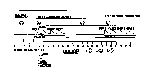
Figures 8a-c show a typical three elec-
trode measurement cycle, which takes 71.43 ms,
i.e. 14 cycles per second, and is divided into
nine intervals. During the first ("calibra-
tion") interval (0 - 4 ms) an internal
18
SUBSTITUTE SHEET (RULE 2B)
CA 02312743 2000-06-02
WO 00/19886 PCT/US98/Z0850
calibration of the instrument takes place.
Calibration of the console is adjustment of the
console's electronics so its performance and
behavioral characteristics are consistent be-
tween consoles. Calibration of the electrical
offsets is to eliminate probe variation due to
different probes and temperature variation as
well as to reduce the need for factory calibra-
tion. This calibration step enables less
costly and lower power circuitry to be used.
Electrode circuitry calibration is carried out
by applying a test signal to the probe, and
then measuring this value with an analog to
digital converter and adjusting the offset
using a digital to analog converter until the
correct value is obtained. The calibration is
carried out under microprocessor control. This
method is a successive approximation type of
search which reduces the calibration time from
2" iterations to n+1 iterations. This is de-
picted schematically as three disconnected
terminals in a circle in Fig. sa.
During the 2"d, 4th, and 6th ("current
measurement") intervals (4-10.5 ms: 18.5-25 ms;
32.5-39 ms), a current (the inrush current) is
injected respectively from one of the three
possible probe tip electrode configurations (in
which one electrode is at anode~potential and
two are cathodes). The temperature of one of
the three LEDs is determined at the same time.
The forward bias voltage of a semiconductor
diode is temperature dependent. This tempera-
ture dependence is due to the variation in the
semiconductor bandgap. The optical output of
an LED is also temperature dependent. To make.
accurate measurements of the backscattered
light from the cervix, the output of the light
19
SUBSTITUTE. SHEET (RULE 28)
CA 02312743 2000-06-02
WO 00/19886 PGTNS98/20850
source needs to be either constant or known.
The optical output of an LED can be determined
by the LED's temperature and drive current if
that LED has been characterized. The need for
determining the temperature of the LED light
source is critical as the short term environ-
mental temperature changes are likely to exceed
20° C. The optical output of uncompensated LEDs
are likely to vary by more than 20% under these
conditions leading to a very inaccurate measure
of backscattered light. The temperature of an
LED junction can be determined by measuring its
bandgap related potential, that is, the forward
bias with a known current thus avoiding the
need for a separate temperature sensor for each
LED. The present invention's novel approach of
leaving the LED's optical output unregulated
and compensating the detector's gain is superi-
or to prior techniques for compensating LED
output, e.g. Mroczka Janusz at al., "Methods of
temperature stabilization of light-emitting
diode radiation", Rav: Sci. Instrum. Vol: 65,
No.4, April 1994, as it removes the chance of
instability in the LED'servo loop. Tempera-
Lure, however, is not the only factor than can
affect the output intensity of the LED. Aging
of the LED affects its output as well. There-
fore an alternative embodiment is depicted in
Figs. 6 and 7 in which another photodetector ~.
is positioned adjacent to the LED dice and
receives light from each of the LED's directly
without light fiber connections. An advantage
of this alternative embodiment is that the in-
tensity of the light may be measured and cor-
rected using signals that are detected while
the LEDs are being pulsed.rather than using
data from a separate measurement as is done for
suesmcn~ sH~t tRU~ zs~
CA 02312743 2000-06-02
wo oon9sss PCT/US98/20850
the bandgap. Where instantaneous correction is
not desired during each pulsing sequence the
average intensity could be corrected using
accumulated data.
During the 3rd, 5th and 7th ("optical
measurement") intervals (10.5-18.5 ms, 25-32.5
ms, 39-47 ms), one of the three LEDs whose
temperature hae been determined emits light the
backacattering of which is simultaneously
detected.
During the 8th ("probe orientation') in-
terval (47-48.5 ms), the proper orientation of
the probe against the tissue surface is
checked.
During the 9th ('discharge") interval
(48.5-71.5 ms.), the surface under examination
is discharged, the data analysis algorithm is
executed, and the user interface is updated.
During each of the three current measure-
meet intervals four square current pulses of
approximately 250 ms duration are employed,
separated by 1.8 ms. Three measurements are
made of the decay amplitude of each of the
first and fourth current pulses during the time
prior to the second pulse or prior to the end
of the current measurement interval. Thus a
series of 18 electrical measurements of pulse
decay are made in each 71.43 ms cycle. A set
of parameters is generated to parameterize the
shape of the inrush current and voltage decay
curves in each interval such as with a multiple
exponential best fit.
Alternative shape parameterizations in-
clude transforming the data with ordinate and
abscissa operators such that they become
piecewise straight line segments. Such
operators include taking logs so as to produce
21
suesTnvr~ sH~r (AUK 2s~
CA 02312743 2000-06-02
WO 00/19886 PCTNS98/20850
log/log displays, using inverse time as the
abscissa or any other transformations that
provide good fit to the data. Parameters asso-
ciated with the transformed functions can then
be associated with the degree of tissue
abnormality. Typical operations that can be
applied to the curves and variables that can be
extracted for use as discriminants are as fol-
lows:
1. The slope and intercept of the log
voltage/invarse time plot of the curves.
2. The slope and intercept of the long
voltags/log time plot of the curves.
3. The subtraction, addition, multiplica-
tion or division of or by a function to dimin-
ish the a priori known obscuration effect of
some artifact of, or noise source within the
system.
4. The slope of the voltage vs. current
curve at the start of the relaxation curve.
5. The relationship between the param-
eters of the inrush current curve and the
parameters of the relaxation curve.
6. The use of integrals of segments of
the curves based on time intervals.
7. The use of integrals of segments of
the curves based on voltage intervals.
8. The use of integrals of segments of
the curves based on current intervals.
9. The magnitudes of the offsets.
The discriminants as listed under item 6
above are the ones presently preferred.
A total of 21 tissue classification
parameters (18 electrical and 3 optical) are
extracted from the digitized optical and
electrical data, in addition to various
parameters extracted for the detection of poor
22
SUBSTrtUTE SHEET (RULE 2~
CA 02312743 2000-06-02
WO 00/19886 PCTNS98/20850
contact conditions. Some of the electrical
parameters are functions d~rivsd from various
portions of the measured relaxation curves.
These parameters are then passed to the
processor chip for classification. With 21
parameters processed per observation, the total
rate of parameter processing is 294 per
second. Assuming that 1000 observations are
processed per patient, the total number of
parameters under consideration is approximately
20,000.
Thus the apparatus of the present inven-
tion categorizes biological tissue by having a
probe tip able to select a tissue surface area
by contact and applying a group of sequential
current pulses from the probe tip to each of a
succession of selected tissue surface areas.
The sequential pulses occur within groups that
occur at a rate fast enough so that they are
applied to substantially the same tissue sur-
face area. A circuit then derives values for a
group of parameters that indicate the response
to the group of sequential current pulses
applied to each selected tissue surface area. ,
A memory stores a catalog of tissue types that
are associated with respective subsets of
groups of parameter values. The processor then
compares the group of parameter values that in-
dicate the response of the selected tissue sur-
face area with the stored subsets of groups of
parameter values to categorize the tissue
surface area.
The parameters in the parameter group are
not necessarily associated on a one-to-one
basis with the sequential current pulses in the
current pulse group. As shown in figures 8a-c,
the successive groups of sequential current
23
SUBSTIT~1TE SHEET (RULE 26~
CA 02312743 2000-06-02
WO 00/19886 PCT/US98/20850
pulses may be s~parated in time from each other
by a time interval substantially greater than
the time interval between the sequential cur-
rent pulses within an individual group.
Also as seen in figures 8a-c during any
current pulse for which a tissue response is
desired multiple measurements of the tissue
potential are taken during decay of the poten-
tial following application of the current
pulse. Furthermore, the system permits at
least two parameter values to be derived during
the potential decay following each current
pulse for which a tissue response is desired
thereby allowing a more sophisticated param- .
eterization of the current decay than a simple
exponential. Enough measurements are made dur-
ing the current decay so that each of the
parameters may be derived from several of the
multiple measurements taken during the decay of
the current pulse for which a tissue response
is desired. These multiple parameters are then
available so that the processor can categorize
any tissue surface in~accordance with at least
two parameter values derived during the poten-
tial decay following each of at least two cur-
rent pulses. In general these two current
pulses are separated by at least one other
current pulse which is not used by the pro-
cessor to categorize the tissue.
In the above description which is based on
,a three electrode probe configuration, the
aforementioned pulses are applied by three
electrodes. This is done so that non-overlap-
ping current pulses flow between different
groups of electrodes and corresponding current
pulse applications and measurement cycles occur
for different groups of electrodes. Similarly,
24
suesnrurs s~ cRU~ ash
CA 02312743 2000-06-02
WO 00/19886 PCTNS981Z0850
corresponding parameter valuos derived follow-
ing the current pulses for different groups of
electrodes are combined for the categorization
of the tissue surface area by the processor.
Other electrode configurations, for example,
two or four, would require modifications to the
sequence as described.
It is preferred that the optical and elec-
trical measurements on the same tissue be in-
terspersed and that the charge dissipation in
the tissue volume underneath a selected tissue
surface is not complete by the time the next
sequential current pulse is applied. This re-
sults in the categorization of the selected
tissue being dependant upon the particular
order of the electrical measurements. This
more complex probing by electrical pulses
creates a more subtle response to the probing
and allows greater discrimination of tissue
characteristics. Navsrthaless, the pulses are
preferably separated in time from each other by
a time interval substantially greater than the
time interval between sequential current pulses
within an individual group so that the cate-
,gorizations of successive selected tissue sur-
face areas are substantially independent of
each other.
Aveg of test results
The timing of the various events aids the
diagnostic abilities of the invention. In par-
ticular it is believed that by measuring only
the decay characteristics of the first and
fourth current pulse in each current measure-
ment interval, two different physical char-
acteristics of the tissue under examination are
characterized. The first pulse provides the '
response of the tissue to a current pulse after
SUBSTmT~E SHEET (RULE 28)
CA 02312743 2000-06-02
WO 00/19886 ~ PGTNS98/20850
the tissue has had an opportunity to discharge
from the previous measurement interval: Indeed
the first pulse of the first measurement inter-
val has had the longest time to recover and
perhaps to recover completely. It has been
conjectured that the different timing of the
pulse recovery times permits tissue at differ-
ent depths below the surface to influence the
measured parameter values. The cumulative
effect of these different recovery times is
determined in the present invention by averag-
ing the responses. Thus some information is
lost, but a wider range of effects influence
the final result. In an alternative embodiment
this averaging is not performed and the greater
information content is utilized.
Allowing tissue chancre from Srior tests to
dissi e.
The timing of the various electrical
measurements into intervals separated by opti-
cal measurement intervals allows a short re-
covery time after each current measurement in-
terval. Furthermore the lengthy discharge in-
terval permits a more total recovery of the
tissue so that individual cycles can maintain
independence from one another. To aid in the
complete discharge, during the discharge inter-
val the three electrical probe tip elements are
made active cathodes and kept at low impedance.
This is quite contrary to the normal construc-
tion of measuring electrodes where the imped-
ance is kept high so that the current charact-
eristics of the object being measured are
effectively isolated from the current flow in
the measuring instrument. Essentially the
benefit of isolation is traded off for the
rapidity of recovery of the tissue for the next
26
SUBSTITUTE SHEET (RULE 26)
CA 02312743 2000-06-02
WO 00/1988b PGTNS98/20850
measurement cycle.
Reducinc overall observation time by order of
test yarformance.
The order of performance of the optical
and the electrical tests also has the benefi-
cial effect of reducing the overall observation
time required far each measurement. Thus the
inactive period between electrical measurements
is used for the optical measurements and vice
versa. The measurement of LED bandgap poten-
tial and the subsequent compensation for temp-
erature variation characterized by the bandgap
potential requires little computational band-
width and does not interfere with the rapidity
of measurement necessary to characterize each
electrical decay curve. In this example, only
eight readings are shown as being taken of the
current flow into the electrodes (the inrush
current) during the early part of the 250 ms
applied pulse. When it is desired to make
additional use of the inrush current readings,
it will be appropriate to take current readings
throughout the 250 ms pulse.
Figure 9 depicts an individual voltage
relaxation curve. As indicated an initial
offset voltage is determined by eight consecu-
tive observations sampled at 9 acs intervals.
The height of the square wave pulse is simi-
larly measured by eight consecutive observa-
tions sampled at 9 ass intervals. During the
voltage decay samples are taken at 9 ~s inter-
vale, but not all are recorded. Fig. 9 also
shows the corresponding current relaxation
curve. In this example, only eight readings
are shown as being taken of the current flow
into the electrodes (the inrush current) during
the early part of the 250 ms applied pulse.
27
SUBSTITUTE SHEET (RULE Z~
CA 02312743 2000-06-02
WO 00/19886 PGT/US98r10850
When it is desired to make additional use of
the inrush current readings, it will be appro-
priate to take current readings throughout the
250 ms pulse.
~~,~, ir~i~, values from two different decay
curves.
The use of the first and fourth electrical
measurement in each set of four as distinct
variables without averaging allows recovery of
the maximum amount of information from the
electrical measurements. This information is
utilized in the statistical analysis of the
electrical and optical data.
During the optical measurement intervals
data is collected and the optical system mea-
sures the bandgap potential of a first LED by
applying a small current to the LED and measur-
ing the potential across it. This provides a
readout of the temperature of the LED and per-
mite correction for temperature variation to be
made.
The results of the tests are displayed to
the physician operating the probe by a series
of display lights. These are depicted in
Figure 3. A summary diagnosis of the tissue
under the probe tip and user error status is
provided by the diagnosis lamps ,ø,~ on the back
~,~ of the probe (seen in Figure 3). These
diagnosis lamps face the clinician in normal
use. The pattern of lights signaling different
conditions is as follows:
' CONDITION LED 1 LED 2
LED 3 LED 4 LED 5
Color Green Red
Blue Blue Blue
System OR on on/off on/off
28
SUBSTITUTE SHEET (RULE 2~
CA 02312743 2000-06-02
wo OOn9886 PCT/US98/20850
on/off on/off
System Error off off off
off off
Unable to Diagnose on off off
off off
Operator Error on on off
off off
Normal Tissue on off on
off off
Low Grade Lesion on off on
on off
High Grade Lesion on off on
on on
The meaning of these conditions is as
follows:
i. High-grade Lesion: (inclusive of
CIN2, CIN3, HGSIL, microinvasive and
invasive cancer)
ii. Low Grade Lesion: (inclusive of CIN1,
LGSIL, atypia, ASC~1S, HPV, necrosis)
iii. Normal: (inclusive of OSE, columnar,
immature metaplasia, mature
metaplasia and nabothian follicle,
regenerative tissue, atrophic tissue)
iv. Unable to Diagnose: (this category
includes data outside the scope of
the algorithm or within overlapping
boundaries between tissue groupings)
v. Operator Error: (inclusive of lift
off, bad angle, slip error and
saturation)
A 6th signaling category indicates whether the
29
SUBSTITUTE SHEET (RULE 2~
CA 02312743 2000-06-02
wo oon9ss6 Pc~rius9snosso
device is working within specifications.
The particular color regime has been
chosen because green is conventional for OK and
system on, red is conventional for error or
malfunction and blue to maximize peripheral
vision stimulation, viz. the outer retina has a
higher concentration of rod cells, which have
greatest sensitivity to blue light. If the
operator is focused on the tip of the probe
then the indicator LEDs will be sighted by
peripheral vision. Thus, the method of signal-
ing a diagnosis is via four approaches, namely,
the display on the console, LED indicators on
the rear of the probe, audible tones via head-
IS phones and a summary printout of the diagnosis.
P'or this purpose, the console display mimics
that of the LED output with the addition of
labeling. In this way the console will serve
as an alternative display of diagnosis and as a
reference to the meaning of the LED configura-
tion on the rear of the probe. The audible
signal also follows the same pattern as the LED
output, however, using tones, for example, the
tones will shift to a higher pitch for a more
significant classification. The printout sun-
marizes the diagnosis.
The algorithm first checks for poor con-
tact, and if detected, the operator is signaled
via the probe handle lights and the console,
and no diagnosis is attempted. As the process
is repeated at a rate of 14 times par second,
the operator receives instantaneous feedback on
the probe position and may adjust device posi-
tinning accordingly. The poor contact check
includes the following conditions: (i) the
probe being at an angle to the cervix: (2) the
probe partially or fully lifting off the
SUBSTITUTE SHEET (RULE 26)
CA 02312743 2000-06-02
WO 00/19886 PCTNS98n0850
cervix, or lift-off: (3) the probe moving too ,
quickly across the cervix for accurate measure-
ments to ba pertoraad, or slip: and (4j the
probe is positioned over a junction b~tween
tisane types. The angle and junction condi-
tions are detected through an imbalance in the
electrical parameters, while the lift-of! con-
dition is detectad by means of out of range
electrical and optical readings.
If the data pass the poor contact check,
then diagnosis into one of i7 tissue types in
the following table is attempted:
High
Grade
Squamous
Intraepithelial
Lesions
(HSIL)
/Invasive
Cancer
(IC)
1 Carcinoma
Z Cervical Intraepithelial Nsoplasia (CZN)
3
a* CIN 3 with an immature metaplasia component
3 CIN Z
3* CIN Z with an iasature metaplasia cosponsnt
Low
Grade
Squamous
Intraepithelial
Lesions
(ISIL)
4 Acatowhite epithelium with or without HPV
stigmata
5 Acetowhite epithelium with vessels and HPV
stigmata
6 Acetowhite epithelium within immature~metaplasia
(atypic)
Original
Squamous
Epithelium
(OSE)
with
8PV
Sticpnata
7 OSE with HPV stigmata Acetowhite epithelium
+
8 oSE i~ith HP'V stigmata Acetowhite epithelium
+ +
8a OSE with aPV stigmata Acetowhite epithelium
+ + wit
vassals _
Sb OSE with HPV stigmata Acetowhite epithelium
+ + wit
surface contour changes
9 OSE with HPV stigmata with micropapilliary
changes(no
Acetowhite)
31
suesmurE sHE~ tRU~ zs~
CA 02312743 2000-06-02
wo OOn 988b PCT/US98/20850
Normal:
l0 Original squamous epithelium
11 Colusnar epithelium
12 Immature Metaplasia, physiologic
13 Interaediate mataplasia
14 Mature metaplasia
Regenerate squamous epithelium (post treatment)
.-
is Cervicitis (acute/ subacute)
10 17 Atrophy
An initial validity check on the data is
pertorm~sd to ensure that the multivariate data
15 distribution is within the limits of all valid
classifier data. If the result signals one out
of range, then no diagnosis will be made and
the operator is signaled.
A most probable tissue type is then
ZO selected. A further validity check is per-
formed to ensure that the multivariate data
distribution is within the limits of all valid
classifier data for the selected tissue type.
Again, if the reading proves to be an outlier,
no diagnosis is performed and the operator is
signaled. The probability estimate (certainty
of assignment to a particular tissue type) is
then assessed against a pre-defined decision
threshold. If the probability estimate is
below the threshold, no diagnosis is per-
formed. Again, because the measurements occur
at the rata of 14 per second, the operator
receives instant feedback. If the estimate is
above the decision threshold, a diagnosis is
made, the tissue grouped into pre-selected
categories, for example, Cancer or High Grade
32
SUBSTITUTE 8HEET (RULE 26)
CA 02312743 2000-06-02
WO 00/19886 PGT/US98I20850
Abnormality (BSIL), Low Grade Abnormality
(LSIL) and Normal, and the operator is signaled
with the result.
There are two levels at which the probe
S classification algorithm involves a predeter-
mined decision-making process affecting the
"trade off" between the sensitivity and speci-
ficity of the test. The pre-determined deci-
sion thresholds define Receiver Operating Char-
acteristic (ROC) curves. The ROC curve is a
graphical description of test performance
representing the relationship between the true
positive fraction (sensitivity) and the false
poaitive fraction (1 - specificity). An
increase in the decision threshold will cause
an overall increase in device specificity at
the expense of sensitivity, and vies versa.
The first level decision threshold
concerns the probability estimate used for the
classification of tissue into one of 17 types.
The second level decision threshold concerns
the grouping of tissue types into categories,
whereby the grouping can bs adjusted, depending
on the desired outcome of the screening test.
~~Appropriats adjustment of the decision thresh-
old allows the configuration of an optimal
trade off between sensitivity and specificity,
with a particular focus on the cut-off between
low grade changes acrd minor atypic.
safety and Reliability of the System
A number of features have been developed
to ensure the safety of the patient and long
term reliability of the probe system. These
include calibration procedures, temperature
compensation and electrical safety precautions.
It is necessary to calibrate each probe
during manufacture in order to ensure that
33
suesT~urs sir (suuE ~
CA 02312743 2000-06-02
WO 00/19886 PCT/US98/20850
optical and electrical output signals.are the
same for each device. Optical calibration is
performed in a turbid solution of stable
optical characteristics with an optical
spectral distribution chosen to simulate that
of cervical tissue, and electrical calibration
is performed using a stable electrolyte solu-
tion. An optical calibration check is also
performed at the beginning of each clinical
session. The operating temperature of the
probe is 5 to 50 degrees Celsius. Temperature
compensation of the LEDs is necessary since the
optical measurements are extremely sensitive to
the ambient temperature. Stability of opera-
tion across the required temperature range is
achieved by continuous automatic measurement of
the temperature and compensatory adjustment.
Electrical safety of the patient has been a
prime consideration in the design of the
ZO device, and a rn~mber of design techniques have
bean employed, including electrical isolation
from the mains voltage, double insulation for
all parts not applied to the patient, a small
voltage employed for the delivered pulses,
"watchdog" monitoring systems including contin-
ual voltage monitoring of the delivered pulses,
hED protection circuitry and the employment of
low voltages throughout the probe and console:
The development of the classification
algorithm is an ongoing'process and the clini-
cal database used as the basis of algorithm
construction should be continually refined.
This process may proceed as follows:
Data for algorithm development is
collected for several thousand women. The
database includes a number of data subsets for
each tissue type and for Poor Contact condi-
34
SUBSTtME SHEET (RULE 28)
CA 02312743 2000-06-02
WO 00/19886 PCT/US98/20850
tions, including contact problems induced by
excess cervical mucus or blood.
Data collection for algorithm development
proceeds by means of a data collection system
incorporating a link from the console to a com-
puter for the download of digitized data, and a
video mixer, recorder and printer. Probing is
performed, followed by foraal colposcopy with
aqueous acetic acid-staining of the cervix, and
the session is recorded on video. A colpo-
photograph is taken after acetic acid staining,
and the colposcopist marks the diagnosis of all
tissue types present on the photograph.
Patient history and current status information,
including Pap smear, colposcopy and biopsy re-
sults are recorded on a clinical record form
and subsequently entered into the probe data-
bass.
Following the data collection session, the
ZO data are analyzed in the laboratory by viewing
the probe session video concurrently with a
display of optical and electrical parameters.
Colposcopy and biopsy.rssults from participat-
ing clinics are subject to a uniform review
'process in order to reconcile colposcog~ic and
histological diagnoses. Briefly, video images
taken during the colposcopy cession and his-
tology results, if available, are reviewed by
an independent colposcopist. Referral to a
second colposcopist is performed in cases of an
initial abnormal diagnosis and in cases of
doubt. Where a reference diagnosis cannot be
established, data are excluded from the algor-
ithm database.
The 17 tissue classification categories
are used in the establishment of the Reference
Diagnosis. Tissue type classification is based
SUBSTITUTE SHEET (RULE 2B)
CA 02312743 2000-06-02
WO 00/19886 PCT/US98/Z0850
on the colposcopic classification of
Copplsson, Pixlsy and Rsid (Copplason M, st al.
"Colposcopy: A scientific and practical
approach to the cervix, vagina and vulva in
health and disease", Third Ed. Thomas, 1986),
and the Rsid and Scalzi abnormality grading
aystsm (Raid R et al., "An improved colposcopic
index for differentiating benign papillomaviral
infections from high grade cervical intra-
epithelial neoplasia" Am J Obstst Gynecol 1985:
153 (6) , 611-8) .
The present invention is designed to bs a
screening, rather than a diagnostic tool.
Therefore, the tiasus types are grouped into
categories which are of use for the clinician
when making the referral decision. These cate-
gorise are: Probe Cancer or High Grade
Abnormality; Probe Low Grade Abnormality; and
Probe Normal. Note that the tissue types
identified as original sguamous epithelium with
HPV stigmata (tissue types 7 to 9) may poten-
tially be grouped into either of the output
categories of Probe Low Grade Abnormality or
Probe Normal, depending upon the desired
'screening result. The two options effectively
correspond to alternative operating points on
the device receiver operating characteristic.
As previously described, the programmable
gain synchronous detector ~ receives the raw
optical signal and provides output to the
tissue type classifier ,~,: Synchronous detec-
tion is a demodulation process in which the
original signal is recovered from a noisy
transmission path by multiplying the modulated
signal by the output of a synchronous
oscillator locked to the carrier. This tsch-
nique traditionally is used in the communi-
36
suesmu~ sH~r ~u~ ~s~
CA 02312743 2000-06-02
WO 00/19886 PC"f/US98/20850
rations field for demodulation of amplitude
modulated signals. Many sources of inter-
ference are present when making measurements of
the backscattared light from the cervix. Thane
sources of interference are both electrical and
optical in nature. Notably, the luminous in-
tensity of colposcope light is far greater than
of the light source used by the probe. Until
synchronous detection was employed, the signal
chain would often saturate. Synchronous datec-
tion has allowed the reduction of interference
by limiting the bandwidth of the processing
chain while using modest levels of probe light.
i~ig. 10 is a block diagram of the syn-
chronous detection system employed in the pre
sent invention. The synchronous oscillator ~
. provides both the drive for a typical LED $;1
and the synchronizing signal for the detector.
The oscillator's frequency is 4 k8z and is thus
away from the frequencies of the most common
noise sources. The photodiode ~ is used to
detect the backscattarad light from the cervix.
Tha photodioda is located in the probe along
with a low gain transimpedance amplifier. The
'gain of this stage is kept low to avoid satura-
tion by ambient light sources. Ths return sig-
nal is accompanied by two common noise sources.
The first is lighting ripple from an illumina-
tion source like a colposcope, the second is
random thermal noise. The high pass filter $~
is used to remove the steady-state light. It
is also effective for reducing the low fre-
quency ripple component from the colposcope
illumination, thus avoiding saturation of the
following signal processing stages. The pro-
grammable gain amplifier ~,~, is used to nor-
malize variations in the LED's optical output.
37
suesn~urs sH~r ~RUt~ ~
CA 02312743 2000-06-02
WO 00/19886 PCTNS98/20850
The multiplier $~ correlates the real signal
component of the photodiode signal while ran-
domizing the noise component. The low pass
filter ~, takes the multiplied signal and pro-
s vides an average. This helps separate the cor-
ralatad signal from the uncorrelated signal
(noise). The low-pass filter also sate the
bandwidth of the signal processing chain. The
lover the cut-off frequency the more narrow the
bandwidth and hence the greater the rejection.
However, if the bandwidth is made too narrow
than the system will take a long time to
respond. Rather than the traditional inte-
grator or first-order low-pass filter, a high
order Hessel filter has been used in the
probe's synchronous detector, thus giving
excellent out-of-band noise rejection as well
as good transient performance.
Although the invention has bean described.
ZO in terms of specific embodiments, it is in-
tended that the patent cover eguivalent substi-
tutions for any of the elements of these em-
bodiments, and that the protection afforded by
this patent be determined by the legitimate
scope of the following claims:
38
SU8ST1TUTE SHEET (RULE 2B)