Note : Les descriptions sont présentées dans la langue officielle dans laquelle elles ont été soumises.
CA 02316777 2000-06-27
1 -
SPECIFICATION
Iontophoresis Device and Method of Assembling The Same
Technical Field
The present invention relates to an iontophoresis device
suitable for percutaneous and mucosal applications of drugs
and in particular to iontophoresis device of the type, which
is activated before practically using as well as a method of
assembling the same.
Background Art
Recently, there have been developed a variety of dosage
forms in the field of pharmaceutical preparations for external
use(external pharmaceutical preparations)and the development
has gradually become a matter of great concern. The reason for
this is as follows: The administration of a drug, which may
have a local or systemic pharmacological action, through the
skin or the mucous membranes has many advantages. For instance,
the sustained-release effect of the drug can be expected; such
administration is not greatly influenced by the metabolism due
to the first-pass effect in the liver unlike the oral
administration and permits the effective use of the drug; and
drugs accompanied by, for instance, liver disorders can
relatively safely be administered.
However, the normal skin naturally has a protective effect
against external stimulations and this makes the absorption
and penetration of a drug through the skin relatively difficult .
CA 02316777 2000-06-27
.- 2 -
For this reason, in the existing circumstances, a drug is not
absorbed in an amount sufficient for ensuring a sufficient effect
even if the drug is administered in a dosage form for external
use. Moreover, in the administration method, which makes use
of absorption routes through biological membrane other than
the skin, such as mouth, rectum, oral cavity and nose as well
as the sublingual route, it is difficult to penetrate into or
transmit through the related biological membrane depending on
the kinds of drugs and therefore, there have been known a large
number of drugs having low bioavailability. Accordingly, there
has been desired for the development of an absorption-promoting
method, which can sufficiently enhance the permeability,
penetrability and absorbency of a drug against the skin and
other biological membranes, can ensure a sufficient
pharmacological efficacy of the drug and is substantially free
of, for instance, its local and systemic toxicity and is highly
useful and safe.
Assuch absorption-promoting methods, there have recently
been known, chemically promoting methods, which make use of
absorption-promoting agents, and physically promoting methods
in which iontophoresis or phonophoresis is employed. Among them,
the iontophoresis has unexpectedly attracted special interest
recently and has been expected as an administration method,
which can solve the foregoing problems.
The iontophoresis is a method for the administration of
a drug by applying a voltage to the skin or a mucous membrane
to electrically induce the migration of an ionic drug and to
thus administrate the drug through the skin or a mucous membrane .
CA 02316777 2000-06-27
- 3 -
In general, an iontophoresis device is provided with a pair
of electrodes for iontophoresis , i . a . , an anode and a cathode
and the device is so designed that these electrodes are arranged
on or attached to the skin at a predetermined distance apart
from one another and an electric current generated by a current
generator is guided to these electrodes to thus carry out
treatments.
Moreover, this iontophoresis device has a structure
comprising a combination of these electrodes and a layer, which
stores a drug therein, and a variety of additives for maintaining
the drug efficacy are optionally enclosed in the layer in
addition to a predetermined amount of the effective component
in order to keep a desired blood concentration in the body over
a long period of time.
The iontophoresis device of this type is, for instance,
disclosed in Japanese Un-Examined Patent Publication Nos. Sho
62-268569 , Hei 2-131779 , Hei 3-268769 andHei 3-45271 andTOKUHYO
Nos. Hei 3-504343 and Hei 3-504813.
However, if a drug (such as physiologically active
peptides), which suffers from a problem of the solubility in
water, is used in these iontophoresis device, the predetermined
amount of the drug may be reduced due to the partial decomposition
thereof with time. Moreover, if the drug is administered in
a high concentration, the drug may be diluted during storing.
If a peptide drug is percutaneously administered by the
iontophoresis, it is common that the drug is not maintained
in an iso-electric environment, but is kept in an acidic or
basic environment . For this reason, the stability of additives ,
CA 02316777 2000-06-27
- 4 -
which are incorporated into the device to assist the development
of the pharmacological efficacy of the biologically active
substance, is greatly influenced by such acidic or basic
environment and accordingly, the drug efficacy may be reduced.
Moreover,it hasbeen recognizedthat when physiologically
active peptides are stored in the form of solutions, members
constituting the pharmaceutical preparation may be adsorbed
on the peripheral site and it is thus quite difficult to maintain
a desired concentration of the drug over a long period of time.
As other problems , it has been reported that in a device ,
which is designed in such a manner that an electrically
conductive layer containing a drug in the form of a solution
is directly in contact with the electrodes immediately after
the electrical charging, the drug is electrolytically
decomposed on the electrode surface during electrically
charging the device . Accordingly, it would be doubtful whether
the decomposed drug through its internal absorption adversely
affects the human body.
There have been proposed a variety of methods for the
solution of such a problem. For instance, Japanese Un-Examined
Patent Publication No. Sho 63-102768 and U.S. Patent No.
5,310,404 disclose a method, which comprises the steps of
arranging a capsule or porch enclosing water or an electrolyte
solution above the electrode structure and breaking the capsule
or porch immediately before the practical use to thus impregnate
the drug-support layer therewith. This method is excellent in
that the drug can be stored in a stable condition (dry state) ,
but it is still insufficient since it takes a long time for
CA 02316777 2000-06-27
- 5 -
uniformly permeating the moisture into the whole drug-support
layer and the drug efficacy may be reduced due to the dilution
of the drug.
In addition, Japanese Patent No. 2,542,792 discloses a
method in which a drug-support layer and an electrode layer
containing an electrolyte are separately disposed in distinct
compartments , which are hinged to one another and then piling
one upon another at the hinged portion to thus activate the
device. This method permits the improvement in the long-term
stability of a drug, but any means for activating the device
upon application is not sufficiently devised and therefore,
the method may include a lot of causes for artificial errors
and cannot achieve sufficiently uniform distribution of the
drug after the activation of the device.
Moreover, Japanese Un-Examined Patent Publication No.Hei
3-94771 discloses a device, which is so designed that a selective
ion-permeable membrane (such as an ion-exchange membrane) is
arranged such that the membrane is adjacent to the skin side
of a water-support portion thereof , while a drug is dried and
adhered to the side of the selective ion-permeable membrane,
which is in contact with the living body, to thus prevent any
dilution of the drug and to realize the administration of a
trace amount of the drug to a local site in a high concentration.
Japanese Un-Examined Patent Publication No. Hei 9-201420
discloses a device for iontophoresis, in which an electrode
structure layer, a solvent-support layer and a drug-support
layer containing a dried physiologically active substance are
put in a layer structure in this order and a water-impermeable
CA 02316777 2000-06-27
- 6 -
separator layer ispositioned between thesolvent-supportlayer
and the drug-support layer. This device is so designed that
thesolvent-support layer isautomatically broughtinto contact
with the drug-support layer by pulling out the separator layer
upon activation. This device is quite excellent in that the
occurrence of any artificial error is prevented when assembling
the device. In this device, however, the solvent-support layer
and the drug-support layer are accommodated in the same package,
the stability of the drug may be reduced due to any leakage
of the solvent from the solvent-support layer and accordingly,
it is difficult to ensure the quality of the device. Moreover,
even if it were technically possible to completely prevent the
leakage of the solvent, the cost required for the development
of such a technique would be very high.
As has been described above, there has not yet been
developed any iontophoresis device, which can ensure the
long-term stability of a drug, can easily and accurately be
assembled immediately before the practical use thereof and
permits the elimination of any artificial error as much as
possible.
Accordingly, it is an ob ject of the present invention to
provide an iontophoresis device, which can ensure the long-term
stability of a drug and can easily be assembled immediately
before the application as well as a method of assembling the
device.
Disclosure of the Invention
The foregoing object of the present invention can be
CA 02316777 2000-06-27
accomplished by providing an iontophoresis device, in which
an electrode portion equipped with a drug-dissolving portion
and a drug portion equipped with a drug-support are provided
with alignment structuresrespectively so that the drug-support
and the drug-dissolving portion are brought into contact with
one another by coinciding the alignment structure of the drug
portion with that of the electrode portion. These alignment
structures are, for instance, openings formed on the electrode
portion and the drug portion, respectively. These portions can
accurately and rapidly be aligned by aligning these openings
of the electrode and drug portions with one another.
In addition, the device is also designed in such a manner
that an electric current-supply portion is provided with the
same alignment structure, the alignment structure is coincided
with that of the electrode portion to thus contact the
current-supply portion with the electrode portion. The
alignment structure for the current-supply portion may be an
electrode terminal. In this case, the alignment structure may
be formed on the terminal connected to the current-supply portion
through a connecting cord.
Alternatively, a connector having the same alignment
structure is disposed and the alignment structures of every
portions are coincided with one another to thus connect them
to the current-supply portion and the connector through the
electrode portion. If some of these alignment structures are
formed from an electrically conductive material, such alignment
structures may be used as electrical connection means.
The drug portion of this iontophoresis device is stored
CA 02316777 2000-06-27
_ g _
as a package separated from the parts such as those for the
current-supply and electrode portions, prior to the practical
use . Thus , the device is designed such that the electrode and
drug portions are mechanically connected or the electrode and
current-supply portions are electrically connected to one
another . A means for holding this arrangement used herein is ,
for instance,electrode terminalsof the current-supply portion
or conductive snap connector or an auxiliary stand for assemblage .
As has been described above, the device according to the present
invention may be a set of units in which the electrode portion
equipped with an alignment structure and the drug portion
equipped with the same alignment structure are separately
packaged. In addition, the current-supply portion or the
connector having an identical alignment structure and an
auxiliary stand for alignment are also packaged separately from
the drug portion, in this set of units.
The electrode portion (electrode unit) used herein
comprises a member holding a conductive layer , a wiring connected
to the conductive layer and an alignment structure formed on
at least one of the wiring and the member. The alignment structure
is an opening formed on at least one of the wiring and the member.
On the other hand, the drug portion (drug unit) comprises a
drug-support, a peelable cover for protecting the drug-support
and an alignment structure formed on the cover. In this case,
the alignment structure is an opening formed on the edge of
the cover.
The method of assembling the iontophoresis device
according to the present invention comprises the steps of peeling
CA 02316777 2000-06-27
- 9 -
off a cover material disposed on an electrode portion; coinciding
an alignment structure of the electrode portion with that of
a drug portion to thus dispose a drug-support of the drug portion
on a drug-dissolving portion; peeling off a cover of the
drug-support on the side of the drug-dissolving portion; and
fixing the drug-support to the electrode portion. In this device,
a cover is also positioned on the drug-support opposite to the
side of the drug-dissolving portion. In this case, however,
if an opening is formed on the cover, the assemblage of this
device can be completed without peeling off the cover. On the
other hand, if any opening is not formed on the opposite cover,
the assemblage of the device is completed after peeling off
at least part of the cover. More specifically, the opposite
cover may completely be peeled off or the part of the cover
other than the portion provided with the alignment structure
may, for instance, be peeled off.
The electrode portion and the drug portion can be aligned
with one another, while making use of the alignment structure
disposed on an auxiliary stand, a current-generating portion
or a connector. In this case, the both alignment structures
of the electrode and drug portions are constituted by openings ,
while the alignment structure of the auxiliary stand is
constituted by an alignment rod capable of being inserted into
the opening. On the other hand, the alignment structures for
the current-generating portion and the connector may be
electrode terminals thereof.
Thus , an iontophoresis device and a method of assembling
the same can be provided, which can ensure the long-term
CA 02316777 2000-06-27
- 10 -
stability of a drug, whose assembling operations are easy and
accurate upon application and which can eliminate any cause
of artificial errors as much as possible.
Brief Description of the Drawings
Fig. 1 is a diagram showing the cross sectional structure
of an iontophoresis device according to the present invention,
immediately before the practical use.
Fig . 2 is a diagram showing an embodiment of a drug portion
( drug unit ) , wherein ( a ) , ( b ) and ( c ) are a view of the surface ,
an internal view and a cross sectional view of the drug unit,
respectively.
Fig. 3 is a diagram illustrating an embodiment of the
structure of a current-generating portion Ia, in which (a),
( b ) and ( c ) are a view of the surface, a view of the back face
and a cross sectional view of the current-generating portion,
respectively.
Fig. 4 is a diagram illustrating an embodiment of the
structure of an integrated electrode portion (electrode unit)
Ib-1, in which ( a ) , ( b ) , ( c ) and ( d ) are a view of the surface ,
an internal view, a view of the back face and a cross sectional
view of the electrode portion, respectively.
Fig. 5 is a diagram showing an embodiment of the structure
of a separate type electrode portion (electrode unit) Ib-2,
in which ( a ) , ( b ) and ( c ) are a view of the surface , an inner
view and a view of the back face of the electrode portion,
respectively.
Fig. 6 is a diagram showing an embodiment of the structure
CA 02316777 2000-06-27
- 11 -
of a conductive snap connector Id, in which (a) and (b) are
a view of the surface and a cross sectional view thereof ,
respectively.
Fig. 7 is a diagram showing an embodiment of the structure
of an auxiliary stand for assemblage Ie-1, wherein (a) and (b)
are a view of the surface and a cross sectional view,
respectively.
Fig. 8 is a diagram showing an embodiment of the structure
of an auxiliary stand for assemblage Ie-2, wherein (a) and (b)
are a view of the surface and a cross sectional view,
respectively.
Fig. 9 is a diagram showing an embodiment of the structure
of an auxiliary stand for assemblage Ie-3 , wherein ( a ) and ( b )
are a view of the surface and a cross sectional view,
respectively.
Fig. 10 is a diagram showing an embodiment of the structure
of an auxiliary stand for assemblage Ie-4, wherein (a) and (b)
are a view of the surface and a cross sectional view,
respectively.
Fig. 11 is a diagram showing the configuration of an
iontophoresis device in which the integrated electrode portion
Ib-1 is incorporated according to the present invention after
the completion of its assemblage, wherein (a) and (b) are a
view of the surface and a view of the back face of the device,
respectively.
Fig. 12 is a diagram showing the configuration of an
iontophoresis device in which the separate type electrode
portion Ib-2 is incorporated according to the present invention
CA 02316777 2000-06-27
- 12 -
after the completion of its assemblage, wherein (a) and (b)
are a view of the surface and a view of the back face of the
device, respectively.
Fig. 13 is a diagram showing an embodiment in which the
current-generating portion Ia is connected to the electrode
portion through a connecting line 30 , wherein ( a ) , ( b ) and ( c )
are a connecting cord, a view of the surface and a view of the
back face, respectively.
Fig. 14 is a diagram illustrating an embodiment of the
method of assembling an iontophoresis device, which makes use
of an integrated electrode, in which (a) shows assembling
processes and (b) shows a process in which the auxiliary stand
Ie-4 for assemblage is used.
Fig. 15 is a diagram illustrating another embodiment of
the method of assembling an iontophoresis device, which makes
use of an integrated electrode, in which (a) shows the first
half of the assembling process and (b) shows the second half
of the process, respectively.
Fig. 16 is a diagram illustrating a further embodiment
of the method of assembling an iontophoresis device , which makes
use of an integrated electrode, in which (a) shows the first
half of the assembling process and (b) shows the second half
of the process, respectively.
Fig. 17 is a diagram illustrating an embodiment of the
method of assembling an iontophoresis device, which makes use
of a separate type electrode , in which ( a ) shows the f first half
of the assembling process and ( b ) shows the second half of the
process, respectively.
CA 02316777 2000-06-27
- 13 -
Fig. 18 is a schematic diagram showing an iontophoresis
device as a comparative example , in which ( a ) , ( b ) and ( c ) are
a view of the surface, views of the inner portion and back face
and a cross sectional view of the device, respectively.
Fig. 19 is a graph showing changes, with time, of salmon
calcitonin in the serum observed in Test Example 1.
Fig . 20 is a graph showing changes , with time , of the rate
of the salmon calcitonin remaining in the drug portion , observed
in Test Example 2.
Best Mode for Carrying Out the Invention
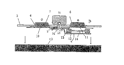
Fig. 1 is a diagram showing the cross sectional structure
of an iontophoresis device according to the present invention,
immediately before the practical use. In this figure, every
parts are depicted separately to make , easier, the understanding
of these parts which are in fact in a laminated relation or
come in close contact with one another.
In this figure, a donor electrode-printed portion 6 is
positioned on one side of a backing layer 4 and a reference
electrode-printed portion 7 is positioned on the other side
of the layer 4. An adhesive film 3 such as a medical adhesive
tape is disposed on the periphery of the backing layer 4 for
securing a pharmaceutical preparation to an application site.
The both electrode-printed portions 6 and 7 are connected to
a current-generating portion Ia through a conductive snap
connector Id. The donor electrode-printed portion 6 on the
backing layer 4 is provided with a conductive layer 10 ( a
drug-dissolving portion) on the donor electrode side, while
CA 02316777 2000-06-27
- 14 -
the reference electrode-printed portion 7 is provided with a
conductive layer 10 on the reference electrode side. A
drug-support 14 is removably connected to the drug-dissolving
portion 11. An adhesive layer 13 is formed on the periphery
of the drug-support 14. Thus, the drug-support 14 is fixed to
the backing layer 4 or the donor electrode-printed portion 6
through the peripheral adhesive layer 13. On the other hand,
a liner 12 is disposed on the peripheral adhesive layer 13 and
on the side facing the skin 40.
The liner 12 is peeled off from the iontophoresis device
having such a structure upon the practical use and thus the
drug-support 14 is exposed. The device, which is in such a
condition, is applied to the skin 40. At this stage, the drug,
which is in a dry state and supported by the drug-support 14,
is dissolved in the water supplied from the drug-dissolving
portion 11. Then a power supply for the current-generating
portion Ia is switched on to thus put the iontophoresis device
in operation.
In this respect , the liner 12 is provided with an opening
15 as an alignment structure. Moreover, the electrode portion
Ib is also provided with an opening 5 as an alignment structure .
In the application of the device, the drug-support 11 can easily
and accurately be positioned on the drug-dissolving portion
11 by aligning these openings with one another . At this stage ,
an electrode terminal of the current-generating portion Ia or
the connector Id is inserted into the opening to thus permit
rapid positioning operations.
Examples of structures of each part of such an iontophoresis
CA 02316777 2000-06-27
- 15 -
device will be described in more detail below.
Fig . 2 is a diagram showing an embodiment of a drug portion
( or drug unit ) , wherein ( a ) , ( b ) and ( c ) are a view of the surface ,
an internal view and a cross sectional view of the drug unit ,
respectively. The drug unit Ic of this example is formed by
sandwiching a porous drug-support 14 in between a liner 17 on
the electrode side, serving as a protective cover, and a liner
12 on the skin side. The liner 17 on the electrode side is provided
with a perforation 24 for folding the liner, while the liner
12 on the skin side is provided with two insertion openings
for conductive snap connectors as will be detailed below
and a perforation 16 for pulling out the liner after the
completion of the assemblage. Both of these liners are formed
from a film having a low adsorptive affinity for the drug such
15 as polyethylene terephthalate. Moreover, the drug is adhered
to and supported on the drug-support 14 by means of , for instance,
spray coating or impregnation and then dried. Adhesive layers
13 are arranged on the both sides and the periphery of the
drug-support 14 for the purpose of adhesion thereof to the
electrode portion and the skin. The adhesive layer 13 is coated
on the support in a stripe-like pattern for ensuring ventilation .
In this connection, the liners 12 and 17 are subjected to a
silicone treatment on the side, which comes in contact with
the drug-support 14 in order to prevent any adsorption of the
drug and to improve the releasability thereof . Further the liners
may likewise be subjected to a treatment for inhibiting any
diffusion of a drug solution to the peripheral adhesive layer
13.
CA 02316777 2000-06-27
- 16 -
Then materials or the like for each part of the drug unit
will be described below. The peripheral adhesive layer 13 for
the drug-support can be formed by the use of an adhesive as
will be detailed below in connection with an adhesive film 3.
This layer is formed by pattern coating ( such as intermittent
coating, stripe coating, intermittent stripe coating) and
desirably has a structure through which the air easily passes .
The width of the pattern coating is not restricted to any
particular one insofar as they can ensure good balance between
the adhesion and the air permeability, but the width desirably
ranges from 0.1 mm to 20 mm.
The drug-support 14 may be any one insofar as it can support
a drug consisting of a physiologically active substance and
permits the permeation of the drug therethrough. Moreover, if
the drug is a physiologically active peptide or a protein, a
hydrophilic porous material may be used, which can support dried
drugs and has low adsorptivity. The hydrophilic film formed
from such a hydrophilic porous material includes a thin film
having high wettability by water such as a hydrophilized
hydrophobic (or water-repellent) polymer thin film or a
hydrophilic substance-containing hydrophilic polymer film.
Examples of hydrophilized hydrophobic polymer thin films
are thin films formed from hydrophilized fluoroplastics (such
as hydrophilic DURAPORE available from Millipore Company and
hydrophilic poly(tetrafluoroethylene) available from Toyo
Roshi Co., Ltd.), thin films such as those formed from
hydrophilic polyther sulfone (such as Supor available from
Gelman Sciences Inc.) and hydrophilized cellulose derivatives
CA 02316777 2000-06-27
- 17 -
(such ashydrophilized cellulose monoacetate and hydrophilized
cellulose triacetate).
Examples of hydrophilic substance-containing hydrophilic
polymer thin films include a variety of polymers obtained by
adding appropriate surfactants and impregnating therewith and
then drying, for instance, hydrophilized cellulose acetate
films ( such as Asymmetric Ultra Filter available from sartorius
Company and cellulose acetate type ones available from Toyo
Roshi Co. , Ltd. ) , hydrophilized polycarbonate films (such as
Isopore Membranes available from Nihon Millipore Ltd.),
hydrophilized poly (tetrafluoroethylene) films (such as
Omnipore Membranes available from Nihon Millipore Ltd.),
hydrophilized polysulfone films (such as HT Tuffryn available
from Gelman Sciences Inc. ) and hydrophilized nonwoven fabrics
( such as films obtained by coating polyester nonwoven fabrics
with cellulose acetate (e. g., coated type membranes available
from Toyo Roshi Co. , Ltd. ) ) . The hydrophilic films also include,
for instance, nylon films ( such as Biodyne available from Nihon
PALL Ltd.).
Incidentally, drugs unstable to water should desirably
be included in or adhered to the drug-support in their dried
state in order to improve the stability of these drugs and to
inhibit any leakage and deterioration thereof. On the other
hand, in case of drugs stable to water, they may be supported
on the drug-support in their gel-like conditions. In such a
gel-like drug-support, suitably used are water-soluble
polymers and hydrogel thereof . A method for preparing such a
gel-like drug-support comprises the step of mixing and kneading
CA 02316777 2000-06-27
- 18 -
a gelling agent such as a water-soluble polymer and a drug
solution. Moreover, the electrical conductivity of the gel-like
drug-support can be enhanced by addition of an electrolyte such
as sodium chloride, potassium chloride, sodium carbonate,
phosphoric acid or sodium citrate; or a pH-buffering agent such
as acetic acid, sodium acetate, phosphoric acid, sodium
phosphate, citric acid or sodium citrate. Moreover, the kneaded
mixture is formed into a product to such an extent that it has
a self shape-maintainability and then spreaded into a sheet
or a film. If the kneaded mixture has an insufficient self
shape-maintainability, a mesh-like support may be introduced
into the gel. The thickness of the gel layer desirably ranges
from 0.1 to 2 mm and particularly preferably 0.3 to 0.8 mm.
If it is too thin, the gel strength is considerably low, while
if it is too thick, the movement of the drug is inhibited and
accordingly, the rate of drug absorption is reduced.
The liners 12, 17 as the protective members may be any
one insofar as they are formed from a water- impermeable material ,
but are desirably those capable of being processed through
molding ( such as thermal molding and vacuum forming ) . Examples
of such water-impermeable materials usable herein are aluminum
foils , polyester films , polypropylene films and polyethylene
films as well as laminated films thereof . In addition, it is
desirable to use these materials after subjecting them to an
adsorption-inhibitory treatment such as a treatment with
silicone or Teflon. This treatment would facilitate the peeling
off thereof from the adhesive layer.
Drugs usable herein are any medicine used in any therapeutic
CA 02316777 2000-06-27
- 19 -
field, which is soluble or dispersible in water and, in
particular, physiologically active substances having a
molecular weight ranging from 1 X 10² to 1 X 10⁶ can widely be
used . Examples of drugs are narcotics , analgesics , anorexics ,
anthelmintics, drugs for asthma, anticonvulsants,
antidiarrheals, antineoplastic agents, drugs for Parkinson's
disease, antipruritics, sympatholytic agents, xanthine
derivatives, drugs for angiocardiac diseases such as calcium
channel blockers, antipyretics, ~3-blockers, antiarrhythmic
agents, hypotensive drugs, diuretics, vasodilators for blood
vessels including systemic, coronary, peripheral and cerebral
vessels, drugs for hemicrania, drugs for drunkness and motion
sickness, antiemetics, central nervous system stimulants,
drugs for cough and common cold, decogestants, diagnostics,
drugs for hormonotherapy, parasympatholytic agents,
parasympathomimetic agents, psychostimulants, sedatives,
tranquilizers, anti-inflammatory agents, anti-arthritic
agents, anti-spasmodics, antidepressants, drugs for treating
psychosis, drugs for treating dizziness, anti-anxiety agents,
narcotic antagonists, carcinostatic agents, hypnotics,
immunosuppressors, muscle relaxants, antiviral agents,
antibiotics,anorexics,antiemetics,anti-cholinergic agents,
antihistamic agents, contraceptives, antithrombotic agents,
bone-absorption suppressorsand osteogenesis-promoting agents.
However, the present invention is not restricted to these
specific drugs. These drugs may be used alone or in any
combination.
Specific examples of these drugs include steroids such
CA 02316777 2000-06-27
- 20 -
as estradiol, progesterone, norgestrel, levonorgestrel,
norethindrone,medroxy-progesterone acetate,testosterone and
esters thereof; vitro group-containing compounds and
derivatives such as nitroglycerin and isosorbide dinitrates,
nicotine, chlorpheniramine, terfenadine, triprolidine and
hydrocortisone; oxicam derivatives such as piroxicam; acetic acid
or propionic acid derivatives such as indometacin, flurbiprofen,
felbinac and diclofenac, ketoprofen; mucopolysaccharidases
such as thiomucase,buprenorphine,fentanyl,naloxone,codeine,
lidocaine, dihydroergotamine, pizotyline, salbutamol and
terbutaline; prostaglandins such as misoprostol, enprostil,
omeprazole and imipramine; benzamides such as metoclopramine,
scopolamine and clonidine; dihydropyridines such as nifedipine,
verapamil, ephedrine, pindolol, metoprolol, spironolactone,
nicardipine HC1 and calcitriol; thiazides such as
hydrochlorothiazide and flunarizine; sydnone imines such as
molsidomine; sulfated polysaccharides such as heparin
fractions and proteins; and peptides such as insulin and
homologues thereof; calcitonins and homologues such as
elcatonin, protamin and glucagone; globulins, angiotensin I,
angiotensin II, angiotensin III, lypressin, vasopressin,
somatostatin and homologues thereof; growth hormones and
oxytocin; as well as , if necessary, pharmaceutically acceptable
salts thereof with acids or bases. Preferred are, for instance,
narcotics, hormones, proteins, analgesics, or other low
molecular weight cations. More preferably, examples of drugs
include peptides or polypeptides such as insulin, calcitonin,
calcitonin-related genetic peptides, vasopressin,
CA 02316777 2000-06-27
- 21 -
desmopressin, protirelin (TRH), adrenocorticotropic hormones
(ACTH), luteinizing hormone-release hormones (LH-RH), growth
hormone-release hormone (GRH), nerve growth factors (NGF) and
other release factors, angiotensins,parathyroid hormone(PTH),
luteinizing hormone (LH), serumal gonadotropin, hypophyseal
hormones (such asHGH, HMG, HCG), growth hormones, somatostatin,
somatomedin,glucagon,oxytocin,gastrin,secretin,endorphin,
enkephalin, endothelin, cholecystokinin, neurotensin,
interferon, interleukin, transferrin, erythropoietin,
superoxide dismutase (SOD), filgrastim (G-CSF),
vasoactive-intestinal-polypeptides (VIP), muramyl dipeptides,
corticotropin,urogastrone and atrialsodium uragogue peptides
(h-ANP). However, the present invention is not restricted to
these specific drugs. Among these, particularly preferred are
peptide hormones. It is also possible to optionally use
adsorption-inhibitory agents such as benzalkonium chloride,
BSA (bovine serum albumin) and monolauric acid.
In the present invention, at least one of the foregoing
drugs and salts thereof may be supported on the drug-support.
In addition, the amount of the drug is determined depending
on a particular drug in such a manner that upon administration
thereof to a patient, a predetermined effective blood
concentration is maintained over an effective period of time
and the size of the iontophoresis device as well as the area
of the drug-delivery surface thereof are determined in
proportion thereto.
Fig. 3 is a diagram illustrating an embodiment of the
structure of a current-generating portion Ia, in which (a),
CA 02316777 2000-06-27
- 22 -
( b ) and ( c ) are a view of the surf ace , a view of the back face
and a cross sectional view of the current-generating portion,
respectively. The current-generating portion Ia is a plastic
molded body having therein a built-in current-control circuit.
A current-control switch 1 is arranged on the current-generating
portion, while a female or male electrode terminal 2 (one each
of the terminal on the sides of the anode and cathode ) is arranged
below the current-generating portion. This current-generating
portion Ia is preferably designed such that no physical burden
due to the size and weight thereof is given to a patient.
More specifically, the current-generating portion is
constituted by a self-oscillator circuit provided with a
built-in small-sized cell, an appropriate high
voltage-generating circuitconnected to the oscillator circuit
and a control circuit for operating and controlling these
circuits. It is also possible to incorporate a BOLUS button
for temporarily increasing the injection rate for a drug into
the current-generating portion. This is quite useful function
when an analgesic is administered to a patient and the patient
desires for a temporary increase in the dose thereof in
proportion to the degree of his pains.
Moreover, the control circuit is, for instance, designed
in such a manner that the circuit permits the manual on/off
switching in order to allow the on-demand medication regime
and the on/off switching at a period adapted for the biological
circadian rhythm and the pattern at intervals of 24 hours . In
addition, the control circuit may be equipped with a built-in
microprocessor and therefore, the circuit permits the
CA 02316777 2000-06-27
23 -
modification of the level of the current and the wave form such
as pulses and sinusoidal waves to be applied over a predetermined
time. Moreover, the control circuit may comprise a biosensor
or a certain kind of feedback system for monitoring the
biosignals of a patient, evaluating the treating method and
adjusting the amount of the drug to be administered to the patient
in response to the results of the evaluation. It is also possible
to incorporate one or more programs predetermined by the maker
of the drug, a physician or a patient into the control circuit .
Fig. 4 is a diagram illustrating an embodiment of the
structure of an integrated electrode portion Ib-1, in which
(a), (b), (c) and (d) are a view of the surface, an internal
view, a view of the back face and a cross sectional view of
the electrode portion, respectively. The integrated electrode
portion Ib-1 has a backing layer 4 consisting of a film of a
polyolefin such as polyester or polypropylene or a molded body
of such a film laminated with an aluminum layer. Printed
electrode portions 6 , 7 are arranged on the molded backing layer
4 and they are formed by printing silver ( on the anode side )
and silver chloride (on the cathode side). Moreover, two
insertion openings 5 ( one each of the opening on the sides of
the anode and cathode) for conductive snap connectors are
positioned on the printed electrode portion at the center of
the backing layer.
Conductive layers 10, 11 are formed on the integrated
electrode portion Ib-1 in such a manner that they are adjacent
to the printed electrode portions 6, 7 and the material used
for forming these layers is a water-retentive material such
CA 02316777 2000-06-27
- 24 -
as a nonwoven fabric or a hydrophilic polymer, which comprises
an electrolyte. In this respect, the conductive layer 11 on
the donor side ( in this example, the layer on the anode side )
also serves as a moisture supply source for the drug accommodated
in the drug portion Ic upon activation . Moreover, the conductive
layers are packaged with a water-impermeable cover material
9 through easily peeled heat seal in order to prevent any moisture
evaporation during storage. Further an adhesive film 3 such
as a medical adhesive tape is applied onto the periphery of
the backing layer 4 for the purpose of fixing the pharmaceutical
preparation to a drug-application site and a liner 8 is fitted
to the adhesive film during storage.
Fig. 5 is a diagram showing an embodiment of the structure
of a separate type electrode portion Ib-2, in which (a), (b)
and ( c ) are a view of the surface, an internal view and a view
of the back face of the electrode portion, respectively. The
separate type electrode portion Ib-2 has a backing layer 4
consisting of a film of a polyolefin such as polyester or
polypropylene or a molded body of such a film laminated with
an aluminum layer. Printed electrode portions 6, 7 are arranged
on the molded backing layer 4 and they are formed by printing
silver ( on the anode side ) and silver chloride ( on the cathode
side). Moreover, an insertion opening 5 for each conductive
snap connector is positioned on the printed electrode portion
6, 7.
Conductive layers 10, 11 are formed on the separate type
electrode portion Ib-2 in such a manner that they are adjacent
to the printed electrode portions 6, 7 and the material used
CA 02316777 2000-06-27
- 25 -
for forming these layers is a water-retentive material such
as a nonwoven fabric or a hydrophilic polymer, which comprises
an electrolyte. In this respect, the conductive layer 11 on
the donor side ( in this example, the layer on the anode side )
also serves as amoisture supply source for the drug accommodated
in the drug portion Ic upon activation. Moreover, the conductive
layers are packaged with a water-impermeable cover material
9 through easily peeled heat seal in order to prevent any moisture
evaporation during storage. Further an adhesive film 3 such
as a medical adhesive tape is applied onto the periphery of
the backing layer 4 for the purpose of fixing the pharmaceutical
preparation to a drug-application site and a liner 8 is fitted
to the adhesive film during storage.
Incidentally, these electrode portions may have a known
electrode structure. For instance, usable herein are materials
such as platinum black, titanium, carbon, aluminum, iron, lead,
carbon-containing conductive rubber and conductive resins,
with platinum electrodes, silver electrodes, silver chloride
electrodes or the like being particularly desirable.
In addition, the cover material 9 is not restricted to
any particular one insofar as it is formed from a
water-impermeable material. For instance, the cover material
is formed from a film laminated with an aluminum layer. If a
highly sealed condition by heat sealing is required, the cover
material is laminated with a plurality of films such as those
described above in connection with the liner or it is coated
with another polymer resin. This makes the peeling off of the
cover material easy. For instance, there can be used an easily
CA 02316777 2000-06-27
26 -
peelable laminate film. It is desirable that the laminate film
have a peel strength at 180 degrees of not more than 2000 g.
A pressure-sensitive adhesive is used as an adhesive
material for the adhesive film 3 ( the adhesive layer 13 at the
periphery of the drug support). Any pressure-sensitive adhesive
may be used herein insofar as they can maintain the iontophoresis
device on the surface of the skin or mucous membrane of a patient ,
while the device is brought into close contact therewith, they
have an adhesive force sufficient for ensuring good adhesion
of the drug support to the drug-dissolving portion and they
are physiologically acceptable for the skin. Specific examples
thereof are acrylic adhesives comprising homopolymers or
copolymers of alkyl acrylates whose alkyl moiety has 4 to 18
carbon atoms, such as acrylic acid, methyl acrylate, ethyl
acrylate, 2-ethylhexyl acrylate, isooctyl acrylate, decyl
acrylate, lauryl acrylate and stearyl acrylate; methacrylic
adhesives comprising homopolymers or copolymers of alkyl
methacrylates whose alkyl moiety has 4 to 18 carbon atoms , such
asmethyl methacrylate,ethyl methacrylate,butyl methacrylate,
2-ethylhexyl methacrylate, isooctyl methacrylate, decyl
methacrylate, lauryl methacrylate and stearyl methacrylate;
silicone type adhesives such as those comprising
polyorganosiloxane and polydimethyl-siloxane; and rubber type
adhesives such as those comprising natural rubber,
polyisobutylene, polyvinyl ether, polyurethane,
polyisobutylene, polybutadiene, styrene-butadiene copolymer,
styrene-isoprene copolymer andstyrene-isoprene-styrene block
copolymer. Moreover, the adhesive material may, if necessary,
CA 02316777 2000-06-27
- 27 -
comprise a tackifier and a softening agent.
A material for the backing layer 4 herein used may be an
effective component-impermeable material.Examplesthereof are
films , sheets and foams of synthetic resins such as polyethylene ,
polypropylene,polyethylene terephthalate,polyvinyl chloride,
polyvinylidene chloride, plasticized vinyl acetate copolymer,
plasticized vinyl acetate-vinyl chloride copolymer,polyamide,
cellophane, cellulose acetate, ethyl cellulose, polyester,
polycarbonate, polystyrene, polyurethane, polybutadiene,
polyimide, poly-acrylonitrile, polyisoprene, polystyrene
derivatives, ethylene-vinyl acetate copolymer,
ethylene-polyvinyl alcohol copolymer,fluoroplastics,acrylic
resins, epoxy resins, which may be used alone or in the form
of a laminate of at least two of them.
In addition, the films, sheets, foams or the like of these
synthetic resins may be laminated with metal foils such as
aluminum and tin foils; nonwoven fabrics and synthetic paper
or may be covered with deposited aluminum layers and ceramic
coatings. Moreover, if closed package by, for instance, heat
sealing is required, they may be laminated with a heat-sealable
material.
The electrode portion may be deposited on the backing layer
by, for instance, a method comprising the steps of mixing an
electrode material with, for instance, a print ink for electric
wirings , applying the print ink to a material for the backing
layer and then drying the same; a method comprising the steps
of spreading an electrode material and then fixing the material
to the backing layer; a method comprising the step of depositing
CA 02316777 2000-06-27
- 28 -
an electrode material onto the backing layer; or a method in
which the electrode portion is formed by photo-etching an
electrode material applied onto the backing layer. In addition,
an insulating layer may additionally be applied onto a part
of the printed electrode layer, which may come in contact with
the skin of a patient.
The conductive layer may simply comprise water or may
comprise at least one member selected from the group consisting
of soft porous materials such as ion-exchangeable polymers,
foaming materials and sponge and water-absorptive polymers.
Moreover, the conductive layer may comprise an electrolyte such
as sodium chloride, potassium chloride, sodium carbonate,
phosphoric acid or sodium citrate; or a pH-buffering agent such
as acetic acid, sodium acetate, phosphoric acid, sodium
phosphate , citric acid or sodium citrate , for the improvement
of the electric conductivity thereof.
Specific examples of the preferably used conductive layer
in general include nonwoven fabric, paper, gauze, absorbent
wadding, polyethylene or polypropylene having open cells,
polyvinyl acetate, porous films and foams of, for instance,
polyolefin foams, polyamide foams and polyurethane, natural
polysaccharides such as karaya gum, tragacanth gum, xanthane
gum, starches, gum arabic, locust bean gum, gellan gum, guar
gum and carrageenan; gelatin, pectin, agar, sodium alginate
or polyvinyl alcohol and partially saponified products thereof ;
polyvinyl formal, polyvinyl methyl ether and copolymers
thereof; polyvinyl pyrrolidone and copolymers thereof; aqueous
or water-soluble cellulose derivatives such as sodium
CA 02316777 2000-06-27
- 29 -
carboxymethyl cellulose, methyl cellulose, hydroxyethyl
cellulose, hydroxymethyl cellulose, hydroxypropyl methyl
cellulose, hydroxypropyl cellulose and cellulose acetate
phthalate; carboxyvinyl polymer, polyacrylamide and
polyacrylamide derivatives, casein, albumin, chitin,chitosan,
polyacrylic acid, sodium polyacrylate, poly-HEMA, poly-HEMA
derivatives, methoxyethylene-maleic anhydride copolymer,
N-vinyl acetamide, N-vinyl acetamide and acrylic acid and/or
acrylic acid salt copolymers, as well as crosslinked products
thereof, water-soluble polymers optionally plasticized with,
for instance , ethylene glycol or glycerin and hydrogels thereof .
However, the present invention is not restricted to these
specific ones . In addition, the foregoing materials may be used
in combination of at least two of them. Moreover, it is also
possible to use, if necessary, benzalkonium chloride, BSA
(bovine serum albumin) and adsorption-inhibitory agent such
as monolauric acid.
Furthermore, the conductive layer may also comprise an
ion-exchangeable polymer for the removal of ions competitive
with a desired drug. Such an ion-exchangeable polymer usable
herein is appropriately selected from anion-exchange polymer,
cation-exchange polymer or ampholytic ion-exchange polymer,
depending on the ionic properties of each particular drug. In
addition, the ion-exchangeable polymer may be incorporated into
the conductive layer by, for instance, a method comprising the
step of dispersing fine powder of an ion-exchangeable polymer
in the foregoing polymer to thus form the mixture in a gel-like
form or a method, which makes use of a product of such an
CA 02316777 2000-06-27
30 -
ion-exchangeable polymer previously formed into a film, but
the present invention is not restricted to these methods at
all.
The capacity of the conductive layer on the donor electrode
side (drug-dissolving portion) is not particularly restricted
to a specific range, but depends on, for instance, the size
of the electrode portion and the optimum amount of water required
for dissolving a drug accommodated in the drug portion, or the
water content of the absorptive member of the drug-dissolving
portion. In this respect, however, if the amount of water is
too large, it may cause leakage of the drug-dissolving liquid,
while if it is too small, the drug present in the drug portion
cannot completely be dissolved and the drug efficacy is
correspondingly reduced. Therefore, the amount of water is
desirably on the order of the maximum water absorption of the
drug support. If a hydrogel is used in the drug-dissolving
portion, the syneresis thereof particularly preferably ranges
from 10 to 100 mg/cmz. Moreover, the hydrogel should have such
a gel strength that the gel is never broken during the assemblage
of the device and during the application thereof to the skin
and therefore, the hydrogel desirably has a gel strength ranging
from 400 to 1500 g/cm2.
The amount of water required for dissolving a drug present
in the drug support is in advance controlled in the
drug-dissolving portion. Thus, a precise amount of water can
certainly and rapidly be supplied to the drug support at any
time upon practical use and this makes the therapeutic effect
accurate. Moreover, this can also simplify the treating
CA 02316777 2000-06-27
- 31 -
operations and reduce the treating time.
Fig. 6 is a diagram showing an embodiment of the structure
of a conductive snap connector Id, in which (a) and (b) are
a view of the surface and a cross sectional view thereof ,
respectively. This connector Id is provided with two electrode
terminals 19 (male and female) on an electrode terminal-fixing
table 18 and they are designed in such a manner that they can
be connected to the electrode terminals 2 (female and male)
of the current-generating portion Ia after the assemblage of
the device.
The current-generating portion Ia is connected to the
electrode portion Ib such that the latter is sandwiched between
the electrode terminal on the side of the current-generating
portion and that on the side of the conductive snap connector
Id. The electrode terminal on the conductive snap connector
side comes in contact with the printed electrode portion ( either
of the anode and cathode ) of the electrode portion due to the
connection. Accordingly, the current-generating portion and
the electrode portion can electrically be charged and the
electrical connection can thus be established.
In addition, if they are connected, while inserting the
drug portion upon the assemblage of the device, the electrode
terminal also serves as a means for mechanical connection for
the purpose of positioning or aligning the electrode portion
with the drug portion. Thus, the electrode terminals of the
current-generating portion and the conductive snap connector
are important as means for assembling the device.
In respect of the modes of the connection of the
CA 02316777 2000-06-27
- 32 -
current-generating portion to the electrode portion, the device
may be operated in a cordless mode or a remote control mode
using a cord. In case of the former, a small-sized
current-generating portion is directly connected to the
electrode portion when it is intended to carry out an easy and
quick treatment. Besides, in~ case of the latter, the
current-generating portion is connected to the electrode
portion through an exclusive connecting cord when it is intended
to carry out a treatment while operating the device at hand.
In this connection, connection means are fitted to the both
sides of the connecting cord for connecting the
current-generating portion to the conductive snap connector.
In this embodiment , electrode terminals ( both anode and
cathode terminals ) are incorporated into a plastic molded body
so that it serves to connect the terminals , to each other, of
the current-generating portion and the conductive snap
connector. In this respect, the connection means is not
restricted to an electrode terminal and the shape and the
connection mode thereof may arbitrarily be changed. Preferably,
the connection means on the conductive snap connector side has
such a structure that the drug portion and the electrode portion
are in line with each other and they can firmly maintain a desired
arrangement.
Fig. 7 is a diagram showing an embodiment of the structure
of an auxiliary stand for assemblage Ie-1, wherein (a) and (b)
are a view of the surface and a cross sectional view, respectively.
The auxiliary stand for assemblage Ie-1 is designed in such
a manner that it possesses a space 21 for accommodating the
CA 02316777 2000-06-27
33 -
electrode portion , whose shape corresponds to that of the backing
layer 4 of the electrode portion and that it has two rods 20
used for positioning upon the assemblage of the device . Materials
for the auxiliary stand for assemblage are not restricted to
any specific one insofar as they are those capable of being
shaped and/or processed such as paper, metals, wood and plastic
films (such as polypropylene, Teflon and polyvinyl chloride
films), but preferred are plastic films having high
shape-retention ability and a thickness of not less than 0.5
mm.
This auxiliary stand for assemblage is devised to make,
easy, the operations required when a patient assemble this device .
In this embodiment , the stand is provided with a space 21 for
accommodating the electrode portion, whose shape corresponds
to that of the backing layer 4 of the electrode portion and
therefore, the electrode portion can be disposed on the precise
position on the auxiliary stand. The electrode-accommodating
space 21 is also important in that it can prevent any damage
of the electrode portion possibly encountered when the device
is assembled.
In addition, the alignment rod 20 makes it easy to align
the electrode portion with the drug portion upon the assemblage
of the device and is effective for eliminating the occurrence
of any artificial error.
Fig. 8 is a diagram showing an embodiment of the structure
of an auxiliary stand for assemblage Ie- 2 , wherein ( a ) and ( b )
are a view of the surface and a cross sectional view, respectively.
The auxiliary stand for assemblage Ie-2 is designed so as to
CA 02316777 2000-06-27
- 34 -
have a space 23 for accommodating the current-generating portion,
whose shape is in conformity with that of the current-generating
portion Ia. The space 23 is provided with a means 22 for fixing
the current-generating portion to the auxiliary stand Ie-2.
Fig. 9 is a diagram showing an embodiment of the structure
of an auxiliary stand for assemblage Ie-3, wherein (a) and (b)
are aview of the surface and a cross sectional view, respectively.
The auxiliary stand for assemblage Ie-3 is designed so as to
have a space 23 for accommodating the current-generating portion,
whose shape is in conformity with that of the current-generating
portion Ia and two alignment rods 20. The functions of the
alignment rods 20 and the space 23 are the same as those discussed
above in connection with the foregoing auxiliary stand.
Fig. 10 is a diagram showing an embodiment of the structure
of an auxiliary stand for assemblage Ie-4, wherein (a) and (b)
are a view of the surface and a cross sectional view, respectively.
The auxiliary stand for assemblage Ie-4 is provided with a space
23' for accommodating the conductive snap connector Id, whose
shape is adapted for that of the connector. The function of
the space 23' is the same as that described above in connection
with the foregoing space 23.
In this connection, the auxiliary stand for assemblage
may have a structure combined with those described above
depending on the shape and the procedures for assemblage of
the device and the shape thereof can further be modified.
Materials for the auxiliary stand for assemblage are not
restricted to any specific one insofar as they are those capable
of being shaped and/or processed, such as paper, metals, wood
CA 02316777 2000-06-27
35 -
and plastic films (such as polypropylene, Teflon and polyvinyl
chloride films ) , but preferred are plastic films having a high
shape-retention ability and a thickness of not less than 0.5
mm.
Fig. 11 is a diagram showing the configuration of an
iontophoresisdevice,in which the integrated electrode portion
Ib-1 is incorporated, according to the present invention, after
the completion of its assemblage, wherein (a) and (b) are a
view of the surface and a view of the back face of the device,
respectively. As will be seen from this figure, a
current-generating portion Ia is disposed on the surface of
the electrode portion and a conductive snap connector Id is
disposed on the back face thereof. Thus, the electrode portion
is fixed to and sandwiched between the current-generating
portion Ia and the conductive snap connector Id.
Fig. 12 is a diagram showing the configuration of an
iontophoresis device, in which the separate type electrode
portion Ib-2 is incorporated, according to the present invention,
after the completion of its assemblage, wherein (a) and (b)
are a view of the surface and a view of the back face of the
device, respectively. Inthis embodiment, a current-generating
portion Ia is disposed on the surface of the wirings connected
to the electrode portion, while a conductive snap connector
Id is arranged on the back face thereof . Thus, the wirings are
fixed to and sandwiched between the current-generating portion
Ia and the conductive snap connector Id.
Fig . 13 is a diagram showing an embodiment , in which the
current-generating portion Ia is connected to the electrode
CA 02316777 2000-06-27
36 -
portion through a connecting line 30 , wherein ( a ) , ( b ) and ( c )
are a connecting cord, and a view of the surface and a view
of the back face of the device, respectively. In case of this
embodiment, a cord-connecting portion 31 (on the side of the
electrode portion), which is connected to one end of the
connecting cord, is arranged on the surface of the device and
a conductive snap connector Id is arranged on the back face
of the device. Thus, the electrode portion is fixed to the device
by the cord-connecting portion 31 ( on the side of the electrode
portion) and the conductive snap connector. A cord-connecting
portion 32 (on the side of the current-generating portion) is
disposed on the other end of the connecting line 30 and thus
the cord-connecting portion is connected to the
current-generating portion. The cord-connecting portion 32(on
the current-generating portion side) is equipped with an
electrode terminal 2' . The use of this connecting cord permits
the operations of this device at a place distant apart from
the device.
Now, we will hereunder explain Examples of the method of
assembling the iontophoresis device using these component
parts.
(Example 1)
Fig. 14 is a diagram illustrating an embodiment of the
method of assembling an iontophoresis device, which makes use
of an integrated electrode, in which (a) shows assembling
processes and (b) shows a process in which the auxiliary stand
Ie-4 for assemblage is used.
As shown in Fig . 14 ( a ) ~, a cover material 9 of an integrated
CA 02316777 2000-06-27
- 37 -
electrode portion is first peeled off to thus expose a
drug-dissolving portion 11. Then a current-generating portion
Ia, a drug portion Ic and the integrated electrode portion Ib-1,
which are independent and separated from one another, are put
in order using a conductive snap connector Id so that the
integrated electrode and the drug portion are in line with each
other and arranged and in contact with each other, as shown
in Fig . 14 ( a ) ~ . Next , a liner 17 of the drug port ion on the
electrode portion side (which has been folded along a
perforation ) is peeled of f as shown in Fig . 14 ( a ) ~ . Subsequently,
a drug support 14 of the drug portion is connected to the
drug-dissolving portion 11 of the integrated electrode portion
as shown in Fig. 14(a)~. Thus the moisture present in the
drug-dissolving portion 11 penetrates into the drug support
14 so that the drug is dissolved and activated. Thereafter,
a liner 12 of the drug portion on the skin side is pulled out
from the conductive snap connector Id as shown in Fig. 14 (a)
Further a liner 8 for an adhesive film is peeled off as
shown in Fig. 14 (a)~. At this stage, the device can be applied
to an affected part of a patient to thus initiate the treatment
thereof. In this regard, the liner 12 of the drug portion on
the skin side is provided with a perforation 16 for pulling
out the same through insertion openings 15 ( two portions ) for
the conductive snap connector as shown in Fig . 2 and accordingly,
the liner can easily be pulled out.
In this Example , the device can likewise be assembled using
the auxiliary stand Ie-4 for assemblage as shown in Fig. 14(b) .
The iontophoresis device according to this Example permits the
CA 02316777 2000-06-27
38 -
improvement of the long-term stability of a drug and easy and
precise operations for assemblage . Thus the device permits the
elimination of any artificial error as much as possible and
the supply of water required for the dissolution of the drug
to the drug support in high precision. Moreover, the conductive
snap connector simultaneously serves as a means for aligning
the electrode portion and the drug portion and a means for
electrically connecting the electrode portion to the
current-generating portion and therefore, the device can easily
be applied to any application site of a patient after the
assemblage thereof.
(Example 2)
Fig. 15 is a diagram illustrating another embodiment of
the method of assembling an iontophoresis device, which makes
use of an integrated electrode, in which (a) shows the first
half of the assembling process and (b) shows the second half
of the process, respectively.
An electrode portion is positioned using an alignment rod
of an auxiliary stand Ie-1 for assemblage as shown by an
20 arrow ~ in Fig . 15 ( a ) and then a cover material 9 of the electrode
portion Ib-1 is peeled off as indicated by an arrow ~ in Fig.
15 ( a ) to thus expose a drug-dissolving portion 11 . Then a drug
portion Ic and the electrode portion Ib-1 are arranged and in
contact with one another using the alignment rod 20 while they
are in line with each other, as indicated by an arrow ~ in Fig.
15 ( a ) . Subsequently, a liner 17 of the drug portion Ic on the
electrode portion side (which has been folded along a
perforation) is peeled off as indicated by an arrow ~ in Fig.
CA 02316777 2000-06-27
- 39 -
15 ( a ) , whereby a drug support 14 of the drug portion can easily
be connected to the drug-dissolving portion 11 of the integrated
electrode portion as indicated by an arrow ~ in Fig. 15(a).
The moisture present in the drug-dissolving portion 11
penetrates into the drug support 14 and thus the drug is dissolved
and activated. Thereafter, the pharmaceutical preparation is
detached from the auxiliary stand as indicated by an arrow
in Fig. 15(a).
Then a conductive snap connector Id and a
current-generating portion Ia are disposed in the same
configuration used in Example 1 as indicated by an arrow
in Fig . 15 ( b ) . After pulling out a liner 12 of the drug portion
on the skin side immediately before the application of the device
to the skin as indicated by an arrow ~ in Fig . 15 ( b ) , a liner
8 for an adhesive film is peeled off as shown by an arrow
in Fig. 15(b). At this stage, the device can be fitted to an
application site of a patient to thus initiate the treatment .
The iontophoresis device according to this Example makes the
assemblage thereof upon application easier and more accurate
and therefore, it permits the elimination of any artificial
error as much as possible and the supply of water required for
the dissolution of the drug to the drug support in high precision .
(Example 3)
Fig. 16 is a diagram illustrating a further embodiment
of the method of assembling an iontophoresis device, which makes
use of an integrated electrode, in which (a) shows the first
half of the assembling process and (b) shows the second half
of the process, respectively.
CA 02316777 2000-06-27
'- 40 -
A current-generating portion Ia is incorporated into a
space 23 for accommodating the current-generating portion on
an auxiliary stand Ie-2 for assemblage so that an electrode
terminal 2 (female) looks upward as indicated by an arrow
in Fig. 16(a) and fixed to the stand by means 22 for fixing.
Then an electrode portion Ib-1 is disposed while it coincides
with a recess of the auxiliary stand Ie-2 as indicated by an
arrow ~ in Fig. 16(a) and thereafter a cover material 9 of the
electrode portion Ib-1 is peeled off to thus expose a
drug-dissolving portion 11 as indicated by an arrow ~ in Fig.
16(a) . Subsequently, the electrode portion Ib-1 is brought into
contact with a drug portion Ic using a conductive snap connector
Id as indicated by arrows ~ and ~ in Fig . 16 ( a ) in such a manner
that they are in line with each other and thereafter a liner
17 of the drug portion Ic on the electrode portion side (which
has been folded along a perforation) is peeled off as indicated
by an arrow ~ in Fig . 16 ( a ) . Then a drug support 14 of the drug
portion is connected to the drug-dissolving portion 11 of the
integrated electrode portion as shown by an arrow ~ in Fig.
16(a), whereby the moisture present in the drug-dissolving
portion 11 penetrates into the drug support 14 and the drug
is thus dissolved.
Thereafter a liner 12 of the drug portion on the skin side
is pulled out from the conductive snap connector Id as indicated
by an arrow ~ in Fig. 16(b), then a liner 8 for an adhesive
film is peeled off immediately before the application of the
device as indicated by an arrow ~ in Fig. 16(b) and finally
the device is detached from the auxiliary stand. Thus, the
CA 02316777 2000-06-27
- 41 -
iontophoresis device can be applied to an application site
without any pre-treatment to thus initiate the treatment . The
iontophoresis device according to this Example makes the
assemblage thereof upon application easier and more accurate
and therefore, it permits the elimination of any artificial
error as much as possible and the supply of water required for
the dissolution of the drug to the drug support in high precision .
(Example 4)
Fig. 17 is a diagram illustrating an embodiment of the
method of assembling an iontophoresis device, which makes use
of a separate type electrode , in which ( a ) shows the first half
of the assembling process and (b) shows the second half of the
process, respectively.
First of all, a current-generating portion Ia is
incorporated into a space 23 for accommodating the
current-generating portion on an auxiliary stand Ie-2 for
activation in such a manner that two electrode terminals (male)
look upward, as indicated by an arrow ~1 in Fig. 17(a). Then
the current-generating portion Ia is fixed to the auxiliary
stand Ie-2 by a fixing means 22 as shown by an arrow 0 in Fig.
17(a). Next, electrode portions Ib-2 (anode and cathode
portions) are disposed on the auxiliary stand Ie-2 such that
each of the portions coincides with a recess on the stand.
Thereafter, a cover material 9 on the electrode portion is peeled
off to thus expose a drug-dissolving portion 11, as indicated
by an arrow ~3 in Fig . 17 ( a ) . The cover material 9 of the electrode
portion can be peeled off after a drug portion and the electrode
portion are put on top each other, on the auxiliary stand.
CA 02316777 2000-06-27
- 42 -
Subsequently, the electrode portion Ib-2 is brought into contact
with the drug portion Ic using a conductive snap connector Id
in such a manner that these portions are in line with each other,
as indicated by arrows ~ and 05 in Fig . 17 ( a ) and then a liner
17 of the drug portion on the electrode portion side (which
has been folded along a perforation ) is peeled off as indicated
by an arrow ~ in Fig . 17 ( a ) . Then a drug support 14 of the drug
portion is connected to the drug-dissolving portion 11 of the
separate type electrode portion as shown by an arrow ~7 in Fig.
17 ( a ) . As a result , the moisture present in the drug-dissolving
portion 11 penetrates into the drug support 14 and the drug
therein is dissolved and activated.
Then a liner 12 of the drug portion on the skin side is
pulled out from the conductive snap connector as shown by an
arrow ~ in Fig. 17(b) and thereafter, a liner 8 for an adhesive
film is peeled off immediately before the application of the
device, as shown by an arrow ~ in this figure. Finally, the
device is removed from the auxiliary stand. At this stage, the
device can be fitted to an application site to thus initiate
a treatment.
In this respect , the order of the procedures for assembling
the device is not restricted to that described above and may
be hanged depending on the mode of usage thereof by a patient ,
while taking measures suited to the occasion. For instance,
the device may have a structure in which a bonding function
is imparted to the cover material 9 of the electrode portion
and the cover material is peeled off and pulled out . Moreover,
it is also possible that the device is not removed from the
CA 02316777 2000-06-27
- 43 -
auxiliary stand, while the latter is used as an auxiliary means
for fitting the device to an application site and then the stand
is removed from the device after the completion of the
application. As has been discussed above, the iontophoresis
device according to this Example permits, at a time, the
achievement of the integration of the electrode portion ( anode
and cathode portions), precision of activation (positioning)
and operability after the activation ( easy handling ability )
by the use of the conductive snap connector and the
current-generating portion. Accordingly, the device can
sufficiently show the desired functions. Moreover, the
electrode portion can separately and independently be produced
and therefore, the separate type electrode portion is superior
to the integrated electrode portion in production facilities
and quality control.
(Comparative Example 1)
Fig. 18 is a schematic diagram showing an iontophoresis
device as a comparative example , in which ( a ) , ( b ) and ( c ) are
a view of the surface, views of the inner portion and back face
and a cross sectional view of the device, respectively. This
Comparative Example relates to a device in which an electrode
portion and a drug portion are unified through a backing and
is designed in such a manner that the electrode portion and
the drug portion connected through a hinge are folded at the
hinged portion upon application after removing a liner 17 on
the electrode portion side to thus assemble the device. In this
connection, internal structures of every portions are the same
as those discussed above in connection with Examples.
CA 02316777 2000-06-27
'- 44 -
(Test Example 1)
Determination of Blood Concentration of Salmon Calcitonin
In this Example, the following are newly produced and used
in respect of Example 1 and Comparative Example 1: In Example
1 and Comparative Example 1, 1.0 g of a 1.5~ agar gel containing
a citric acid buffering solution ( 33 mM, pH 5 ) was introduced
into the conductive layer adjacent to 2 . 5 cm2 of a silver-printed
portion (anode), while 1.0 g of sodium chloride-containing
polyvinyl alcohol (UF-250G available from Unitika Ltd.) was
introduced into a silver chloride-printed portion (cathode)
to form an electrode portion. Moreover, a drug portion was
prepared by dropwise addition of 20 IU of salmon calcitonin
to 3.46 cm2 of a drug support film (BIODYNE+ available from
Nihon PALL Ltd.) and then drying the film.
After assembling the iontophoresis devices provided with
the parts thus produced, according to the procedures used in
Example 1 and Comparative Example 1, each device was fitted
to the abdominal region of an SD rat ( body weight : 250 g ) and
the device was electrically charged by passing an electric
current from the current-generating portion at a pulsed,
depolarized voltage of 12 V, through a donor electrode as an
anode and a reference electrode as a cathode . In this connection,
four male persons each assembled the iontophoresis devices of
Example 1 and Comparative Example 1. Sera were obtained by
intrajugularly collecting blood from the rats with the elapse
of time. The concentration of salmon calcitonin in the sera
were determined using a radioimmunoassay kit ( Peninsula Salmon
Calcitonin Quantitative Analysis Kit). The result s thus
CA 02316777 2000-06-27
' ~- 45 -
obtained are plotted on Fig. 19.
As will be seen from the results shown in Fig. 19, the
blood concentrations of salmon calcitonin observed after 5
minutes were found to be 2056 t 139 pg/ml (averaged value ~
standard deviation) for Example 1 and 18671548 pg/ml, the
tendency of the changes in the blood concentration observed
for Example 1 and Comparative Example 1 were approximately
identical to each other and there was not observed any
significant difference therebetween. However, the blood
concentration of salmon calcitonin observed for Comparative
Example 1 varied widely as compared with that observed for
Example 1 and this clearly indicates that the artificial errors
upon the assemblage of the devices exert considerable influence
on the blood concentration of salmon calcitonin. Consequently,
these results clearly indicate that the assembling method of
Example 1 makes the assembling operations easy and precise when
practically using the device and permits the elimination of
any artificial error as much as possible and the precise supply
of water required for the dissolution of the drug to the drug
support.
(Test Example 2)
Evaluation of Stability with Time of Salmon Calcitonin
Incorporated into Drug Portion
The devices of Example 1 and Comparative Example 1 used
in Test Example 1 were packaged under the conditions specified
in the following Table 1 and allowed to stand at 25~ , 65% RH
to thus evaluate the stability, with time, of salmon calcitonin.
CA 02316777 2000-06-27
- 46 -
Table 1
Structure of Drying Conditions for
the Content Agent Allowing to Stand
Example 1 Drug portion alone Present 25~, 65% RH
Comparative Provided with Present 25'x, 65% RH
Example 1-A integrated electrode
and drug portions
Comparative Provided with Absent 25'C, 65% RH
Example 1-B integrated electrode
and drug portions
In this Test Example, a composite aluminum packaging
material ( available from OKADA SHIGYO K . K . ) and 1. 0 g of a product
(available from OZO Chemical Co., Ltd.) were used as the
packaging material and the drying agent, respectively. The
results thus obtained are plotted on Fig. 20.
The results plotted on Fig. 20 indicate that, when the
water-containing electrode portion and the drug portion in a
dried condition are packed in the same package, the former
adversely affects the stability, with time, of the drug in the
drug portion. In addition, when the electrode and drug portions
are packed in the same package and a drying agent is used therein,
the drug stability is improved to some extent, but it was found,
in one out of four cases , that the water in the electrode portion
was exhausted after allowing it to stand over 6 months . On the
other hand, the drug exhibited quite excellent stability in
the device of Example 1.
From the foregoing, it would be recognized that it is
practically difficult to ensure the long-term stability of a
drug using a device provided with integrated electrode and drug
portions . Moreover, in case of a device provided with electrode
and drug portions separated from one another, a drying agent
CA 02316777 2000-06-27
- 47 -
may be used and therefore, the long-term stability of a drug
would further be improved.
In addition, the iontophoresis device according to the
present invention has technical features described above and
therefore, the three portions, i.e., the current-generating
portion, the drug portion and electrode portion can separately
be stored before the operation of the device. For this reason,
if a drug having insufficient stability to water ( such as
physiologically active peptides) is incorporated into the drug
portion, it is not feared that the drug is decomposed with time
due to the evaporation of water present in the electrode portion
provided with the built-in drug-dissolving portion.
Moreover, the device does not require any package for the
complete elimination of the evaporation of water originated
from the electrode portion and therefore, the device is also
advantageous from the economical standpoint. Furthermore, an
agent for improving the stability of the drug portion such as
drying agent can be used in the device according to the present
invention, while such an agent cannot be used in case where
the drug portion and the electrode portion are united, because
of the influence thereof on the drug-dissolving portion and
accordingly, the long-term stability of the drug is further
improved.
Moreover, the device of the present invention is designed
in such a manner that the drug support of the drug portion
automatically contacts with the drug-dissolving portion of the
electrode portion and the drug present in the drug support is
thus activated, by assembling, upon operating the device, the
CA 02316777 2000-06-27
48 -
separatelystored three portions, i.e.,the current-generating
portion, the drug portion and the electrode portion with the
aid of the conductive snap connector or the auxiliary stand
for assemblage such that there is no aberration of position
between the electrode and drug portions and by peeling off and
pulling out the liner of the drug portion from the device. For
this reason, the present invention makes the assembling
operations easy and precise when practically using the device
and permits the elimination of any artificial error as much
as possible and the precise supply of water required for the
dissolution of the drug to the drug support.
In addition, the conductive snap connector also serves
as a means for electrically connecting the electrode portion
to the current-generating portion and therefore, the treatment
of a patient can be initiated simply by fitting the device,
after the assemblage, to an application site of the patient.
The operability of the device would be further improved if using
an auxiliary stand for assemblage as a means for helping the
application of the device.
From the foregoing, the iontophoresis device according
to the present invention exhibits excellent pharmacological
effect and permits the achievement of an improved compliance
of patients . Moreover, the device can ensure sufficient safety
in both operations and functions and thus has high reliability.
Industrial Applicability
The iontophoresis device according to the present
invention is effective for ensuring the long-term stability
CA 02316777 2000-06-27
- 49 -
of a drug and the method of assembling the device according
to the present invention is advantageous in that the device
can easily be assembled. Therefore, the present invention is
suitably used for the iontophoresis in the medical field.