Note : Les descriptions sont présentées dans la langue officielle dans laquelle elles ont été soumises.
CA 02319527 2000-08-O1
- WO 99/38544 PCT/US99/01889
-1-
CALCIFICATION-RESISTANT MEDICAL ARTICLES
Field of the Invention
The invention relates to medical articles
having at least a portion designed to contact a
patient's bodily fluids and/or tissues, where the
medical articles are constructed from biocompatible
material that resists calcification. The invention
further relates to methods of producing these medical
articles.
BACKGROUND OF THE INVENTION
Various medical articles have been designed
particularly for contact with a patient' s bodily fluids .
This contact can be sufficiently long such that
calcification of the medical article becomes a concern.
Relevant medical articles include, for example,
catheters and prostheses. Catheters include
percutaneous devices that penetrate the skin to provide
access to a bodily system.
Prostheses, i.e., prosthetic devices, are used
to repair or replace damaged or diseased organs, tissues
and other structures in humans and animals. Prostheses
must be generally biocompatible since they are typically
implanted for extended periods of time. Specifically,
prostheses include artificial hearts, artificial heart
valves, annuloplasty rings, ligament repair material,
vessel repair structures, surgical patches constructed
of mammalian tissue and the like. Prostheses can be
constructed from natural materials, synthetic materials
. or a combination thereof.
Calcification, i.e., the deposit of calcium
salts especially calcium phosphate (hydroxyapatite), can
occur in and on some materials of a medical article
while contacting the patient's bodily fluids.
CA 02319527 2000-08-O1
- WO 99/38544 PC'T/US99/01889
-2-
Calcification can affect the performance and structural
integrity of medical articles constructed from these
calcification sensitive materials, especially over
extended periods of time. For example, calcification is
the primary cause of clinical failure of bioprosthetic
heart valves made from porcine aortic valves or bovine
pericardium. Calcification is particularly severe at
stress points where suture passes through tissue.
Calcification also significantly of fects the performance
of prostheses constructed from synthetic materials, such
as polyurethane.
The importance of bioprosthetic animal heart
valves as replacements for damaged human heart valves
has resulted in a considerable amount of interest in the
effects of calcification on these xenotransplants.
Bioprosthetic heart valves from natural materials were
introduced in the early 1960~s. Bioprosthetic heart
valves typically are derived from pig aortic valves or
are manufactured from other biological materials such as
bovine pericardium. Xenograft heart valves are
typically fixed with glutaraldehyde prior to
implantation to reduce the possibility of immunological
rejection. Glutaraldehyde reacts to form covalent bonds
with free amino groups in proteins, thereby chemically
crosslinking nearby proteins.
Generally, bioprosthetic heart valves begin
failing after about seven years following implantation,
and few bioprosthetic valves remain functional after 20
years. Replacement of a degenerating valve prosthesis
subjects the patient to additional surgical risk,
especially in the elderly and in situations of emergency
replacement. While failure of bioprostheses is a
problem for patients of all ages, it is particularly
pronounced in younger patients. Over fifty percent of
CA 02319527 2000-08-O1
- WO 99138544 PCT/US99101889
-3-
bioprosthetic valve replacements in patients under the
age of 15 fail in less.than five years because of
calcification.
Similarly, calcification of polyurethane
bladders in artificial hearts and of leaflets in
polyurethane valves is potentially clinically
significant. Other prostheses made from natural and/or
synthetic materials also display clinically significant
calcification.
As a result, there is considerable interest in
preventing the deposit of calcium on implanted
biomaterials, especially heart valves. Research on the
prevention .of calcification has focused to a
considerable extent on the pretreatment of the
biomaterial prior to implantation. Detergents (E. g.,
sodium dodecyl sulfate), toluidine blue or
diphosphonates have been used to pretreat tissues in an
attempt to decrease calcification by reducing calcium
nucleation. Within a relatively short time, these
materials tend to wash out of the bioprosthetic material
into the bodily fluids surrounding the implant, limiting
their effectiveness.
Other approaches to reducing calcification
have employed a chemical process in which at least some
of the reactive glutaraldehyde moieties are inactivated.
Still other approaches have included development of
alternative fixation techniques, since evidence suggests
that the fixation process itself contributes to
calcification and the corresponding mechanical
deterioration. In addition, since nonviable cells
present in transplanted tissue are sites for calcium
deposition, various processes have been developed to
remove cellular material from the collagen - elastin
matrix of the tissue prior to implantation.
CA 02319527 2000-08-O1
WO 99/38544 PCT/US99/01889
-4-
A significant advance toward reducing
calcification of bioprostheses was the determination
that A1'3 rations and other multivalent rations inhibit
calcification. Biocompatible materials were treated
with an acidic, aqueous solution of A1C13 prior to
implantation. While some of the Al'3 rations wash away
after being removed from the treatment solution, a
significant quantity of rations remain joined with the
treated materials for extended periods, presumably due
to some type of association of the rations with the
bioprosthetic material.
The associated A1'3 rations are found to
contribute to significant inhibition of calcium
deposition. Furthermore, this effect persists over a
significant period, at least several months in a
juvenile animal. Treatment with Fe'3 salts is observed
to produce similar reductions in calcification.
Physiologically normal calcification of
skeletal and dental tissues and pathological
calcification, such as calcification of bioprostheses,
have important similarities including the initial
deposit of apatitic mineral. These mineral deposits
contain calcium and phosphates, and mineral growth takes
place at nuclei provided by initial deposits.
Nucleation in bone development takes place at structures
that have a high concentration of calcium binding
phospholipids and high activity of phosphatases,
especially alkaline phosphatase. Alkaline phosphatase
activity is particularly high in children, which may
contribute to the severe calcification problem for
bioprostheses implanted into young patients.
Phosphatase activity is found to be inhibited
by incubation with A1C13 and FeCl3. This observation
suggests that the effect of Al'' and Fe'' rations in
CA 02319527 2000-08-O1
- WO 99/38544 PCT/US99/01889
-5-
reducing calcification may be due to the inhibition of
the phosphatase activity. Alternatively or in addition,
the ions may act by substitution into the hydroxyapatite
crystal lattice which could prevent growth by
destabilizing the crystal.
SUMMARY OF THE INVENTION
Medical articles can include one or more
portions of biocompatible material with deposits of
anticalcific elemental metal. The anticalcific
elemental metal provides a source of anticalcific metal
ions upon oxidation of the metal. Reduction of
calcification can result in less deterioration of the
article with a corresponding prolonged period of
effective function. Use of an anticalcific elemental
metal can result in the gradual and relatively long term
release of anticalcific metal ions. A variety of
methods can be used to deposit the anticalcific
elemental metal. Anticalcific elemental metal can be
combined with other anticalcific agents to obtain
further reductions in calcification.
Approaches based on anticalcific elemental
metal can deliver effective quantities of anticalcific
elemental metal ions to tissue without damaging the
tissue . The approach can be used to deliver the ions to
portions of tissue particularly sensitive to
calcification, for example, by attaching metal coated
fabric near the sensitive portion of the tissue. The
release rate can be adjusted by changing conditions to
accelerate or decelerate the corrosion of the
anticalcific metal.
In a first aspect, the invention features a
medical article including a biocompatible material, the
biocompatible material including anticalcific elemental
metal. The biocompatible material is suitable for
CA 02319527 2000-08-O1
- WO 99/38544 PCT/US99/01889
-6-
contact with a patient's internal bodily fluids and
tissues. Preferably, the biocompatible material is
positioned within the medical article such that the
biocompatible material is in a low blood flow area when
the medical article is used for its intended purpose.
The anticalcific elemental metal can include a metal
such as aluminum, iron, magnesium or combinations
thereof. The biocompatible material can include greater
than about 0.01 mg of the elemental metal per gram of
dry biocompatible material.
In certain embodiments, the biocompatible
material includes a fabric where the fabric has a
deposit of the anticalcific elemental metal. The
medical article can include a heart valve prosthesis
where the heart valve prosthesis has an orifice (orifice
ring) with an interior and an exterior. The orifice
forms a passage for the flow of blood with the blood
flow contacting the interior of the orifice. The fabric
forming a sewing cuff is secured to the exterior of the
orifice. The calcification of the medical article
preferably is reduced at least about 30 percent
following about one month of implantation within a
patient, the reduction being determined in comparison
with a comparable medical article lacking the elemental
metal. The medical article further can include a
deposit of an anticalcific metal compound. In certain
embodiments, the medical article comprises a heart valve
prosthesis, the heart valve prosthesis including tissue
forming an annulus with the interior of the annulus
defining a blood flow path, and wherein the
biocompatible material comprises fabric located on the
outside of the annulus.
In another aspect, the invention features a
method including distributing a medical article as
CA 02319527 2000-08-O1
WO 99/38544 PCT/US99/0~889
described above for use under the supervision of a
health care professional.
In another aspect, the invention features a
medical article including tissue, the tissue including
a deposit of anticalcific, elemental metal. The medical
article can be a heart valve prosthesis . The tissue can
include crosslinked and/or uncrosslinked tissue. The
tissue preferably includes a deposit of at least about
0.01 mg of elemental metal per gram of tissue.
In another aspect, the invention features a
method of producing a medical article including a
biocompatible material, the method including depositing
anticalcific elemental metal on at least a portion of a
substrate to form the biocompatible material. In these
embodiments, the deposition is performed with the
biocompatible material contacting a solution comprising
oxidized forms of the anticalcific metal. The
deposition step can include chemical reduction to
deposit elemental metal from the solution. The
deposition can include electroplating. The substrate
can include fabric and/or tissue. The method further
can include attaching fabric to additional components to
form the medical article.
In another aspect, the invention features a
method of producing a medical article including a
biocompatible material, the method including depositing
anticalcific elemental metal on at least a portion of a
substrate to form the biocompatible material. The
biocompatible material is suitable for contact with a
3~0 patient's internal bodily fluids and tissues and can be
located on the medical article such that the
biocompatible material is removed substantially from any
blood flow when the medical article is used for its
intended purpose. The biocompatible material can be a
CA 02319527 2000-08-O1
- WO 99/38544 PCT/US99/01889
_g_
sewing cuff including fabric, and the deposition step
can include vapor deposition.
In another aspect, the invention features a
medical article including a biocompatible material, the
biocompatible material including elemental metal such as
iron, magnesium, zinc, gallium, lanthanum or beryllium.
The biocompatible material can be suitable for contact
with a patient's internal bodily fluids and tissues.
The biocompatible material can be removed substantially
from any blood flow when the medical article is used for
its intended purpose.
In another aspect, the invention features
suture including a thread in an unwoven configuration
coated with an anticalcific elemental metal.
Other features and advantages of the invention
are apparent from the following detailed description of
the invention and the claims.
BRIEF DESCRIPTION OF THE DRAWINGS
Fig. lA is a sectional view of a mechanical
heart valve prosthesis taken through the center of the
sewing cuff.
Fig. 1B is a side view of a heart valve
prosthesis with polymer leaflets.
Fig. 2 is a side view of the mechanical heart
valve prosthesis of Fig. 1 with an attached aortic
graf t .
Fig. 3 is a perspective sectional view of an
annuloplasty ring prosthesis with the cut away portions
of the ring indicated with dashed lines.
3~0 Fig. 4 is a perspective view of a tissue heart
valve prosthesis.
Fig. 5 is a plot of aluminum concentration in
parts per million in a bovine serum solution as a
CA 02319527 2000-08-O1
- WO 99138544 PCT1US99/01889
_g_
function of time where the solution contains one of
three fabric samples.
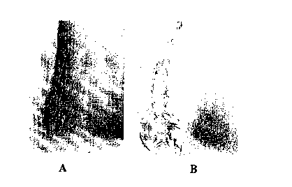
Fig. 6 is a collection of two photographs (40x
magnification) of tissue following 21 days of subdermal
implantation in the back of a rat where the tissue was
stained such that calcium appears dark: A) one surface
was covered with aluminum deposited fabric; B) control
tissue sample.
Fig. 7 is a collection of four photographs of
tissue following 26 days of subdermal implantation in
the back of a rat where the tissue was stained such that
calcium appears dark: A) one surface was covered with
aluminum deposited fabric; B) one surface was covered
with plain polyester fabric; C) one surface was covered
with aluminum/silver deposited fabric; D) one surface
was covered with plain polyester fabric.
DETAILED DESCRIPTION OF THE PREFERRED EMBODIMENTS
To impart a medical article with resistance to
calcification, the medical article can be supplied with
a deposit of elemental metal that gradually forms metal
ions upon oxidation. Deposits of anticalcific elemental
metal can provide a long lasting source of metal ions
that inhibit calcification. The quantity and type of
metal deposits can be selected to provide a desired
degree of calcification inhibition. The deposition of
anticalcific elemental metal can be combined with other
approaches to provide further improved calcification
inhibition.
A variety of medical articles can be used to
contact bodily fluids of a patient. Relevant medical
articles generally incorporate a biocompatible material
that is intended to contact the patient's biological
fluids and/or tissues. Bodily fluids include, for
example, blood, plasma, serum, interstitial fluids,
CA 02319527 2000-08-O1
- WO 99/38544 PCT/US99/01889
-10-
saliva and urine. The patient can be an animal,
especially a mammal, and preferably is a human.
Any degree of inhibition of calcium deposition
is useful, given the association between calcification
and deterioration of prostheses. A preferred degree of
inhibition results in a reduction of calcium deposition
by at least about 30 percent, preferably at least about
50 percent and more preferably at least about 75 percent
after about a one month period in contact with a
patient's bodily fluids and/or tissues, when compared
with a comparable medical article without deposits of
anticalcific elemental metal. The deposit of elemental
metal should not inhibit the mechanical functioning of
the medical device or provide a toxic level of metal
ions within the patient's fluids given the rates of
dissolution and the excretion of the metal by the
patient. Association of anticalcific metal with suture
should reduce the severe calcification associated with
passing suture through tissue.
In certain embodiments, biocompatible material
with deposits of anticalcific elemental metal is located
on the medical article such that this biocompatible
material is removed substantially from blood flow when
the medical article is used for its intended purpose.
In other words, when the medical article is in position
for use in contact with a patient's bodily fluids or
tissues, biocompatible material associated with the
medical article does not contact any blood flow except
possibly for a small portion of the biocompatible
material such as an edge of the material at a seam. In
other embodiments, the biocompatible material with
anticalcific elemental metal can be located completely
in a low blood flow area where the biocompatible
material experiences effectively no vascular blood flow.
CA 02319527 2000-08-O1
- WO 99/38544 PCT/US99/01889
-11-
Such medical articles can include additional portions of
biocompatible material with deposits of anticalcific
elemental metal.
Various methods can be employed for
associating elemental metal with the biocompatible
material of a medical article. Vapor phase methods
basically involve the accumulation of metal onto the
surface of the biocompatible material from a gas phase.
Other methods involve the reaction of metal solutions
with a chemical reductant. In addition, elemental metal
can be deposited by electrochemical reduction.
Particular methods may be more suitable for
the deposition of metal into and/or onto certain types
of biocompatible material. Using the various methods
described below, a large variety of materials car be
produced with associated anticalcific elemental metal.
In preferred embodiments, elemental metal is directed
specifically to or near portions of a medical article
that are particularly sensitive to calcification.
A. Biocompatible Articles
Relevant biocompatible articles include
medical articles that contact bodily fluids for extended
periods of time. The biocompatible articles can be made
from the biocompatible materials described below.
Relevant articles include, for example, implanted
devices and percutaneous devices. Medical articles of
particular interest are those susceptible to failure due
to calcification.
Implanted devices broadly include articles
that are fully implanted in a patient, i.e., are
completely internal. Implanted devices include, for
example, prostheses such as transplant organs, heart
valve prostheses, pericardial patches, vascular grafts,
CA 02319527 2000-08-O1
- WO 99/38544 PCT/US99/01889
-12-
biological conduits, annuloplasty rings, bone, skin,
ligaments and tendons.
Percutaneous devices include articles that
penetrate the skin, thereby extending from outside the
body into the body. Percutaneous devices include
without limitation catheters of various types.
Catheters can be used for accessing various bodily
systems such as the vascular system, the
gastrointestinal tract, or the urinary system.
Suture can be used, for example, to secure
sections of living tissue such as when closing a wound,
to fasten together components within a medical article
and/or to attach a medical article to living tissue.
Suture can be made from a variety of materials such as
collagen, polyesters, polypropylene, polyamides (nylon),
cat gut, coated cat gut, polydioximone,
polycaprolactone, polyhydroxy butyrate, polylactic acid
and polyglycolic acid. Therefore, in certain
applications, suture can be considered a component of a
larger medical article. In other applications, suture
can be considered an independent medical article. Since
its structure allows for a variety of uses, suture
cannot be classified exclusively as an implanted device
or as a percutaneous device . Other articles also may be
useful both as an implanted device and as a percutaneous
device.
Certain medical devices when used for their
intended purpose are located away from major blood
vessels. Other medical devices when used for their
intended purpose are associated with major blood
vessels. In general, medical devices associated with
major blood vessels have portions associated with high
blood flow and other portions in regions of low blood
flow, which are not in contact with blood flow through
CA 02319527 2000-08-O1
- WO 99/38544 PC1'/US99/01889
-13-
the vessel. Similarly, these medical devices can have
portions of biocompatible material substantially removed
from the blood flow that have an edge or the like within
the vessel in a region of high blood flow or in a region
with turbulent flow.
B. Biocompatible Materials
As noted above, the medical articles of
interest include biocompatible materials. Many medical
articles include several different types and/or separate
portions of biocompatible material that are fabricated
to form the medical article. Preferably, the
anticalcific elemental metal associated with a portion
or portions of biocompatible material is located at or
near sections of the medical article susceptible to
calcification. Tissue and polyurethane prosthetic
valves are particularly susceptible to calcification.
Appropriate biocompatible materials include
natural materials, synthetic materials and combinations
thereof. Natural, i.e., biological, material for use in
the invention includes relatively intact (cellular)
tissue as well as decellularized tissue. These tissues
may be obtained from, for example, natural heart valves;
portions of natural heart valves such as roots, walls
and leaflets; pericardial tissues such as pericardial
patches; connective tissues; bypass grafts; tendons;
ligaments; skin patches; blood vessels; cartilage; dura
mater; skin; bone; umbilical tissues; and the like.
Natural tissues are derived from a particular
animal species, typically mammalian, such as human,
bovine, porcine, seal or kangaroo. These natural
tissues generally include collagen-containing material.
Natural tissue is typically, but not necessarily, soft
tissue. Appropriate tissues also include tissue
equivalents such as tissue-engineered material involving
CA 02319527 2000-08-O1
WO 99/38544 PCTNS99/01889
-14-
a cell-repopulated matrix, which can be formed from a
polymer or from a decellularized natural tissue.
Biological tissues can be fixed by
crosslinking. This provides mechanical stabilization,
for example, by preventing enzymatic degradation of the
tissue. Glutaraldehyde is typically used for fixation,
but other fixatives can be used, such as epoxides,
formaldehyde and other difunctional aldehydes.
Biological materials can be used in either crosslinked
or uncrosslinked form, depending on the type of tissue,
the use and other factors.
Relevant synthetic materials include, for
example, polymers and ceramics. Appropriate ceramics
include, without limitation, hydroxyapatite, alumina and
pyrolytic carbon. Polymeric materials can be fabricated
from synthetic polymers as well as from purified
biological polymers. Appropriate synthetic materials
include hydrogels and other synthetic materials that
cannot withstand severe dehydration.
Appropriate synthetic polymers include without
limitation polyamines le.g., nylon), polyesters,
polystyrenes, polyacrylates, vinyl polymers le.g.,
polyethylene,polytetrafluoroethylene,polypropylene and
poly vinyl chloride), polycarbonates, polyurethanes,
poly dimethyl siloxanes, cellulose acetates, polymethyl
methacrylates, ethylene vinyl acetates, polysulfones,
nitrocelluloses and similar copolymers. These synthetic
polymeric materials can be woven into a mesh to form a
matrix or substrate. Alternatively, the synthetic
polymer materials can be molded or cast into appropriate
forms.
Biological polymers can be naturally occurring
or produced in vitro by, for example, fermentation and
the like. Purified biological polymers can be
CA 02319527 2000-08-O1
- WO 99/38544 PCT/US99/01889
-15-
appropriately formed into a substrate by techniques such
as weaving, knitting, casting, molding, extrusion,
cellular alignment and magnetic alignment. For a
description of magnetic alignments see, for example, R.
T. Tranquillo et al., Biomaterials 17:349-357 (1996),
incorporated herein by reference. Suitable biological
polymers include, without limitation, collagen, elastin,
silk, keratin, gelatin, polyamino acids, cat gut
sutures, polysaccharides (e. g., cellulose and starch)
and copolymers thereof. The biological polymers can be
resorbable.
Biocompatible materials can include a
combination of the various natural materials and
synthetic materials described above. The biocompatible
materials also can include metal portions. Mechanical
heart valves are relevant products, which generally are
made from metallic and/or ceramic components, along with
a sewing cuff and/or a vascular graft.
The biocompatible materials are combined to
form the medical article. For example, a mechanical
heart valve can include mechanical and ceramic
components that are located within the blood flow path
along with additional components for securing the valve .
Referring to Fig. lA, the cross section of one
embodiment of a mechanical heart valve 100 is depicted.
Heart valve 100 includes an orifice 102 that forms a
blood flow path through the interior 104 of orifice 102.
Heart valve 100 is depicted as a bileaflet valve with
two leaflets or occluders 106, 108 that pivot between an
open position and a closed position such that the blood
flow path through orifice 102 is correspondingly open or
closed.
Sewing cuff 110, which generally can be made
of fabric, is located at the exterior 112 of orifice 102
CA 02319527 2000-08-O1
- WO 99/38544 PCT/US99/01889
-16-
substantially out of the path of blood flow. The sewing
cuff is a potential site for calcification. Sewing cuff
108 can be surgically sutured to heart tissue to secure
valve 100. The invention also includes other designs
and/or types of mechanical heart valves.
Alternatively, heart valve prostheses can be
based on synthetic polymer leaflets, as depicted in Fig.
1B. Heart valve prosthesis 120 includes a stent 122
that provides support for the leaflets. Stent 122 can
be made from a variety of materials including, for
example, polymers, metals and combinations thereof.
Suitable synthetic polymers for use in forming stmt 122
include, for example, thermoplastics such as
polyolefins, polyesters, polyamides, polysulfones,
acrylics, polyacrylonitriles, acetal polymers such as
Delrin~, polyethers such as polyetheretherketone (PEEK),
and polyaramides. Leaflets 124 and stent 122 can be
made from synthetic polymers, which optionally can be
bioresorbable, being made from polymers such as
polyamino acids and/or polysaccharides. Preferred
nonresorbable polymers for incorporation into leaflets
124 include, for example, polyurethanes,
polyether/polyurethane block copolymers, silicone
elastomers, polytetrafluoroethylene and sulfur
crosslinked 1-hexene/methyl hexadiene copolymer.
Prosthesis 120 includes a fabric sewing cuff 126.
Referring to Fig. 2, heart valve prosthesis
130 is attached to a vascular graft 132 to configure the
valve as an aortic valued graft . Vascular graft 132 can
3b replace a portion of the blood vessel leading to valve
130. Heart valve prostheses, configured to replace
different natural valves such as pulmonary valves,
aortic valves, mitral valves and tricuspid valves,
CA 02319527 2000-08-O1
- WO 99/38544 PCT/US99/01889
generally include similar, appropriately located sewing
cuffs 134 substantially outside of the blood flow.
Referring to Fig. 3, annuloplasty ring 150 can
include a frame 152 covered with a layer of fabric 154
such as woven or knitted polyester. Fabric 154 can
cover the entire outer surface of annuloplasty ring 150.
Annuloplasty ring 150 can be implanted to support the
base of a native heart valve. Annuloplasty ring 150 is
located substantially outside of the direct blood flow.
An embodiment of a bioprosthetic heart valve
180 including a tissue component 182 is depicted in Fig.
4. The tissue component includes three leaflets 184,
186, 188 that function to open and close the valve and
cylindrical section 190 that defines a blood flow path
through the interior of the cylindrical section 190 with
f low controlled by the leaf lets 184 , 186 , 188 . Leaf lets
184, 186, 188 are attached to cylindrical section 190 at
commissures. Cylindrical section 190 includes an
annular portion and three commi,ssure supports. The
outside of cylindrical section 190 is covered with
fabric 192. Fabric 192 can be attached with suture 194
or using nonsuture fastening approaches. Fabric 192 is
outside of the blood flow in a low flow region when the
valve 180 is in place within the patient. As depicted
in the insert of Fig. 4, suture 194 can include a
coating 196 of anticalcific elemental metal.
C. Deposit of Anticalcific Elemental Metal
The approaches for applying deposits of
anticalcific metal to biocompatible materials can be
~ broadly classified according to whether the deposition
takes place from a vapor phase or from a liquid phase.
Anticalcific metals include, for example, aluminum,
iron, magnesium, zinc, gallium, lanthanum and beryllium
with aluminum, iron and magnesium being preferred.
CA 02319527 2000-08-O1
WO 99/38544 PCT/US99/01889
-18-
Various deposition approaches can be selected for use
with particular types of biocompatible materials. For
example, some methods may use conditions that are harsh
with respect to certain materials such that the
materials would be significantly degraded. In
particular, tissue generally cannot withstand the
conditions used for vapor phase metal deposition.
Vapor phase methods include, for example,
vapor-deposition, sputtering and magnetron sputtering.
Vapor phase techniques generally require varying degrees
of vacuum, i.e., low pressures. Some materials may not
tolerate the low pressures easily. Vapor based methods
are particularly suitable for the deposition of
anticalcific metal onto fabric. This coated fabric can
be incorporated into any of the medical articles
described above such as those depicted in Figs. 1-4.
Vapor deposition can simply involve directing
vaporized metal through an opening toward the substrate
to be metalized. Vapor deposition preferably is
performed using ion-beam-assisted deposition (IBAD)
under high vacuum as described, for example, in U.S.
Patent 5,474,797 to Sioshansi et al., incorporated
herein by reference. IBAD involves an evaporator that
forms a vapor of the desired metal. The metal vapor is
delivered to the substrate by a beam of ions formed from
one or more gases.
Solution based methods for anticalcific metal
deposition include chemical reduction and
electroplating. Suitable chemical reducing agents
include, for example, sodium borohydride, HZ and CO for
reduction of a variety of metals. Gaseous reducing
agents can be bubbled through the solution. Suitable
solvents are generally aqueous although other solvents,
such as alcohol, can be used if the biocompatible
CA 02319527 2000-08-O1
- WO 99/38544 PCT/US99/01889
-19-
material is not damaged by the solvent . When processing
tissue, it is preferred to keep the pH between values of
about 4 and about 11, and more preferably between about
7.0 and about 8.0, to the extent that the pH can be
adjusted within the particular processing approach.
Ionic strength can be adjusted, if desired, by the
addition of inert salts, the identity of which generally
depends on the nature of the deposition process and the
corresponding compositions.
Electrochemical deposition involves the
application of a voltage to a suitable biocompatible
material, such as tissue, in order to electroplate, from
a metal solution, elemental metal in contact with the
biocompatible material. The biocompatible material
functions as the cathode. The required voltage depends
on the counter reaction and the concentrations of ions
in solution. The selection of the metal salt influences
the effectiveness of the plating process.
To determine the amount of metal to deposit,
the rate of dissolution generally is a consideration.
The environment in which the biocompatible material is
placed can influence the rate of dissolution. Given a
particular rate of dissolution, the amount of deposited
metal establishes the length of time over which metal is
available for calcium inhibition.
With any method of deposition, the amount of
deposited metal should not interfere significantly with
important functionality of the biocompatible material.
If the conditions for depositing the elemental metal are
relatively harsh, it may be desirable to limit the
deposition time while accepting a corresponding decrease
in deposited metal. With respect to the deposition, the
amount of anticalcific metal generally is greater than
about 0.01 mg per gram of dry biocompatible material,
CA 02319527 2000-08-O1
- WO 99/38544 PC'T/US99/01889
-20-
and preferably from about 0.05 mg to about 40 mg per
gram of dry biocompatible material, and more preferably
from about 0.1 mg to about 20 mg per gram of dry
biocompatible material. When incorporated into a
medical article, the proportion of elemental metal for
the total quantity of biocompatible material can be less
than the above range since some of the biocompatible
material may not have deposits of elemental metal.
In general, the biocompatible material can be
subjected to deposition of elemental metal prior to,
during or after processing into a biocompatible article .
For example, to form a tissue heart valve prosthesis
with a fabric cover, the tissue component and the fabric
can be separately subjected to deposition of
anticalcific elemental metal using conditions suitable
for each material. Similarly, only the tissue or only
the fabric can be subjected to anticalcific metal
deposition. Following the desired deposition of
elemental metal, the tissue component and the fabric
components can be combined. Alternatively, the tissue
components and the fabric components can be formed into
a biocompatible article followed by the deposition of
anticalcific elemental metal on the article using a
suitable method for both materials.
Multiple elemental metals can be deposited.
For vapor phase techniques, the deposition of multiple
metals can be performed sequentially or simultaneously.
Generally, solution-based methods involve the sequential
deposition of the elemental metals. In addition,
different elemental metals can be incorporated onto
different portions of one or more sections of
biocompatible material for incorporation into a single
medical article.
CA 02319527 2000-08-O1
WO 99/38544 PCT/US99/01889
-21-
Multiple elemental metals can be deposited
such that the different metals are or are not in
electrical contact with each other. If the different
metals are in electrical contact, the oxidation
potential of one metal influences the rate of oxidation
of the other metal. In this way, the rate of oxidation
of one metal can be accelerated or slowed by the
selection of the second metal. The second metal can be
selected also to supply beneficial effects, as described
l0 below.
Combined Ant~icalcification A ents
Multiple anticalcific agents can be combined
to obtain greater anticalcific activity than that
provided by one of the agents alone. The additional
anticalcific agents can be elemental metal, other types
of chemical compositions or combinations thereof. The
deposition of multiple elemental metals has been
described above, where two or more elemental metals can
have anticalcific properties.
All of the considerations described above
apply equally if multiple elemental metals have
anticalcific properties. For example, if the metals are
in electrical contact, one metal generally is stabilized
in its elemental form while enhancing the oxidation of
the other metal. Therefore, the stabilized metal may
not be as effective as an anticalcific agent while the
other metal is present. Even if the elemental metals
are not in direct electrical contact, the presence of
the second elemental metal may influence the oxidation
rate and corresponding effectiveness as an anticalcific
agent. Two or more anticalcific elemental metals can be
combined with one or more additional elemental metals
that lack any appreciable anticalcific effectiveness.
The additional elemental metal or metals can introduce
CA 02319527 2000-08-O1
- WO 99/38544 PCT/US99/01889
-22-
a different activity such as antimicrobial
effectiveness, or can adjust the delivery or adhesion of
the anticalcific elemental metals.
Multiple metals can be placed in successive
layers, the metals can be simultaneously deposited to
create an amorphous surface, and/or they can be
patterned onto the substrate such that each metal
contacts a selected portion of the substrate. Solution
phase techniques generally are not used to pattern the
metals unless the metals are deposited onto portions of
substrate that are later attached to form the pattern.
The order of sequential deposition may be influenced by
the method used to deposit the elemental metals if, for
example, one elemental metal is unstable during the
deposition of the second metal. The placement of the
multiple metals generally is influenced by the effect on
the anticalcific effectiveness resulting from the
particular relationship between the metals.
An anticalcific elemental metal can be
combined with other chemical forms of anticalcific
agents. For example, the biocompatible material can be
treated with a solution of a compound including
anticalcific metal ions such as A1'3, Mg'2 or Fe'3. The
direct application of metal ions can provide a more
immediate anticalcific effect while the elemental metal
provides longer term anticalcific activity. Metal salt
concentrations of the salt solutions generally are
between 0.00001 and O.z molar, and preferably between
0.001 and 0.1 molar. Appropriate salts include, for
example, aluminum chloride, aluminum chlorate, aluminum
lactate, aluminum potassium sulfate, aluminum nitrate,
ferric chloride, ferric nitrate, ferric bromide, ferric
sodium edentate, ferric sulfate, and ferric formate.
CA 02319527 2000-08-O1
- WO 99/38544 PCT/US99/01889
-23-
The metal salts also can be incorporated into
a polymer matrix used in the prosthesis. The metal
salts are preferably added during the polymerization
step so that they are incorporated into the polymer
matrix. In this way, the calcification inhibitor is
released at a controlled rate over an extended period of
time.
In addition, anticalcific metal ions can be
supplied to the biocompatible material reversibly bound
to exogenous storage structures. Preferred exogenous
storage structures for the delivery of A1'3 and Fe'3
include, for example, ferritin and related metal storage
proteins. The ferritin can be attached to tissue and
other substrates by chemical crosslinking and the like.
The delivery of anticalcific metal cations using
exogenous storage structures is described in copending
and commonly assigned U.S. Patent Applications Serial
Nos. 08/595,402 and 08/690,661, both of which are
incorporated herein by reference.
Calcium ion chelators preferably at
concentrations between approximately 0.00001 M and
approximately 0.1 M can be added to the metal salt
solutions prior to treatment. For example, citrate
salts and citric acid have been found to enhance
synergistically the calcification inhibition effect of
A1'' and Fe'3 ions. Similarly, other calcium ion
chelators such as diphosphonate salts, including without
limitation ethanehydroxydiphosphonate (EHDP or
etidronate) and aminopropanehydroxydiphosphonate, also
produce a synergistic improvement in the
anticalcification effect of the Al'3 and Fe'' ions.
Higher or lower concentrations can be used in particular
applications.
CA 02319527 2000-08-O1
WO 99/38544 PCT/US99/01889
-24-
The order of application of multiple
anticalcific agents can influence the effectiveness of
a particular agent. The particular application
techniques can influence the selected order of
application such that one agent is not rendered
ineffective by the deposition of a second agent. These
factors can be examined empirically, if desired.
E. Other Biological Agents
Metals including Au, Ag, Pt, Pd, Ir, Cu, Sn,
Sb, Bi and Zn are known to yield antimicrobial activity,
with silver being preferred. When depositing multiple
elemental metals, one or more of the metal can be
selected for its antimicrobial efficacy. In this way,
the deposits of elemental metal can inhibit
calcification as well as inhibit infection. Electrical
contact of the elemental metals influences their
respective oxidation rates and their corresponding
efficacies.
In addition; metal compounds with
antimicrobial activity can be deposited. These metal
compounds can be deposited by precipitation of the
compound from a solution of a corresponding soluble
metal compound by the addition of a precipitation agent,
generally an appropriate anion or a reducing agent to
form a lower oxidation state metal ion. Deposition of
antimicrobial metal compounds is described further in
copending and commonly assigned U.S. Patent Application
Serial No. 08/974,992, incorporated herein by reference.
In addition, there are certain situations
where other biological activities are desirable. In
these situations, materials can be made by forming a
bioactive coating on a base material, where the
bioactive coating can include, for example, cell
CA 02319527 2000-08-O1
- WO 99/38544 PCf/US99/01889
-25-
adhesion molecules, anticoagulants such as heparin and
hirudin, or growth factors, and combinations thereof.
The order of application of the anticalcific
metal and bioactive coating can be selected based on
compatibility of the application methods. If
appropriate, the anticalcific metal and the bioactive
coating can be added simultaneously. Performance may be
influenced by the order of application of the different
active agents, and in such cases, the order of
application can be selected based on performance
considerations. Empirical evaluation of these factors
can be performed, if desired.
F. Storaae, Packaaina Distribution and Use
Following deposition of the desired
anticalcific elemental metal, the biocompatible
material, possibly formed into a medical article, is
stared. Preferred storage techniques minimize the risk
of microbial contamination. For example, the
biocompatible material can be stored in a sealed
container with an aqueous glutaraldehyde solution. In
a sealed container, the biocompatible material is not
subjected to a continuous supply of fluids. As a
result, corrosion of the anticalcific elemental metal
may be limited.
Due consideration should be given to possible
loss of the anticalcific elemental metal or other active
agents over time. If excessive corrosion is a
possibility, the storage time can be appropriately
limited to keep the corrosion to an acceptable level.
Additives can be added to reduce the corrosion. For
example, antioxidants such as ascorbic acid can be
added.
For distribution, the medical articles are
placed in sealed and sterile containers. The containers
CA 02319527 2000-08-O1
- WO 99/38544 PCT/US99/01889
-26-
generally are dated such that the date reflects the
maximum advisable storage time accounting for possible
degradation of anticalcific and other agents as well as
other factors. The containers are distributed to health
care professionals for use in appropriate medical
procedures such as surgical implantation of a prosthesis
and the like. The surgical implantation of heart
valves, such as those depicted in Figs. lA, 1B and 4, is
of particular interest.
The resulting prostheses with associated
anticalcific metals have advantages with respect to long
term durability. The anticalcific ions can be effective
to reduce calcification of tissue either by depositing
the metal on the tissue or by associating an
anticalcific metal coated material such as fabric with
the tissue. The method can involve relatively large
quantities of anticalcifics. The release rate of the
anticalcific ions can be adjusted by the selection of
metal or combination of metals or by pretreating the
metal. Furthermore, anticalcifics can be associated
with polyurethane heart valve prostheses and suture.
Anticalcific coated fabric and/or suture can be
associated with homografts or commercially available
heart valve prostheses. The medical articles of the
invention can include antimicrobial elemental metal
and/or an antimicrobial metal composition along with the
anticalcific elemental metal to reduce the risk of
infection as well as reducing calcification.
EXAMPLES
Example 1 - Washout Studies
This example involves a determination of the
rate of dissolution of aluminum from a coated fabric
when in contact with blood serum.
CA 02319527 2000-08-O1
WO 99/38544 PCT/US99/01889
-27-
Four pieces each of three types of fabric were
used. Each piece of fabric was about 1 square
centimeter. The first fabric, the control fabric, was
a woven double velour Dacron-polyester fabric obtained
from Meadox Medicals, Inc. (Lot 186116). The second
fabric (A1 fabric) was identical to the control fabric
except for a coating of elemental aluminum applied using
an Ion Beam Assisted Deposition (IBAD) Process such as
described in U.S. Patent 5,474,797, supra. In the IBAD
process, the substrate is mounted on a rotating
substrate holder within a vacuum chamber. A beam of
energetic ions directs evaporated metal atoms at the
substrate surface to form a coating of elemental metal
on the substrate. The IBAD aluminum deposition was
performed by Spire Corp., Bedford, MA.
The third fabric (A1/Ag fabric) first received
an antimicrobial coating including elemental silver,
titanium and palladium using a process developed by
Spire Corp. The three layer, metal coating is described
in U.S. Patent 5,520,664 to Bricault Jr., et al.,
incorporated herein by reference. Then, the silver
coated fabric received a further coating of aluminum
using the IBAD process, as described above. The
aluminum presumably was in electrical contact with the
silver, titanium and palladium metals.
All twelve fabric pieces were weighed after
they were excised. Then, the twelve fabric pieces were
sterilized with steam. Nine-500m1 bottles of bovine
serum (Sigma Chemical, St. Louis, MO) were obtained.
Five milliliters of serum were removed antiseptically
from each bottle and used as a "zero day" control. Each
of nine sterilized fabric samples was transferred
antiseptically under a laminar flow hood into a separate
serum bottle . After the fabric samples were placed into
CA 02319527 2000-08-O1
- WO 99/38544 PCT1US99/01889
-28-
serum bottles, the serum bottles were placed onto a
shaking water bath (Environ ShakerT'", Lab-Line, Melrose
Park, IL) set at. about 37°C and about 100 RPM.
Following 1, 2, 3, 4 and 7 days of incubation,
5 ml samples were removed antiseptically from each
bottle. The liquid samples were placed separately into
25 ml vials. The liquid controls and liquid samples
were subjected to analytical analysis for aluminum
content using an ICP-AES AtomScan 16T"' (Thermo Jarrell
Ash Corp., Franklin, MA). The results are presented in
Table 1 including averages and the standard deviation
(S.D.), and the average results are plotted in Fig. 5.
Table 1
A1 ( A1 (mg/ml)
pm)
1 Time Sample Sample Sample Ave. S.D. Average S.D.
5 (da #1 #2 #3
s)
A1/Ag
Fabric
0 0.1076 0.1244 0.1734 0.14 0.03 2.70E-046.84E-05
1 0.1118 0.1179 0.2095 0.15 0.05 2.93E-041.09E-04
2 2 0.1619 0.1675 0.2150 0.18 0.03 3.63E-045.84E-05
0
3 0.2100 0.1722 0.3790 0.25 0.11 5.07E-042.21E-04
4 0.4316 0.4289 0.5162 0.46 0.05 9.18E-049.93E-05
7 0.4696 0.5601 0.5923 0.54 0.06 1.08E-031.27E-04
A1
Fabric
2 0 0.0713 0.0899 0.0796 0.08 0.00 1.61E-043.21E-07
5
1 0.13 0.1518 0.1491 0.14 0.01 2.89E-042.OlE-05
2 0.15 0.1741 0.1692 0.17 0.01 3.30E-042.36E-05
3 0.18 0.1857 0.1716 O.1B 0.01 3.57E-041.42E-05
4 0.20 0.2274 0.1929 0.21 0.02 4.10E-043.85E-05
7 0.56 0.5870 0.5670 0.57 0.01 1.14E-032.59E-05
Control
Fabric
0 0.21 0.1521 0.0845 0.15 0.06 2.97E-041.25E-04
1 0.18 0.15 0.1376 0.16 0.02 3.13E-044.58E-OS
2 0.0543 0.077 0.1138 0.08 0.03 1.63E-046.OlE-05
3 3 0.1297 0.1716 0.1276 0.14 0.02 2.86E-044.96E-05
5
CA 02319527 2000-08-O1
WO 99/38544 PCT/US99/01889
-29-
4 0.142 0.1506 0.13340.14 0.01 2.84E-041.72E-05
7 0.1197 0.0636 0.02930.07 0.05 1.42E-049.13E-05
Significant concentrations of aluminum were present in
the serum with both the A1 fabric and the A1/Ag fabric
by 4 days within the serum. While the A1/Ag fabric
released greater amounts of aluminum than the A1 fabric
after 4 days within the serum, by seven days the A1
fabric and the A1/Ag fabric released comparable
quantities of aluminum into the serum.
Following seven days of incubation, the fabric
samples were removed from the serum and dried with a
lyophilizer. The fabric samples along with comparable
pieces that had not been placed in serum were analyzed
for aluminum content. To analyze the fabric samples,
the fabric pieces were hydrolyzed in nitric acid. Then,
measurements were made using ICP-AES, as described
above. The results are presented in Table 2, where the
weights were measured before serum contact.
Table 2
Sample wt (mg) Al(ppm) Al(mg/g}
A1/Ag Fabric
No Serum
Contact
1 5.24 0.61 2.90
2 6.61 0.73 2.75
3 6.19 0.74 2.99
2.88(Avg.)
- 0.12(S.D.)
Post Serum
Contact
1 7.88 0.70 2.23
2 12.08 1.08 2.23
CA 02319527 2000-08-O1
- WO 99/38544 PCT/US99/01889
-30-
3 10.60 0.63 1.50
1.99(Ave)
0.42(S.D.)
A1 Fabric
No Serum
Contact
1 7.76 2.72 8.76
2 10.85 4.01 9.23
3 10.05 3.81 9.48
9.16(Ave)
0.36(S.D.)
Post Serum
Contact
1 13.26 4.05 7.63
2 10.45 3.59 8.59
3 9.25 2.67 7.21
7.81(Ave)
0.71(S.D.)
Control Fabric
No Serum
Contact
1 13.82 0.04 0.08
2 17.09 0.04 0.05
3 14.98 0.04 0.06
0.07(Ave)
0.01(S.D.)
Post Serum
Contact
1 17.29 0.03 0.05
2 17.53 0.03 0.04
3 17.36 0.02 0.04
0.04(Ave)
0.01(S.D.)
CA 02319527 2000-08-O1
- WO 99/38544 PCT/US99/01889
-31-
The results in Table 2 indicate that A1 was released
into the serum.
Example 2 - In vivo Studies
This example demonstrates an in vivo reduction
of calcification of aluminum coated fabric. Two sets of
experiments were performed using similar procedures.
Samples were prepared from 8mm punches of
porcine aortic root tissue. The tissue samples were
crosslinked in buffered 0.5% glutaraldehyde solutions.
In the first study, twelve samples were used. Six
samples were sewn to aluminum coated fabric, and six
samples were sewn to polyester fabric, as controls.
After sewing the tissue to the fabric, the samples were
placed in buffered glutaraldehyde.
For the second study, a total of thirty six
tissue samples were used. A piece of fabric was sutured
to each tissue sample. Twelve tissue samples were
sutured to plain, polyester fabric. Twelve tissue
samples were sutured to aluminum coated polyester
fabric, where the A1 fabric was prepared as described in
Example 1. The remaining twelve tissue samples were
sutured to aluminum/silver coated fabric, where the
A1/Ag fabric was prepared as described in Example 1.
All the samples were stored for twelve days in a HEPES
buffered saline solution containing 0.5% glutaraldehyde
prior to implantation.
Prior to implantation, all of the samples were
rinsed three times for 2-5 minutes using sterile saline .
The 12 samples in the first study were placed
subdermally in the backs of three juvenile male rats
using color coded suture . The thirty six samples in the
second study were placed subcutaneously in the backs of
six juvenile male rats (two of each type per rat) using
color codes suture. The samples were removed after 21
CA 02319527 2000-08-O1
WO 99/38544 PCT/US99/01889
-32-
days (first study) or 26 days (second study) . Following
removal the samples were placed in 0.9 percent saline
(NaCl in H20) prior to analysis.
For analysis, each tissue sample was sectioned
in half. One half of each sample was cleaned of host
capsule. For the first study, the fabric was removed
from all the samples. For the second study, the fabric
was removed from the control samples while the fabric
was left attached to the other samples. The tissue and
fabric were placed into a polypropylene test tube
(separately if detached) and lyophilized. For elemental
analysis, the dried samples were hydrolyzed in nitric
acid. Elemental analysis was performed by ICP-AES, as
described above. The results of the elemental analysis
are presented in Table 3 (first study) and Table 4
(second study). For the second study, the calculations
were adjusted to remove approximately the contribution
of the fabric, which calcifies significantly less
relative to the calcification of the tissue.
Table 3
Sample Weight CALCIUM ALUMINUM
mg PPS ~J/J PPS ~g/J
Control-Tissue
1 23.6 55.29 58.57 0.0486 0.05
2 22.1 50.8 57.47 0.0507 0.06
3 20.6 30.19 36.64 0.0521 0.06
4 15.8 43.04 68.10 0.0516 0.08
5 24.7 45.65 46.20 0.0496 0.05
6 18.8 48.81 64.91 0.0462 0.06
average= 55.31 0.06
std dev= 11.85 0.01
CA 02319527 2000-08-O1
- WO 99/38544 PCT/US99/01889
-33-
A1-Tissue
1 20:6 32.82 39.83 0.0621 0.08
2 18.2 36.13 49.63 0.0554 0.08
3 19.7 31.86 40.43 0.0564 0.07
4 15.2 23.34 38.29 0.0446 0.07
5 18.2 35.12 48.24 0.0641 0.09
6 22.3 40.64 45.56 0.0627 0.07
average= 43.68 0.08
std dev= 4.76 0.01
A1-Fabric
1 3.5 0.0913 0.65 0.7272 5.19
2 3.3 0.0644 0.49 0.7267 5.51
3 3.6 0.1293 0.90 0.7341 5.10
4 2 0.0461 0.58 0.414 5.18
5 1.9 0.0289 0.38 0.4729 6.22
6 3.4 0.0762 0.56 0.7755 5.70
average= 0.59 5.48
std dev= 0.18 0.48
Table 4
Sample Weight ALUMINUM CALCIUM
mg PPm mg/g PPm mg/g
Control-Tissue
1 31.75 0.0254 0.02 98.04 77.20
2 25.78 0.03 0.03 64.89 62.93
3 18.22 0.03 0.04 59.74 81.97
4 20.43 0.04 0.04 63.08 77.19
5 20.52 0.03 0.03 47.84 58.28
6 25.21 0.03 0.03 68.19 67.62
7 22.44 0.0328 0.04 54.5 60.72
CA 02319527 2000-08-O1
WO 99/38544 PCT/US99/01889
-34-
8 19.74 0.0347 0.04 68.79. 87.12 II
9 26.16 0.031 0.03 72.43 69.22
30.33 0.0325 0.03 67.69 55.79
11 15.24 0.0269 0.04 46.14 75.69
5 12 ~ 24.17 0.0365 0.04 47.59 49.22
average= 0.03 68.58
std dev= 0.01 11.53
A1/Ag
1 23.69 0.88 0.93 33.10 42.11
10 2 20.72 0.08 0.10 42.85 64.22
3 27.22 0.09 0.08 25.94 27.98
4 20.97 0.07 0.08 31.27 46.18
5 27.21 0.12 0.11 35.95 38.79
6 21.69 0.08 0.09 34.39 48.71
7 25.71 0.11 0.10 25.42 29.33
8 26.93 0.07 0.07 45.16 49.32
9 22.03 0.09 0.10 36.62 50.89
10 19.99 0.08 0.10 19.64 30.78
11 22.57 0.08 0.08 25.36 34.21
12 23.06 0.09 0.10 29.93 39.34
average= 0.16 41.82
std dev= 0.24 10.66
A1
1 33.41 0.8159 0.61 54.2 46.14
2 21.83 0.77 0.88 8.96 12.59
3 28.2 0.77 0.68 34.69 35.90
4 20.12 0.58 0.71 43.19 67.15
5 31.78 0.58 0.46 30.68 27.65
6 29.73 0.78 0.65 33.79 32.88
7 24.65 0.5641 0.57 16.3 19.77
CA 02319527 2000-08-O1
WO 99!38544 PCT/US99/01889
-35-
8 18.19 0.0638 0.09 20.99 37.08
9 22.79 0.3745 0.41 31.73 42.31
22.31 0.5504 0.62 34.81 47.63
11 18.12 0.4201 0.58 13.59 24.13
5 12 21.96 0.6577 0.75 35.2 49.11
average= 0.59 36.86
std dev= 0.20 14.91
The results from the first study are at the
10 edge of statistical significance with respect to
demonstrating calcium reduction. The results from the
second study do show clear statistical reductions in
calcification for tissue associated with aluminum coated
fabric. The improvements observed in the second study
relative to the first study may be due to the storage of
the samples for twelve days in a saline buffered
glutaraldehyde solution prior to implantation. The
extended period of time prior to implantation may have
accelerated the corrosion process making more
anticalcification ions present.
The second half of each sample was placed in
10% buffered formalin (first study) or HEPES buffered-
0.5 % glutaraldehyde solution (second study) prior to
histological examination. The histological analyses
were performed using von Kossa stain. Exemplary
photomicrographs from the first study are shown in Fig.
6. Referring to Fig. 6A, the control tissue had a band
of continuous calcification at the outer surface.
- Referring to Fig. 6B, a transition area can be seen
between the calcified and noncalcified tissue on the
fabric coated side of the sample. Calcification was
significantly, if not completely, mitigated in the areas
where the fabric was sutured to the tissue. In the
CA 02319527 2000-08-O1
- WO 99/38544 PCT/US99/01889
-36-
center of the sample, where there was no suture, some
calcification near the surface of the sample occurred.
Exemplary micrographs from the second study
are shown in Fig. 7. It can be seen that the samples
with A1 fabric (Fig. 7A) and A1/Ag fabric (Fig. 7C) have
less calcium, shown in dark, deposited in the fabric
side of the tissue than deposited in the corresponding
control fabric samples (Figs. 7B and 7D).
Example 3 - Pretreated Surface - Washout Studies
This example involves evaluating the degree of
ionization of an aluminum coating following pretreatment
of the aluminum coating.
Washout studies were performed with 24 strips
of fabric each about 2 cm wide and about 3.75 cm long.
Half of the strips were A1 coated fabric and the other
half were A1/Ag coated fabric identical to the
comparable fabric used in Example 1. Three strips of
each type received no pretreatment and served as
controls. Another three strips of each type received 20
cuts each all the way through the fabric to increase the
surface area exposed to oxidation. The cuts were made
ten per edge across about 3/4 of the width of the fabric
strip.
Also, three strips of each type were exposed
to 3.2% peroxyacetic acid for about 5 minutes. The
three strips of each type were exposed together to 12.5
ml of the peroxyacetic acid. The 3.2% peroxyacetic acid
was prepared by diluting 2.5 mls of a stock solution of
32% by weight peroxyacetic acid, Aldrich Chemical Co.,
Milwaukee, WI, to 25m1s with purified water.
The remaining three strips of each type were
exposed to 10% HC1 far 5 minutes. Again, the three
strips of each type were exposed together to 12.5 mls of
acid. The 10% HC1 solution was prepared by diluting 6.5
CA 02319527 2000-08-O1
WO 99/38544 PCT/US99/01889
-37-
mls of concentrated HC1, Fisher Chemical, Fair Lawn, NJ,
with sufficient purified water to fill a 25 ml
volumetric flask. The strips treated with either
peroxyacetic acid or hydrochloric acid were immediately
removed from the respective acid and rinsed three times
each with 150 ml of purified water with the second and
third rinses taking 5 minutes and 10 minutes,
respectively.
The washout study was performed using the 24
fabric strips. Each strip was placed in 1 liter plastic
containers with 500m1s of 0.9% (NaCl) sterile saline
from Baxter, Deerfield, IL. The containers with the
strips and saline were placed on a shaker and incubated
at 37°C. At 0, 3, 5 and 7 days, 5mls of liquid was
removed from each container and analyzed for aluminum
content. The results for the A1 coated fabric are
presented in Table 5, and the results for the Al/Ag
coated fabrics are presented in Table 6.
Table 5 - A1 Fabric
A1 (ppm)
Time Sample Sample Sample Average S.D.
(days) #1 #2 #3
Control
0 0.10 0.10 0.09 0.10 0.00
3 0.12 0.12 0.14 0.12 0.01
5 0.16 0.14 0.13 0.14 0.01
7 0.22 0.21 0.18 0.20 0.00
Cut
0 0.09 0.07 0.08 0.08 0.01
3 0.09 0.08 0.16 0.11 0.04
5 0.10 0.10 0.19 0.13 0.05
7 0.15 0.12 0.22 0.16 0.00
CA 02319527 2000-08-O1
- WO 99/38544 PCT/US99/01889
-38-
Peroxyacetic
Acid
0 0.10 0.09 0.09 0.09 0.01
3 0.12 0.09 0.10 0.10 0.02
0.13 0.13 0.13 0.13 0.00
5 7 0.35 0.38 0.32 0.35 0.03
Hydrochloric
Acid
0 0.10 0.09 0.09 0.09 0.00
3 0.15 0.10 0.11 0.12 0.03
5 0.23 0.17 0.12 0.1? 0.05
7 0.33 0.32 0.28 0.31 0.03
Table 6 - A1/Ag Fabric
A1 (ppm)
Time Sample Sample Sample Average S.D.
(days) #1 #2 #3
Control
0 0.08 0.11 0.07 0.09 0.02
3 0.07 0.13 0.11 0.10 0.03
5 0.16 0.13 0.12 0.14 0.02
7 0.48 0.46 0.43 0.46 0.02
Cut
0 0.08 0.09 0.09 0.09 0.00
3 0.15 0.13 0.11 0.13 0.02
5 0.17 0.14 0.16 0.15 0.01
7 0.54 0.42 0.41 0.46 0.07
Peroxyacetic
Acid
0 0.12 0.08 0.10 0.10 0.00
3 0.21 0.16 0.15 0.17 0.03
5 0.21 0.17 0.21 0.20 0.02
7 0.51 0.55 0.46 0.51 0.04
CA 02319527 2000-08-O1
- WO 99/38544 PC'T/US99/01889
-39-
Hydrochloric
Acid
0 0.13 0.10 0.07 0.10 0.03
3 0.19 0.20 0.10 0.16 0.06
0.23 0.22 0.19 0.21 0.02
5 7 . 0.48 0.46 0.56 0.50 0.05
Note that the acid pretreatments significantly
increase the ionization of the Al coated fabric but not
the A1/Ag coated fabric. Contrary to the results in
Example 1, the A1/Ag fabric resulted in greater aluminum
ionization than observed for the A1 coated fabric . This
difference may be due to the fact that the washout study
in Example 3 was performed using saline rather than
bovine serum. Bovine serum contains biological
chelators such as transferrin, which may accelerate the
delivery of metals into solution.
The embodiments described above are intended
to be exemplary and not limiting. Additional
embodiments are within the claims.