Note : Les descriptions sont présentées dans la langue officielle dans laquelle elles ont été soumises.
CA 02321690 2000-08-30
WO 99/44743 PCT/US99/04520
Disposable Apparatus for Performing Blood Cell Counts
BACKGROUND OF THE INVENTION
1. Technical Field
The present invention relates to apparatus for analyzing biologic fluid
samples
in general, and to containers for holding a biologic fluid sample during
analytical
procedures in particular.
2. Background Information
1o Most analytical methods for evaluating constituents within a biologic fluid
sample require that the sample be substantially diluted prior to evaluation. A
typical
chemical analysis, for example, involves placing a substantially diluted
sample into a
transparent cuvette of known dimensions and constant light path for
evaluation. The
cuvette can be made from glass or a hard acrylic that is ground or otherwise
manufactured to tight tolerances. The tight tolerances, which are necessary to
insure
the accuracy of the light path through the cuvette, also make the cuvette
undesirably
expensive. In hematological analyses, a substantially diluted sample is
typically passed
through a flow cell within an optical flow cytometer or through an impedance
orifice in
an impedance type flow cytometer. Most flow cytometers require mechanical
2o subsystems to dilute the sample, to control the sample flow rate through
the flow cell,
and multiple sensors to evaluate the diluted sample. A special problem
associated with
hematology measurements is the wide dynamic range of particles that must be
enumerated. The red blood cells (BBC's) are the most numerous at about 4.5 x
106
per microliter (pl), followed by the platelets at about 0.25 x 10⁶ per ~1, and
the white
blood cells (WBC's) at 0.05 x 10⁶ per pl. Since all cells or particles must be
enumerated during a full analysis, the range of cells/particles necessitates
at least two
dilution levels. The ability to perform multiple dilutions undesirably adds to
the
complexity of the machine. A person of skill in the art will recognize
disadvantages
associated with flow cytometers including plumbing leaks and inaccuracies due
to fluid
3o control nuscalibration. In both of the aforementioned analyses, the
operator (or the
apparatus itself] must purge the biologic fluid sample from the apparatus and
thoroughly clean the apparatus to avoid contaminating subsequent analyses. The
CA 02321690 2000-08-30
WO 99/44743 PCT/US99/04520
2
substantial dilution required in both analyses also increases the likelihood
of error, the
complexity of the analysis, and the per analysis cost.
Other analytical methods minimize the above described problems by employing
a disposable sample analytical chamber. In one chemical analytical method, the
biologic fluid sample is placed in a flexible sealed pouch where it remains
during the
analysis. This approach avoids the need for plumbing, flow controls, and
cleaning the
container, but requires a large diluent volume and is restricted to standard
measurements of light transmission. In the above method, the light path
dimensions
are controlled by the analytical instrument, which form the flexible pouch
into a cuvette
of the desired thickness at the time of measurement. Other, similar 'vet
chemical",
systems employ a rigid analytical cuvette of specifically manufactured
thickness. Other
methods for performing a chemical analysis on a biologic fluid sample employ
single or
multiple test film substrates. The test film substrates also avoid the
problems
associated with dilution, flow controls, etc., but still require precise
sample
measurement and placement and are also limited to those analyses that employ
light
reflectance. The test film substrate methods are further limited by requiring
that the
associated disposable always have identically located analyticat regions; if
the desired
information is not present in the predetermined analytical areas, then the
test film
substrate will not yield useful information. Hematological analytical methods
which
2o employ a disposable sample analytical chamber include the HemaCueTM and the
QBCTM. The HemaCueTM system is a method for measuring hemoglobin using a small
cuvette. The HemaCueTM method is particularly useful for its intended propose,
but it
is unable to measure particulate constituents of whole blood. The QBCTM
system, a
registered trademark of Becton Dickinson and Company of Franklin Lakes, New
2s Jersey, USA involves placing a hematological fluid sample within a
cylindrical tube and
centrifuging the tube and sample for a given period of time. The centrifuge
process
separates fluid sample constituents into layers according to their density. A
float
disposed within the tube facilitates evaluation of constituents within each
layer.
Specific hematological tests may be performed in a disposable test system
employing a
3o scannable optical window in a device produced by Biometric Imaging. In this
device, a
substantially undiluted sample of whole blood is placed into a capillary of
known and
constant dimension where it is subjected to a laser scan which identifies some
sub-
CA 02321690 2000-08-30
WO 99/44743 PCT/US99/04520
3
types of WBC's. The Biometric Imaging method is also limited in that it is
unable to
measure any other constituents of whole blood.
Serologic or immunologic analyses measure soluble substances in blood,
usually proteins such as specific immunoglobulins. These tests are often
performed by
admixing the sample with a sensitized particulate, such as latex, which will
agglutinate
in the presence of the protein of interest. Another method for performing a
more
quantitative immunological analysis is to use enzymatically linked color
changes, such
as ELISA. All of these methods are performed on apparatus specialized for
their use.
Another common specialized test is urinalysis. The analysis of urine is
1o generally divided into two separate phases: the determination of the bulk
and/or
chemical properties of the sample and the analysis of particulates within the
sample.
These analyses require distinctly different disciplines and are usually done
separately.
There are large and complicated machines that can perform both types of
analyses, but
they are extremely expensive and require moderate maintenance and operator
skill.
None of the above described analytical methods is capable of performing
hematological, chemicaUimmunochemical, and serologic analyses on sample
constituents within the same instrument. As a result, it has been necessary to
purchase
apparatus devoted to performing chemical analyses and apparatus devoted to
performing hematological analyses. It has also been necessary to train
technicians to
operate the various types of apparatus, and to provide laboratory space and
maintenance for them. It has also been impossible to combine hematological and
chemical analyses in the same apparatus for those analyses where it would be
advantageous to combine the analyses. In an analysis to determine anemia, for
example, it is preferable to perform both hematological analyses (e.g.,
hemoglobin,
hematocrit, and reticulocyte count) and chemical or immunochemical analyses
(e.g.,
iron or ferritin, and/or vitamin B 12 or folate determinations) on the sample.
None of
the above described methods permit hematological and chemical analyses on a
single
sample of blood in a single disposable sample chamber. As a result, the
laboratory
technician must separate and transport the various samples to their separate
3o instruments which are often in separate laboratories, thereby increasing
the ine~ciency
of the process as well as the potential for loss or misidentification of the
sample. Also,
CA 02321690 2000-08-30
WO 99/44743 PCf/US99/04520
4
the results of the analyses may not be available at the same time which
increase the
difficulty of interpreting the analysis results.
What is needed is a single container for holding a biologic fluid sample that
can
be used for multiple analyses including but not limited to hematological,
chemical,
immunological, serological, and urine analyses, one in which multiple analyses
can be
performed on the same sample in one instrument which presents a common
operator
interface, one that is operable with substantially undiluted biologic fluid
samples, one
whose method of sample introduction into the container is similar for each set
of
analyses, and one that can be used e$'ectively as a disposable.
DISCLOSURE OF THE INVENTION
It is, therefore, an object of the present invention to provide a container
for
holding a biologic fluid sample which permits analysis of multiple
constituents within
the same sample, and in particular analysis of constituents residing
individually or in
is groups, using quantitative image analysis.
It is another object of the present invention to provide a container for
holding a
biologic fluid sample for analysis which is operable for analyses which
require
information related to the bulk and/or chemical properties of the sample and
those
which require information related to the particulates content of the sample.
2o It is another object of the present invention to provide a container for
holding a
biologic fluid for analysis which is operable in multiple analytical
disciplines including
but not limited to hematology, chemical/immunochemical,
serology/immunological,
and urinalysis.
It is another object of the present invention to provide a container which can
25 include analytical chambers of varying dimensions whose thickness can be
correlated to
a spatial coordinate in order to encompass a wide dynamic range of contained
particulates.
It is another object of the present invention to provide a container for
holding a
biologic fluid sample that requires only a single instrument to sense for
information
3o within the sample or associated with the container and interpret the sensed
information
for use in multiple analyses, thereby decreasing the training and quality
control
requirements of the laboratory.
CA 02321690 2000-08-30
WO 99/44743 PCTNS99/04520
It is another object of the present invention to provide a container for
holding a
biologic fluid sample for analysis that can be used effectively as a
disposable.
It is another object of the present invention to provide a container for
holding a
biologic fluid sample for analysis that does not require substantial dilution
of the
sample before analysis.
It is another object of the present invention to provide a container for
holding a
biologic fluid sample which is operable with minimal quantities of blood or
other
biologic fluid.
It is another object of the present invention to provide a container for
holding a
biologic fluid sample that facilitates safe handling of the biologic fluid
sample for the
test operator.
It is another object ofthe present invention to provide a container for
holding a
biologic fluid sample that includes an analytical area suitable for imaging by
a digital
camera or other digital imaging device/image dissector which produces output
suitable
for quantitative analysis.
It is another object of the present invention to provide a container for
holding a
biologic fluid sample that has the capability of retaining an untreated or a
substantially
undiluted sample prior to analysis and releasing said sample into the
analytical region
when needed.
2o It is another object of the present invention to provide a container for
holding a
biologic fluid sample that carries indicia which provides information to the
instrument
of use in performing the analysis(es) at hand.
According to the present invention, a container for holding a biologic fluid
sample for analysis is provided which includes a chamber and a label. The
chamber
includes a first wall, a transparent second wall, and a plurality of features
including
features spatially located within the chamber. The transparent second wall
permits a
fluid sample quiescently residing within the chamber to be imaged through the
second
wall. The plurality of features, including those spatially located within the
chamber,
are operable to enable the analysis of the biologic fluid. The features may,
for
3o example, include regions where chemical constituents are analyzed, chambers
of
varying height and size where particulates may be analyzed, and regions which
allow
the calibration or quality control of the analysis. The features may also
include
CA 02321690 2004-04-29
information that is useful to set-up, adjust, andlor calibrate the analytical
device to the
task at hand; e.g., filter alignment, lens adjustment, etc. The label directly
or indirectly
contains information regarding the features and the spatial location of the
features
within the chamber. The sample is analyzed by an analytical device that
utilizes the
information communicated through the label.
The preferred analytical device for use with the present invention container
includes a Reader Module, a Transport Module, and a Programmable Analyzer. The
Reader Module includes optics which are operable to image a field within the
container, and apparatus to access information through the label attached to
the
container. The Transport Module includes apparatus for moving the container
relative;
to the Reader Module, or vice versa. The Programmable Analyzer is programmed
with
instructions to coordinate the operation of the Reader Module and Transport
Module
according to a variety of analysis algorithms. Which analysis algorithms are
used is
determined by reading the container label.
In another aspect, the present invention provides a container for holding a
biologic fluid sample for analysis, said container comprising: fluid holding
chamber
having a first wall and a transparent second wall, and wherein the fluid
holding
chamber has a mapped interior so that positions within the fluid holding
chamber are
identifiable by a coordinate address; at least one feature operable to enable
a
determination of the volume of a field of the fluid sample, the at least one
feature
located within the chamber at a predetermined coordinate address; and a label
attached
to said container, said label operable to supply the predetermined coordinate
address
within the fluid holding chamber.
In another aspect, the present invention provides a container for holding a
biologic fluid sample for analysis, said container comprising: a fluid holding
chamber
having a first wall and a transparent second wall, and wherein the fluid
holding
chamber has a mapped interior so that positions within the fluid holding
chamber are
identifiable by a coordinate address; wherein the fluid holding chamber has at
least a
first through-plane thickness and a second through-plane thickness, extending
between
the first wall and the second wall, wherein the first through-plane thickness
and the
second through-plane thicknesses are each located within the chamber at
predetermined
coordinate addresses, and the first through-plane thickness is greater than
the second
6
CA 02321690 2004-04-29
through-plane thickness; and a label attached to the container, which label is
operable
to supply the predetermined coordinate addresses of the first through-plane
thickness
and the second through-plane thickness within the fluid holding chamber.
In another aspect, the present invention provides a container for holding a
biologic fluid sample for analysis, said container comprising: a fluid holding
chamber
having a first wall and a transparent second wall, and wherein the fluid
holding
chamber has a mapped interior so that positions within the fluid holding
chamber are
identifiable by a coordinate address; wherein the fluid holding chamber has: a
first
through-plane thickness extending between the first wall and the second wall,
wherein
the first through-plane thickness is located within the chamber at a first
predetermined
coordinate address; and a second through-plane thickness extending between the
first
wall and the second wall, wherein the second through-plane thickness is
located within
the chamber at a second predetermined coordinate addresses; a first reagent
located
within the chamber at the first predetermined coordinate address; a second
reagent
located within the chamber at the second predetermined coordinate address;
wherein
the first predetermined coordinate address and the second predetermined
coordinate
address are separated from each other within the chamber by a distance great
enough
such that an analysis of each of the I reagents with the fluid sample can be
completed
without interference from the other of the reagents; and a label attached to
the
container, which label is operable to supply the predetermined coordinate
addresses of
the first through-plane thickness and the second through-plane thickness
within the
fluid holding chamber.
In another aspect, the present invention provides a container for holding a
biologic fluid sample for analysis, said container comprising: a fluid holding
chamber
having a first wall and a transparent second wall, and wherein the fluid
holding
chamber has a mapped interior so that positions within the fluid holding
chamber are
identifiable by a coordinate address; means operable to enable a determination
of the
volume of a field of the fluid sample, the means located within the chamber at
a
predetermined coordinate address; and a label attached to said container, said
label
operable to supply the predetermined coordinate address within the fluid
holding
chamber.
6a
CA 02321690 2004-04-29
An advantage of the present invention container is that it is operable for a
variety of analyses including but not limited to hematological, chemical,
immunochemical, serologic, urinalysis and immunological analyses. In addition,
it is
possible to perform a multitude of those analyses on the same sample, in the
same
analytical device. Some traditional analysis methods pass light into a
cuvette, and
interpret the light traversing through or emitting from the cuvette to provide
analytical
data. Other methods, such as those which utilize film substrates for analyzing
sample;
constituents utilize light reflected from the film layer. The data available
using these
types of methods is relatively uniform and does not contain any spatial
information.
Thus, they are useful for analyzing bulk properties of the sample, meaning
those
properties that are distributed uniformly in solution or in suspension, but it
is
impossible to derive useful data about individual particulate materials within
the
sample. The absence of spatial information limits the number of tests possible
on a
given sample. If a sample is tested for optical density using the above
described
cuvette, for example, the test parameters will provide information about a
particular
6b
CA 02321690 2000-08-30
WO 99/44743 PCT/US99/04520
constituent, but will not provide the information necessary to characterize
cellular
contents. The present invention container, in contrast, includes a analytical
chamber
which includes features that enable the Reader Module to extract both spatial
information and quantitative photometric information from the sample
quiescently
residing within the sample. The ability to analyze both types of information
aliows the
combination of the instrument and the disposable to analyze a large number of
different
constituents, and consequently perform a far greater number of tests.
The ability to perform different discipline analyses, for example
hematological
and chemical analyses, is significant for several reasons. First, the amount
of
1o equipment required to do the same number of analyses is reduced
significantly. It
follows that the cost of procuring and maintaining that equipment is similarly
reduced.
Also, the personnel training required to operate the equipment is reduced.
Another
reason is the versatility provided by a device that can perform different
discipline
analyses. Many clinical offices and laboratories are presently unable to
justify the
t5 office space and expense associated with available test apparatus for each
analytical
discipline. With the versatile present invention, however, it will be possible
to have
greater in-house analytical ability because of the present invention's
relative minimal
space requirements and low cost.
Another advantage of the present imrention is that a disposable container for
2o holding, analyzing, and disposing of a biologic fluid sample for analysis
is provided.
The present invention container is independent of the analytical device,
inexpensive,
readily loaded, and easily handled by an automated analytical device. These
characteristics make the present container a desirable disposable. As a
disposable, the
present invention obviates the need to clean the sample chamber after each use
and
25 therefore the opportunity for contamination from the prior sample. The
disposable
nature of the present invention container also facilitates safe handling of
the biologic
fluid sample for the test operator by minimizing contact with all fluids.
Another advantage of the present invention container is that it uses a
relatively
small volume of biologic fluid rather than a large volume of significantly
diluted
3o biologic fluid. A person of skill in the art will readily recognize the
advantages of
avoiding the plumbing and fluid controls associated with most flow cytometers
which
CA 02321690 2000-08-30
WO 99/44743 PCTNS99/04520
8
require relatively large volume of diluted sample, as well as the advantages
of avoiding
the dilution steps, the dilution hardware, and the need for diluent.
Another advantage of the present invention is that it can hold an untreated or
substantially undiluted sample prior to analysis and selectively release that
sample into
s the analytical region when needed. As a result, those analyses which are
time
dependent can be performed using the present invention.
These and other objects, features and advantages of the present invention will
become apparent in light of the detailed description of the best mode
embodiment thereoiy
as illustrated in the accompanying drawings.
BRIEF DESCRIPTION OF THE DRAWINGS
FIG.1 is a diagrammatic view of the present invention container.
FIG.2 is a sectional view of the container shown in FIG. l, sectioned along
line
A-A.
FIG.3 is a sectional view of the container shown in FIG.1, sectioned along
line
B-B.
FIG.4 is the sectional view of FIG.3, showing the valve actuated open.
FIG S is a diagrammatic view of a present invention container having two
chambers.
2o FIG.6 is a diagrammatic illustration of a field within the chamber.
FIG.7 is a diagrammatic view of a chamber.
FIGS.BA-8F are sectioned diagrammatic views of chambers having a variety of
features.
FIG.9 is a diagrammatic view of a present invention container having two
chambers.
FIG.10 is a diagrammatic view of a chamber.
BEST MODE FOR CARRYING OUT THE PRESENT INVENTION
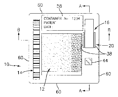
Referring now to FIGS. 1-4, a container 10 for holding a biologic fluid sample
3o includes at least one chamber 12, a label 14, a reservoir 16, a channel I
8, and a valve
20. The container I 0 holds a biologic sample in a manner that enables
analysis of the
sample by an analytical device (not shown) as will be described below. The
container
CA 02321690 2000-08-30
WO 99/44743
PGT/US99/04520
9
i0 embodiment shown in FIGS. I-3 includes a first piece 22 and a second piece
24
snapped together. The chamber 10 includes a first wall 26 disposed in the
first piece
22 and a transparent second wall 28 held between the first piece 22 and second
piece
24. In some embodiments, the first wall 26 may also be transparent thereby
enabling
light to pass through the container 10 by way of the chamber 12. The chamber
I2 has
a through-plane thickness ("t") at any given point. FIG.6 shows a diagrammatic
illustration of a field within a chamber to better illustrate the relationship
between
volume and through-plane thickness. As used herein, the term "through-plane
thickness" refers to a line of sight which corresponds to the shortest
distance between
1o the interior chamber surface 30 of the first wall 26 and the interior
chamber surface 32
of the second wall 28. The reservoir 16 typically holds 50 p,l of biologic
fluid sample
and preferably includes a cap 34 for sealing the reservoir 16 and a mixing
element 36,
such as a ball, that operates to keep the sample in uniform suspension. The
channel 18
extends between the reservoir 16 and the chamber 12. The valve 20 operates
between
the reservoir 16 and the chamber 12 to selectively allow passage of fluid from
the
reservoir 16 to the chamber 12. As used herein, the term '~ralve" includes not
only a
structure that includes a movable part that selectively prevents flow, but
also any
structure that selectively permits flow. The valve 20 shown in FIGS. l and 3-5
includes a pair of slits 38 adjacent the reservoir 16 is operated by a rod 40
which is a
2o part of the analytical device. The slits 38 allow the rod 40 to separate
the reservoir 16
a small distance from the first piece 22, thereby providing an opening through
which
biologic fluid can pass through the channel 18 and into the chamber 12. The
optimum
valve 20 type will vary depending upon the application. In those embodiments
where
there is more than one chamber 12 (see FIG.S), each chamber 12 is in
communication
with the reservoir I 6 via a channel I 8. The reservoir 16 and valve 20
provide
considerable utility for analyses where time is a consideration as will be
described
below. In some instances, however, it may be advantageous to provide a
container 10
without a reservoir I 6 and/or a valve 20.
Referring to FIGS. 1,5, and 7, each container 10 includes a plurality of
features
3o which are operable to enable the analysis of the biologic fluid sample,
some of which
are located in the chamber 12. The features located within the chamber 12 are
spatially
located, each having an address describable, for example, in x,y,z
coordinates. The
CA 02321690 2000-08-30
WO 99/44743 PCT/US99/04520
advantage of an x,y,z type coordinate address system is that the chamber can
be
mapped in an x,y,z, grid with a locatable origin that orients the analytical
device
relative to the container. The phrase "operable to enable the analysis of the
biologic
fluid" is used to describe the fact that the features either directly or
indirectly provide
s information that enables the analytical device to provide usefiil analytical
information.
For example, most analyses require either the volume or the through-plane
thickness of
the sample be known. Henceforth, the term 'volume" as used herein will refer
to this
requirement since the volume of a given image field of view can be ascertained
using
the through-plane thickness or vice versa. For example, when the sample is
imaged
1o using a fluorescent light source, it is the volume of the field that
provides the useful
information directly since fluorescent signal is a fixnction of colorant per
unit volume.
On the other hand, when a light absorption technique is used for imaging, the
volume
of the field will indirectly provide the necessary useful information, since
absorption is
a function of the through-plane thickness of the field (i.e., the distance the
light travels
through the sample). The through-plane thickness can be readily determined
from the
sensed volume and the known field area of the analytical device. To enable
those
analyses, the features include means for determining the volume of one or more
select
fields within the sample.
Referring to FIGS. 3,4, and 7, in a first embodiment of the means for
2o determining the volume of one or more fields within the sample, the first
wall 26 and
second wall 28 of the chamber 12, or a portion thereof, are in fixed
relationship to one
another and the slope values for each wall 26,28 and a chamber 12 through-
plane
thickness are known and are communicated to the analytical device through the
label
14 (the label 14 is discussed in detail below). The possible configurations of
the walls
26,28 (or a portion of the walls 26,28) include parallel walls (i.e., slope =
0) separated
by a known amount, and walls 26,28 which are at an angle toward one another
(i.e., a
slope ~ 0), separated by a known amount.
A second embodiment of the means for determining the volume of one or more
select fields within the sample includes: 1 ) a known quantity of sensible
colorant for
3o mixture with a known volume of biologic fluid sample; 2) chamber 12 regions
where
particular analyses are best performed; and 3) spatial information locating
those
optimum regions for the analytical device. As used herein, the term colorant
is defined
CA 02321690 2000-08-30
WO 99/44743 PCT/US99/04520
as any reagent that produces a sensible signal by fluorescent emission, or by
absorption
of light at a specific wavelength, that can be quantified by the analytical
device. The
colorant has a known signal magnitude to colorant concentration ratio that is
communicated to the analytical device through the label 14. The colorant
concentration is fixed by virtue of a known volume of biologic fluid sample
being
added to a known quantity of colorant. Alternatively, the signal magnitude to
colorant
concentration is determinable by comparison with a second known material such
as a
pad 44 of material (hereinafter referred to as a "calibration pad" - see FIG.1
) with
stable characteristics which is referenced by the analytical device and used
to calibrate
1o the response of the colorant. If the colorant signal is sensed in a
particular field via the
analytical device, then the volume of that field can be calculated using the
magnitude of
the sensed signal and the known concentration of colorant within the sample.
The
chamber 12 regions where an analysis is best performed refers to those chamber
regions having physical characteristics such as a particular through-plane
thickness that
allow for discrimination of particular constituents within the sample. For
example, a
chamber through-plane thickness of about 25 microns is known to be favorable
for the
formation of rouleaux and lacunae within a sample of whole blood. The absence
of
RBC's in the lacunae makes each lacunae a favorable region to accurately sense
colorant signal. The spatial information locating the optimum regions for the
analytical
2o device refers to the coordinate addresses of the features which, in terms
of the above
rouleaux/lacunae whole blood example, are the regions where lacunae are likely
to
develop. The analytical device contains means for identifying which features,
and
therefore the information available with those features, should be used in
particular
analyses.
Referring to FIGS. l and 8A-8F, a third embodiment of the means for
determining the volume of one or more select fields includes: 1 ) a quantity
of colorant
uniformly dispersed within the biologic fluid; 2) geometric characteristics
within the
chamber 12 which include, but are not limited to, a step 46 of known height
within one
or both walls 26,28, a cavity 48 or protuberance 50 of known height or volume,
or an
object 52 of known volume; 3) chamber 12 regions where particular analyses are
best
performed; and 4) spatial information locating those optimum regions for the
analytical
device. In this embodiment, it is not necessary to know the amount of sensible
CA 02321690 2000-08-30
WO 99/44743 PCT/US99/04520
12
colorant within the sample, nor the total volume of the sample. Rather, the
field
volume determination is done on a comparative basis. A field containing no
geometric
characteristic is sensed and compared against a field containing a known
geometric
characteristic. The known volume of the object 52, cavity 48, or protuberance
50 (or
volume which is determinable from a step of known height and the cross-
sectional area
of the field which is known to the analytical device) displaces a known volume
of
sample. Since the signal from the sensible colorant is a fimction of sample
volume, the
difference in signal sensed between the two fields is attributable to the
sample volume
displaced by the geometric characteristic. Hence, a signal to sample volume
ratio can
to be calculated, and applied to the whole field to ascertain the volume of
the field. Like
the second embodiment of the means for determining the volume of a sample
field, the
chamber 12 regions where an analysis is best performed refers to chamber
regions
having physical characteristics that allow for discrimination of particular
constituents
within the sample. The spatial information locating the optimum regions for
the
~ 5 analytical device also refers to a chamber 12 coordinate system wherein
each feature
within the chamber 12 has a coordinate address. The analytical device contains
means
for identifying which features, and the information available with those
features, should
be used in particular analyses.
A fourth embodiment of the means for determining the volume of one or more
2o select fields includes a chamber 12 having specular surfaces on which a
virtual
reflected image may be detected by the analytical device. The specular
surfaces are the
two wall surfaces 30,32 in contact with the biologic fluid, or the outer
surfaces if the
wall thicknesses are known. The analytical device detects the virtual
reflected image
on one of the specular surfaces 30,32 and then refocuses on the virtual
reflected image
25 formed on the second surface 32,30. The distance the analytical device's
optics must
move between the two images is the through-plane thickness of the chamber 12
in the
particular field. The label 14 communicates the coordinate addresses of the
select
fields within the chamber 12 to the analytical device.
Referring to FIG. 8E, in the second and third embodiments of the means for
3o determining the volume of one or more select fields, one or both of the
first or second
walls 26,28 may be formed from a flexible material that will deflect a
determinable
amount due to capillary forces presented by the sample acting on the wall
2b,28, and
CA 02321690 2000-08-30
WO 99/44743 PCT/US99/04520
13
thereby form a desirable convergent relationship between the first wall 26 and
the
second wall 28.
Referring to FIG.7, for chemicaUimmunochemical analyses of a biologic fluid
sample, the features include a plurality of di$'erent chemical reagents 54,
each located
s at a particular coordinate address, and may also include chamber 12 regions
where
particular analyses are best performed and coordinate addresses locating those
optimum regions. In a first embodiment, a known quantity of each chemical
reagent
54 is disposed at a particular coordinate address, usually in the form of a
coating
bound to one of the chamber walls 26,28. When the biologic fluid sample is
to introduced into the container chamber I2, the biologic sample admixes with
each
reagent 54. The fluid sample may be contiguous in those regions, but there is
no
appreciable reagent mixing between adjacent regions for a period of time
because of
the chamber configuration. Specifically, although the rates of diffusion
vertically and
laterally are equal, the chamber 12 through_plane thickness is small enough
relative to
15 the possible lateral expanse that the chemical reagent 54 will diffuse
vertically and
reach equilibrium at a much faster rate than it will laterally. In fiict,
because vertical
diffusion reaches equilibrium much faster than lateral diffusion, lateral
diffusion may be
considered negligible for a short period of time. The lateral spacing between
the
addresses of the different chemical reagents 54 is such that during that short
period of
2o time in which lateral reagent diffusion is negligible, useful analysis of
any reaction that
may be occurring at a particular address can be performed. The coordinate
addresses
of the various chemical reagents 54 enable the analytical device to access
each reagent
54 and perform meaningful analyses. In those instances where chemical and
hematological analyses are desirable, the above described chamber
configuration can
25 be provided in a particular region of a single chamber I2 and other
configurations
provided elsewhere within that chamber 12. The negligible lateral diffusion of
the
reagent 54 prevents interference with contiguous chamber I2 regions which may
be
devoted to other type analyses. Alternately, the different reagent regions may
be
partially or completely isolated in subcompartments of the chamber by means of
3o intervening partitions 55 formed within one or both of the chamber 12
surfaces (see
FIG.8F).
CA 02321690 2000-08-30
WO 99/44743 PCT/US99/04520
14
Referring to FIGS. l and 5, the label 14 is a mechanism for communicating
information to the analytical device. A practical example of a label 14 is one
which is
machine readable and one which is capable of communicating information
including,
but not limited to: 1 ) type of analysis(es) to be performed; 2) information
concerning
the type of features, and the coordinate addresses of those features located
within the
sample chamber; 3) reagent information; 4) lot information; 5) calibration
data; etc. In
one form, the label 14 may be a magnetic strip or a bar code strip, or the
like, which
directly contains all the information useful to the analytical device in the
performance
of the analysis(es). This type of label 14 is particularly useful in those
instances where
1o the information to be communicated is limited. In those instances where the
quantity
of information to be communicated is considerable, it may be more desirable to
have
the label 14 direct the analytical device to a data file (stored within the
analytical device
or remotely accessible by the analytical device via modem, network link, etc
.)
containing the appropriate information. In this instance, the label 14 can be
said to
indirectly contain the information by providing the necessary path to the
information.
Here again, the label 14 could be a bar code or magnetic strip, which in this
case
communicates a particular code that is interpreted by the analytical device as
being
associated with a certain data file. The same result could be achieved by
incorporating
a physical feature 56 in the container (e.g., a notch, a tab, etc. - see FIGS)
that is
2o interpretable by the analytical device. Other labels 14 which function to
communicate
information to the analytical device can be used alternatively.
The container 10 also preferably includes a human readable label 58 to
facilitate
handling within the laboratory or clinic. The human readable label 58 may
include
information such as the patient's name, a sample taken date, an oi~ce address,
an
2s appropriate warning (e.g., "Biohazard - Handle with Care"), trademarks,
etc. The
sides 60 of the container 10 are suitable to interact with a transport means
(not shown)
contained within the analytical device. The transport means is operable to
move the
container 10 relative to an imaging device (not shown) contained within the
analytical
device.
30 As stated above, the considerable utility of the container 10 enables a
wide
variety of analyses to be performed on a single sample, using a single
analytical device.
CA 02321690 2000-08-30
WO 99/44743 PCT/ITS99/04520
The examples given below are offered so that a complete appreciation of the
present
invention container 10 may be gained.
Example I: Hematological Analyses
5 Referring to FIGS. 1 and 4, to enable an analysis of white blood cells
(WBC's)
within an anticoagulated whole blood sample, the container 10 includes
approximately
0.8 micrograms (p,g) of a sensible colorant disposed within the reservoir 16.
EDTA is
an example of an anticoagulating agent that may be used with the sample and a
fluorescent highlighting supravital stain such as acridine orange, basic
orange-21, or
the like are examples of sensible colorants that may be added to the reservoir
16. For
purposes of evaluating WBC's, it is preferable to have a region within the
chamber 12
that has a plurality of select fields with a through-plane thickness on the
order of 20
microns in magnitude. A chamber 12 through-plane thickness of approximately 20
microns is chosen for a couple of reasons. First, an evaluation volume of
0.02p1,
15 (formed by a particular field of the chamber 12 having a cross-sectional
area of 1
millimeter (mm) and a thickness of 20 microns) typically contains 50-200 WBC's
which is a favorable quantity for evaluative purposes. Second, a through-plane
thickness of 20 microns provides an optimal chamber 12 for rouleaux and
lacunae
formation. The coordinate addresses of select fields are communicated to the
2o analytical device by way of the label 14. In the example, therefore, the
plura(ity.of
features operative to enable analysis of the biologic fluid sample include: 1
) the sensible
reagent disposed within the reservoir 16; 2) the chamber 12 regions) having a
plurality
of select fields with a particular through-plane thickness; and 3) the
coordinates
addresses of those f elds within the chamber 12.
Approximately 20 pl of anticoagulated whole blood is placed into the reservoir
16 by the operator and the cap 34 secured. The container is gently shaken
until the
reagent and whole blood sample are adequately mixed. A mixing ball 36 disposed
in
the reservoir 16 facilitates mixing. The container 10 is inserted into the
analytical
device and the valve 20 is subsequently actuated to release the sample into
the chamber
12 by way of the channel 18. Once the sample is distributed within the chamber
12,
the sample resides quiescently. The only sample motion within the chamber 12
will
possibly be Brownian motion of the sample's formed constituents, and that
motion is
CA 02321690 2000-08-30
WO 99/44743 PCT/US99/04520
16
non-disabling for the present invention. Note that for simple tests such as a
WBC
count where timing is not important, a sample could be deposited into the
chamber 12
directly, thereby obviating the need for a reservoir 16 and valve 20.
Immediately after the sample has been inserted into the chamber, the sample
will appear opaque when examined either with transmitted light, or more
preferably by
epi-illuminated fluorescence. The opaque appearance is caused by the red blood
cells
(RBC's), which form an overlapping mass prior to the formation of the
rouleaux.
After lying substantially motionless for approximately thirty (30) seconds,
within the
chamber 12, the RBC's will have spontaneously clustered into rouleaux, leaving
to lacunae between the rouleaux. It is in these lacunae where the other whole
blood
sample constituents (e.g., WBC's and platelets) can be found and evaluated. If
a
count of WBC's is desired, a square millimeter field of the 20 micron thick
chamber
12, which contains 0.021 of whole blood sample, can be evaluated. A 0.02p1
sample
is chosen to keep the number of WBC's reasonable (a normal whole blood sample
contains approximately 7,000 WBC's per p,l of sample; a 0.02E.i1 sample of
normal
whole blood contains approximately 140 WBC's). A number of these fields would
be
evaluated until enough cells are counted to get a number which has sufficient
statistical
accuracy, which is in practice approximately 1000 cells. If additional WBC
information is sought, the WBC's (lymphocytes, granulocytes, monocytes, etc:)
can be
2o analyzed within the sample using an image dissector such as a CCD camera,
for
example, alone or with analysis software. A differential count could be
determined
from the data collected.
The above example of the utility of the present invention container 10 in
hematological analyses includes a plurality of features operative to enable
analysis of
the biologic fluid sample. In a preferred embodiment, the features not only
include the
plurality of select fields with a through-plane thickness on the order of 20
microns, but
fields of slightly larger and smaller volume as well. The larger/smaller field
volumes
can be created by several ofthe mechanisms described above; e.g., convergent
chamber walls 26,28, or steps 46 within one or both walls 26,28, etc. A range
of field
3o volumes is advantageous because constituent populations quite often vary in
magnitude within the biologic fluid sample. If, for example, the WBC
population
within the sample was abnormally high, a chamber 12 region having a through-
plane
CA 02321690 2000-08-30
WO 99/44743 PCT/US99/04520
17
thickness of 20 microns may have more than an optimal number of WBC's for
evaluative techniques such as counting. Changing to a field of smaller volume
would
decrease the number of WBC's and therefore facilitate the analysis at hand. On
the
other hand, if the WBC population within the sample was abnormally low, a
chamber
12 region having a through-plane thickness of 20 microns may have less than an
optimal number of WBC's for evaluative purposes. Changing to a field of larger
volume would increase the number of WBC's and likewise facilitate the analysis
at
hand. The spatial locations of alternate features (i.e., larger or smaller
through-plane
thickness regions in the above example) are communicated to the analytical
device
to through the label 14.
Example II: Chemical Analyses
Referring to FIG.9, a complete blood count requires that the RBC's be
evaluated for hemoglobin content. In a first embodiment, the hemoglobin
evaluation is
t5 performed in a first chamber 62 which is connected to the reservoir 16 by a
channel 18.
At least two chemical reagents 64,66 are initially stored within the first
chamber 62.
The reagents 64,66 are shown in the first chamber 62 as independent deposits
to
illustrate the use of multiple reagents. Reagents can often be combined into a
single
reagent mixture stored as a single deposit. One of the chemical reagents 64 is
a lysing
2o reagent which breaks down RBC's within the sample and thereby releases the
hemoglobin stored within the RBC's. The other reagent 66 is a hemoglobin
stabilizer
that increases the reliability of the hemoglobin evaluation. In most cases,
the
hemoglobin evaluation is performed after the lysing agent has been introduced
into the
sample for a given period of time, or at particular intervals. Using the
present
25 invention, the period of time begins when the valve 20 is actuated to
permit the sample
to enter the first chamber 62 and a second chamber 68. The remaining analyses
associated with a complete blood count are performed in the second chamber 68.
In
this embodiment, the features operable to enable the analysis of the biologic
fluid
sample are: 1) the first and second chambers 62,68 within the container 10 in
fluid
3o communication with the reservoir 16; 2) the chemical reagents 64,66
disposed in the
first chamber 62; 3) the spatial location of the first chamber 62 and the
spatial location
of the chemical reagents 64,66 within the first chamber 62; and 4) the valve
20
CA 02321690 2000-08-30
WO 99/44743 PCT/US99/04520
18
between the reservoir 16 and the chambers 64,66 that initiates the time
period.
Additional features such as those described heretofore in the '~iematological
Analyses"
example may be present in the second chamber 68.
Referring to FIG.10, in a second embodiment all of the complete blood count
analyses are performed in a single chamber 12. The portion of the biologic
fluid
sample used for the hemoglobin evaluation is contiguous with remaining portion
of the
fluid sample, but that portion is preferably oriented toward one side of the
chamber 12
to minimize potential mixing of the lysing agent with the remaining portion of
the fluid
sample. In addition to orienting the hemaglobin evaluation to one side, it is
also
to preferable to choose a chamber 12 through-plane thickness small enough such
that
vertical di~'usion (and ultimate equilibrium) of the chemical reagents 64,66
within the
biologic fluid sample occurs at a much faster rate than lateral diffusion. The
difference
in diffusion rates is such that lateral diffusion may be considered negligible
for a short
period of time. The lateral spacing between the hemoglobin evaluation site and
the
remainder of the fluid sample is such that during that short period of time in
which
lateral reagent diffusion is negligible, the remainder of the desired analyses
can be
performed without interference from the lysing agent. In this embodiment, the
same
two chemical reagents 64,66 as described above are initially deposited in the
hemoglobin evaluation region of the chamber 12, and actuating the valve 20
begins the
2o time period for the evaluation. The features operable to enable the
analysis of the
biologic fluid sample are: 1 ) the chemical reagents 64,66 disposed in the
aforementioned chamber 12 region; 2) the spatial location of the reagents
64,66 within
the chamber; 3) the chamber configuration functionally operable to separate
the
hemoglobin evaluation region from the remainder of the biologic fluid sample;
4) the
2s valve 20 between the reservoir 16 and the chamber 12 that initiates the
time period;
and 5) any features such as those described above in the 'TIematological
Analyses"
example.
Example III: Urinal~rsis
3o Referring to FIGS, a complete urinalysis requires a chemical analysis and a
particulate analysis of the urine sample. Chemical reagents 70 spatially
located at
particular coordinate addresses within a chamber are used to colorometrically
relate
CA 02321690 2000-08-30
WO 99/44743 PCT/US99/04520
19
information after a given period of time. The particulate analysis involves
detecting,
evaluating and/or enumerating the particles within the sample. In a first
embodiment,
the chemical analysis is performed in a first chamber 72 and the particulate
analysis is
performed in a separate second chamber 74. Both the first chamber 72 and the
second
chamber 74 are in fluid communication with the reservoir 16. In a manner
similar to
that described above, the through-plane thickness and other physical
characteristics of
the first chamber 72 and the second chamber 74 are chosen to facilitate the
chemical
and particulate analyses, respectively. In the first embodiment, the features
operable to
enable the analysis of the biologic fluid sample are, therefore: 1} the
chemical reagents
70 disposed in the first chamber 72; 2) the physical features of the chamber
12 chosen
to facilitate the chemical analysis; 3) the spatial location of the chemical
reagents 70
within the chamber 72, and the spatial location of the chamber 12 physical
features;
and 4) the valve 20 between the reservoir 16 and the chamber 12 that initiates
the time
period. In a second embodiment, the chemical and particulate analyses are
performed
in the same chamber 12. In a manner similar to the hemoglobin evaluation
described
above (see FIG.10) , the chamber 12 region devoted to the chemical analysis is
preferably oriented to one side of the chamber 12 and the through-plane
thickness is
such that interference from the chemical reagents will be negligible if at
all. The
features within the second embodiment operable to enable the analysis of the
biologic
2o fluid sample are: 1 ) the chemical reagents 70 disposed in the chamber 12;
2) the spatial
location of the chemical reagents 70 within the chamber 12; 3) the chamber 12
configuration functionally operable to separate the chemical evaluation region
from the
remainder of the biologic fluid sample; and 4) the valve 20 between the
reservoir 16
and the chamber 12 that initiates the time period.
Although this invention has been shown and described with respect to the
detailed embodiments thereof, it will be understood by those skilled in the
art that
various changes in form and detail thereof may be made without departing from
the
spirit and the scope of the invention.