Note : Les descriptions sont présentées dans la langue officielle dans laquelle elles ont été soumises.
CA 02322212 2007-09-20
5-3050-1
1
APPARATUS AND PROCESSES FOR LASER DEFORMATION OF
DIELECTRIC PARTICLES
BACKGROUND OF INVENTION
The first direct experimental confirmation in a
laboratory that light carries momentum was accomplished by
E. F. Nichols and G. F. Hull in the US (Nichols E. F.,
Hull G. F., Phys. Rev. 13, p.307 (1901)) and P. N. Lebedev
in Russia (Lebedev P. N., Ann. Phys. (Leipzig) 6, p.433
(1901)) around the turn of the century. In both experiments
the radiation pressure of a light source was detected by the
twisting motion of mirrors suspended by thin wires in a high
vacuum. The vacuum was crucial to eliminate the effects of
thermal or radiometric forces. No one at the time imagined
that there would be any practical application for this
minute effect. The first process in which photon momentum
played an important role was the Compton effect, i.e. the
scattering of X-rays against electrons. Another four
decades passed until the invention of the laser in 1960.
The possibility of producing spatially coherent light with
very high intensity brought the effect of radiation pressure
into the macroscopic world. An early pioneer in this field
was A. Ashkin at Bell Laboratories. Ashkin was the first to
use a laser for manipulating transparent, micron sized latex
spheres in the late 60s. These spheres, suspended in a
water solution, were first accelerated with one horizontal
laser beam and then trapped in between two beams in a second
experiment (Ashkin A., "Acceleration and Trapping of
Particles by Radiation Pressure" Phys. Rev. Lett. 24(4)
p.156-159 (1970)).
Since then, this characteristic of light has been
exploited in many ways. One broad field is the trapping and
cooling of atoms and molecules. The fact that this year's
CA 02322212 2007-09-20
53050-1
la
Nobel prize was awarded to Steven Chu, William D. Phillips,
and Claude Cohen-Tannoudji for the development of methods to
cool and trap atoms with laser light shows its absolute
relevance to today's science (Chu S., "Laser Trapping of
Neutral Particles" Sci. Am., p.71 (February 1992);
Phillips W. D., Metcalf H.J., "Cooling and Trapping Atoms"
Sci. Am., p.36 (March 1987); Cohen-Tannoudji C.,
Phillips W. D., "New Mechanisms for Laser Cooling" Physics
Today, p.33 (October 1990); Chu S., "Laser Manipulation of
Atoms and Particles" Science 253, p.861-866 (1991)).
CA 02322212 2000-08-29
PCTIUS99/04845
WO 99/44488
2
The interactions of light with dielectric matter can be divided into two
predominant
forcing mechanisms. The gradient force, which pulls material with a higher
relative index
towards the areas of highest intensity in a laser beam, and the scattering
force, which is a
result of the momentum transfer of photons to the material. Most optical micro-
manipulation
tools rely on gradient forces. However, the "Optical Stretcher" of this
invention uses the
scattering forces to deform elastic dielectric samples.
The characteristics of these two forces (gradient and scattering force) allow
for
different possible trap designs.
One of the first stable traps was the levitation of particles with a vertical
laser beam
(Ashkin A., Dziedzic J.M., "Optical Levitation by Radiation Pressure" Appl.
Phys. Lett.
19(6), p.283-285 (1971); Ashkin A., "The Pressure of Laser Light" Sci. Am.
226, p.63-71
(1972); Ashkin A., Dziedzic J.M., "Optical Levitation in High Vacuum" App.
Phys. Lett.
28(6), p.333-335 (1976); Ashkin A., Dziedzic J.M., "Observation of Light
Scattering from
Nonspherical Particles Using Optical Levitation" Appl. Opt. 19(5), p.660-668
(1980)). The
scattering force is strong enough to balance gravity while the gradient force
keeps the particle
on the optical axis.
A big improvement in this trap was the use of a highly focused laser beam,
first
realized by Ashkin in 1986 (Ashkin A., Dziedizic J.M., Bjorkholm J. E. , Chu
S.,
"Observation of a Single-Beam Force Optical Trap for Dielectric Particles"
Opt. Lett. 11(5),
p.288-290 (1986)), because it is independent of gravity and one can orient the
trap in any
direction in space or even use it in micro-gravity (Gussgard R., Lindmo T.,
Brevik I.,
"Calculation of the Trapping Force in a Strongly Focused Laser Beam" J. Opt.
Soc. Am. B
9(10), p.1922-1930 (1992)). The idea is that extreme focusing leads to a point
in space
(rather than an axis) with a very high intensity. Thus, the gradient force
pulls a dielectric
particle towards this point. The focusing has to be strong enough that the
gradient force
overcomes the scattering force, which is an unwanted effect in this case,
because it pushes the
particle away from the focus. This is usually realized by directing a laser
beam through a
microscope objective with high numerical aperture (NA). An experimental
improvement can
be achieved by using an objective with a central field stop producing a
conical dark field.
This enhances the relative contribution from high NA illumination and
diminishes the
influence of the scattering force. At the same time the particle is trapped
near the focus and
SUBSTITUTE SHEET (RULE 26)
CA 02322212 2000-08-29
WO 99/44488 PCT/US99/04845
3
can be observed with the microscope. This one-beam setup, usually referred to
as the Optical
Tweezer, has been studied extensively (Ashkin A., Dziedizic J.M., Bjorkholm J.
E. , Chu S.,
"Observation of a Single-Beam Force Optical Trap for Dielectric Particles"
Opt. Left. 11(5),
p.288-290 (1986); Ashkin A., "Forces of a Single-Beam Gradient Laser trap on a
Dielectric
Sphere in the Ray Optics Regime" Biophys. J. 61, p.569-582 (1992); Wright W.
H., Sonek
G.J., Berns M. W., "Radiation Trapping Forces on Microspheres with Optical
Tweezers"
App. Phys. Lett. 63, p.715-717 (1993); Gussgard R., Lindmo T., Brevik I.,
"Calculation of
the Trapping Force in a Strongly Focused Laser Beam" J. Opt. Soc. Am. B 9(10),
p.1922-
1930 (1992); Visscher K., Brakenhoff G.J., "Theoretical Study of Optically
Induced Forces
on Spherical Particles in a Single Beam Trap I.= Rayleigh Scatterers" Optik
89(4), p.174-180
(1992); Kuo S. C., Sheetz M.P., "Optical Tweezers in Cell Biology" Trends in
Cell Biology
2, p.116-118 (1992)) and is widely used in biological applications. The power
of the laser
used is in the range of a few mW up to 1.5W for the trapping of glass or latex
beads and the
achieved trapping forces vary from Pico- to Nanonewton depending on the size
and index of
refraction. For detailed reviews see Kuo S. C., Sheetz M.P., "Optical Tweezers
in Cell
Biology" Trends in Cell Biology 2, p.116-118 (1992); Berns M. W., Wright W.H.,
Steubing
R. W., "Laser Microbeam as a Tool in Cell Biology" Int. Rev. Cytol. 129, p.1-
44 (1991);
Block S.M., Optical Tweezers: A new Tool for Biophysics, in Noninvasive
Techniques in
Cell Biology, G.S. Foskett J. K., Editor. 1990, Wiley-Liss.: New York. p. 375-
402; Greulich
K. 0., Weber G., "The Laser Microscope on its Way from an Analytical to a
Preparative
Tool" J. Microsc. 167, p.127-151 (1991); Simmens R. M., Finer J.T.,
"Glasperlenspiel II.=
Optical Tweezers" Curr. Biol. 3, p.309-311 (1993); and Weber G., Greulich
K.O.,
"Manipulation of Cells, Organelles, and Genome by Laser Microbeams and Optical
Traps"
Int. Rev. Cytol. 133, p.1-41 (1992).
Although the Optical Tweezer is a very powerful tool, it also has its
limitations. The
working distance of high NA objectives is very short and does not allow for
additional test
equipment between objective and object. The trapping zone is rather small (on
the order of
the light wavelength). It has been shown that a good trapping efficiency can
only be achieved
for indices of refraction smaller than --1.7 because of the loss of axial
stability (the scattering
force becomes stronger than the backward gradient force) (Svoboda K., Block
S.M.,
"Biological Applications of Optical Forces" Annu. Rev. Biophys. Struct. 23,
p.147-285
SUBSTITUTE SHEET (RULE 26)
CA 02322212 2000-08-29
WO 99/44488 PCT/US99/04845
4
(1994)). Furthermore, focusing the beam down to the theoretical limit of spot
sizes (half the
wavelength of the used light) leads to very high intensities that can endanger
the integrity of
biological objects. Most cells trapped with an optical tweezer do not survive
powers greater
than 20-250 mW because the extreme focusing leads to very high local
intensities. This
depends also on the specific cell type and the used wavelength, of course
(Ashkin A.,
Dziedzic J.M., Yamane T., "Optical Trapping and 'Manipulation of Single Cells
Using
Infrared Laser Beams" Nature 330(24), p.769-771 (1987); Ashkin A., Dziedzic
J.M.,
"Optical Trapping and Manipulation of Viruses and Bacteria" Science 235,
p.1517-1520
(1987); Kuo S. C., Sheetz M.P., "Optical Tweezers in Cell Biology" Trends in
Cell Biology
2, p.116-118 (1992)).
A different setup, that circumvents most of these problems, is a two beam trap
(Ashkin A., "Acceleration and Trapping of Particles by Radiation Pressure"
Phys. Rev. Lett.
24(4) p.156-159 (1970); Roosen G., Imbert C., "Optical Levitation by Means of
Two
Horizontal Laser Beams: A Theoretical and Experimental Study" Phys. Lett.
59A(1), p.6-9
(1976); Roosen G., "La Levitation Optique de Spheres" Can. J. Phys. 57, p.1260-
1279
(1979); Ashlcin A., Dziedzic J.M., "Optical Levitation by Radiation Pressure"
Appl. Phys.
Lett. 19(6), p.283-285 (1971)). Historically the two beam trap was developed
more than a
decade before optical tweezers but has since fallen into disuse. Two
identical, counter-
propagating laser beams with Gaussian beam profiles can stabilize a particle
in the point of
symmetry. The forces on a sphere are the superposition of the forces of two
individual
beams. The gradient force confines the particle to the axis while the
scattering force provides
stability along the axis. This is a stable configuration as long as the
refractive index n of the
particle is higher than that of the surrounding medium. Air bubbles in water,
an example
where this condition is not fulfilled, are pushed out of the beam (like a
rubber ball out of a
water beam). Another condition, which is somewhat unexpected, is that the
diameter of the
particle has to be smaller than the beam radii in the center. For the
situation where the beam
radius is smaller than the particle radius, and thus the ratio is larger than
1, the force closer to
the waist of the beam is smaller than further away due to the divergence of
the beam. This
leads to an amplification of small displacements of the sphere from the center
which means
that the particle is not stably trapped (Roosen G., "A Theoretical and
Experimental Study of
SUBSTITUTE SHEET (RULE 26)
CA 02322212 2007-09-20
53050-1
the Stable Equilibrium Positions of Spheres Levitated by Two
Horizontal Laser Beams" Opt. Comm. 21(1), p.189-195 (1977)).
An improvement of this setup has been demonstrated
in which single mode (SM) optical fibers are used to deliver
5 the laser beams to the trapping area (Constable A., Kim J.,
Mervis J., Zarinetchi F., Prentiss M., "Demonstration of a
Fiber-Optical Light-Force Trap" Opt. Lett. 18(21),
p.1867-1869 (1993)). This avoids the need for additional
optical elements and their alignment, and allows a very easy
and cheap implementation of the trap into custom made
experiments. This trap can also be set up independently
from a microscope. A good review of all these traps and
their applications in biology can be found in Svoboda K.,
Block S.M., "Biological Applications of Optical Forces"
Annu. Rev. Biophys. Struct. 23, p.147-285 (1994).
A new member in the family of light traps is the
Optical Spanner (Padget M., Allen L., "Optical Tweezers and
Spanners" Physics World, p.35-38 (September 1997)). The
basic setup is as for the Optical Tweezer but instead of a
Hermite-Gaussian laser profile, a Laguerre-Gaussian profile
is used. These beams have a circular cross-section, a
helical wavefront, and a Poynting vector that spirals around
the axis. This means that such a beam has an oribtal
momentum in addition to the translational momentum
previously discussed. Since the angular momentum of the
system has to be conserved too, the particle starts to
rotate. In this way particles can not only be translated
but also rotated.
Although these traps are extremely useful for all
kinds of manipulation of objects, they can only translocate
and/or rotate them and are not intended to deform them.
Embodiment of this invention expand the line of optical
CA 02322212 2007-09-20
53050-1
6
tools and allows for a full spectrum of particle
manipulation.
All existing methods to examine the elasticity of
cells have their limitations. In micropipette aspiration
experiments the tip of a micropipette is placed onto a cell
with a micro-manipulator and part of the cell is pulled into
the pipette with an applied negative pressure. This
provides only very local information and can detach the
membrane from the cell which leads to inaccurate
measurements.
Another possibility is the use of an Atomic Force
Microscope (AFM) in tapping mode (Radmacher M., Fritz M.,
Kacher C. M., Cleveland J. P., Hansma P. K., "Measuring the
Viscoelastic Properties of Human Platelets with the Atomic
Force Microscope" Biophys. J. 70, p.556-567 (1996)). The
oscillating AFM tip is scanned across the cell body allowing
the force and the indentation to be measured. Usually, the
Young modulus of the cell is then determined using the Hertz
model which assumes a semi-infinite slab of material and
connects deformations to its material constants. The
problem here is that the spring constant of the AFM
tip/cantilever is rather big compared to the strength of the
cytoskeleton which means that it is not possible to detect
small elasticity differences of cells - the cell is either
compressed or not. This treatment is also very rough, as
many cells do not survive it. AFM also looks at the
elasticity only over small areas of a cell's surface.
Similar to this are "cell poking" experiments where the AFM
tip is replaced by a glass needle (Elson E. L., "Cellular
Mechanics as an Indicator of Cytoskeletal Structure and
Function" Annu. Rev. Biophys. Chem. 17, p.397-430 (1988)).
CA 02322212 2007-09-20
5.3050-1
7
Another major disadvantage, which all these
methods have in common, is that they are not efficient. It
is very inconvenient to position the micropipette or the AFM
tip manually on a cell. Thus, it is effectively not
possible to measure a significant number of cells in a short
period of time which results in lack of good statistics.
A more indirect approach was to shear a whole
pallet of densely packed cells with a rheometer
(Eichinger L., Koppel B., Noegel A. A., Schleicher M.,
Schliwa M., Weijer K., Wittke W., Janmey P. A., "Mechanical
Perturbation Elicits a Phenotypic Difference Between
Dictyostelium Wild-type Cells and Cytoskeletal Mutants"
Biophys. J. 70, p.1054-1060 (1996)). However, this is a
bulk measurement and yields only mean values and not
specific information about a single cell. Another
limitation is that not only the cell elasticity but also
sticking forces and friction between the cells influence the
outcome of the measurement.
SUMMARY OF INVENTION
Some embodiments of the present invention provide
solutions to one or more of the disadvantages and
deficiencies described above.
There exists several optical tools for the
manipulation of dielectric particles such as biological
cells. Until now, manipulation by radiation pressure was
considered to be translation and rotation. This is the
first time that the forces arising from the interaction of
light with matter are used intentionally to deform cells in
a controlled and nondestructive manner. With this novel
tool of embodiments of the present invention, it is possible
to measure the elasticity of deformable objects, such as
cells, with diameters typically between 5-50 microns. As
CA 02322212 2007-09-20
53050-1
8
used herein, "Optical Stretcher" refers to embodiments of
the invention described herein. Cells owe their stability
to a three-dimensional network of filamentous polymers,
known as the cytoskeleton. It is not understood how the
cytoskeleton is able to provide a mechanical stability to
cells, as classical concepts in soft condensed matter
physics fail to explain this phenomenon. The use of the
Optical Stretcher, in combination with.modern techniques in
molecular biology, will clarify the role of the cytoskeletal
constituents. This novel symbiosis of physics and molecular
biology will help to gain a clearer picture of the way the
cytoskeleton works.
Furthermore, the Optical Stretcher may be used for
the analytical detection of single malignant cells. Cancer
cells are shown to modify their morphology compared to
normal cells which changes their elasticity. This novel
tool, the Optical Stretcher, will be useful in detecting
these changes.
The Optical Stretcher is a novel technique to
measure the elasticity of single cells which avoids the
disadvantages mentioned above. One principle idea is to use
two counter-propagating laser beams to trap a single cell,
which is suspended in a buffer solution, by radiation
pressure. The feasibility of the trapping itself was
already demonstrated by A. Ashkin in the late 1960s. Since
then, the trapping of all kinds of particles with sizes
ranging from Angstroms (such as atoms and molecules) to tens
of microns (such as small glass beads or cells) has found
many applications.
This is the first time that this setup is used not
only for trapping, moving, and rotating particles, but for
the deformation of the particle under investigation in a
CA 02322212 2007-09-20
53050-1
9
noninvasive, nondestructive, and controlled way. The idea
of deforming dielectric surfaces with intense light is not
new. The effect was predicted by Askar'yan, Kats and
Kantorovich (Askar'yan G. A., "Radiation Pressure on an
Object with Varying Polarizability Changes. Deformation
Absorption of a Wave by Variable Inhomogenities" JETP
Lett. 9, p.241-243 (1969); Kats A. V., Kantorovic V. M.,
"Bending of Surface and Self-Focusing of a Laser Beam in a
Linear Medium" JETP Lett. 9, p.112-114 (1969)) and later
shown experimentally for a free liquid surface by Ashkin
(Ashkin A., Dziedzic J. M., "Radiation Pressure on a Free
Liquid Surface" Phys. Rev. Lett., 30(4) p.139-142 (1973)).
Yet, no one realized the full potential of this effect and
it has consequently been ignored, until now.
With this novel approach we can measure the
elasticity of cells and circumvent most of the problems
mentioned above. Even though it is a relatively simple
setup, this invention may: investigate single cells without
killing them, apply forces over a wide range, and take time
dependent measurements over a broad frequency range (from
mHz to MHz in principle) just by varying the light
intensity. In addition, it is also possible to measure
large numbers of cells in a short period of time by
incorporating a flow chamber.
Diseases which effect the cytoskeleton, such as
the malignant transformation of cells, can be analytically
detected with the Optical Stretcher in a simple,
inexpensive, and noninvasive way. Thus, it will be superior
to existing techniques and a basis for successfully fighting
these diseases.
In view of the foregoing, in one broad respect
this invention is an apparatus, comprising: a stage capable
CA 02322212 2007-09-20
53050-1
9a
of supporting micron-sized dielectric particles; two or more
sources of laser light directed toward an area of the stage
where the particles are located; a first detector which
determines whether a particle is trapped between the first
and second laser sources; a second detector which determines
deformation of a particle upon increasing intensity of the
laser light; wherein the first and second laser sources may
be adjusted to vary the power of the laser light to both
trap a particle and stretch the particle.
The dielectric particles to be used in the
practice of embodiments of this invention may vary widely
from inanimate particles such as polymer beads such as latex
or polystyrene beads to biological cells such as red blood
cells and nerve cells.
In another broad respect, this invention is an
apparatus comprising a stage capable of supporting
biological cells in an aqueous medium, first and second
sources of laser light directed toward an area of the stage
where the cells are located, a first detector which
determines whether a cell is trapped between the first and
second laser sources, and a second detector which determines
and/or measures deformation of a cell upon increasing
intensity of the laser light whereas the first and second
laser sources may be adjusted to vary the power of the laser
light to thereby either trap a cell or stretch the cell.
In another broad respect, this invention is a
process for the controlled deformation of biological cells.
This process may be comprised of exposing a cell to two
counterpropogating laser beams at an intensity sufficient to
deform the cell, and optionally measuring the deformation of
the cell.
CA 02322212 2007-09-20
5=3050-1
9b
In yet another broad respect, this invention is a
process for the detection of individual cancer cells by
measuring their deformability using the apparatus described
above. The detection of cancer cells essentially works by
measuring suspect cells in suspension one by one. Any cell
that will show an increased (or decreased) deformability
(i.e., will be deformed characteristically more or less)
than comparable controls can thus be identified as cancer
cell.
In still another broad respect, this invention is
a process for the controlled deformation of micron-sized
dielectric particles, comprising: exposing a particle to
two or more counterpropogating laser beams at an intensity
and under conditions effective to deform the particle; and
measuring the deformation of the particle.
In still another broad respect, this invention is
a process for the controlled deformation of dielectric
micron-sized particles comprising exposing a particle to
several laser beams at an intensity sufficient to deform the
particle and optionally measuring the deformation of the
cell.
It is generally believed that the forces to deform
mammalian cells cannot be achieved without causing
simultaneous radiation damage by heating, previous
experiments used focused beams and the higher local laser
intensities destroyed the cells. We have now recognized
that this can be overcome by using optical fiber traps which
have been previously used by to trap glass beads but not to
stretch cells. Also, it has been predicted that laser light
can deform dielectric surfaces. However, that the radiation
pressure of two opposing laser beams stretches a cell is
somewhat counterintuitive. One would expect that the cell
CA 02322212 2007-09-20
5=3050-1
9c
would be squeezed. It is totally unexpected and surprising
that two opposing laser beams do not squeeze a cell, that
radiation pressure stretches cells. This is
counterintuitive and not predicted in literature. While not
wishing to be bound by theory, we can now explain this
effect by the increase of momentum of the laser bean when it
enters the cell. Due to momentum conservation this increase
has to be compensated by a pulling force.
BRIEF DESCRIPTION OF THE DRAWINGS
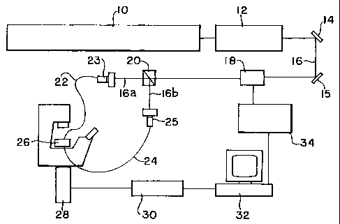
FIG. 1 shows a non-limiting, representative
configuration for the apparatus of an embodiment of the
invention.
FIGS. 2A-2C show a non-limiting, representative
perspective, top and side views of a stage that may be used
in the practice of an embodiment of this invention.
CA 02322212 2000-08-29
WO 99/44488 PCTIUS99/04845
DETAILED DESCRIPTION OF THE INVENTION
The Optical Stretcher is a novel optical tool which uses one or more laser
beams to
pull on transparent, dielectric materials. In particular, a set up with two or
more counter
propagating beams can be used to stably trap and stretch micron-sized
dielectric particles
5 such as biological cells. The fact that the photon pressure of a laser beam
entering or leaving
a medium of higher refractive index pulls on the interface is unexpected, but
can be explained
by momentum conservation.
The type of cells that may be trapped and stretched may vary widely. For
instance,
this invention may be used to trap and deform eukaryotic cells including
mammalian cells,
10 except for muscle and neuronal cells which are too big for the stage shown
herein. A
representative, non-limiting list of such cells for use in the practice of
this invention include
epithelial cells, lymphocytes, macrophages, fibroblasts, PC12 cells,
keratinocytes and
melanoma cells. It may also be possible to use small tissue clusters with a
diameter of about
100 microns.
FIG. 1 shows a setup for our experiment following the one of the two-beam
fiber trap
described in Constable A., Kim J., Mervis J., Zarinetchi F., Prentiss M.,
"Demonstration of a
Fiber-Optical Light-Force Trap" Opt. Lett. 18(21), p.1867-1869 (1993). This
set up is
representative and should not be construed as limiting the scope of this
invention.
In FIG. 1 a first laser 10 such as an Ar+-Laser (Spectra Physics Lasers, Inc.,
Beamlok
2080 RS), with up to 28W power to pump, pumps a laser 12 such as a tunable, cw
Ti-
Sapphire Laser (Spectra Physics Lasers, Inc., 3900S having a wavelength of
about 790
nanometers). While other wavelengths may be employed, we have found that for
the
stretching of live biological cells, a wavelength of from about 700 nanometers
to about 900
nanometers, preferably from about 940 nanometers to about 840 nanometers. Two
mirrors
14, 15 (Newport Corp.) are used for the height adjustment of the laser beam
16. The beam 16
is modulated a modulator 18 such as by an Acousto Optic Modulator (AOM)
(IntraAction,
AOM-802N), split in two 16a, 16b by a beam-splitter 20 (Newport Corp.) and
then coupled
into optical fibers 22, 24. The modulator 18 serves to regulate the laser
power, and may be
controlled by the computer 32 through the modulator driver 24. The fiber
couplers 23, 25
were purchased from Oz Optics Ltd., the single-mode and multi-mode optical
fibers from
Newport. The optical fibers were connected to a stage (a flow chamber) 20. The
flow
SUBSTITUTE SHEET (RULE 26)
CA 02322212 2000-08-29
WO 99/44488 PCT/US99/04845
11
chamber serves as a stage for the trapping and deformation of cells, and from
which said
trapping and detection may be detected (observed manually, electronically,
automatically or
otherwise). In the flow chamber 26 the beams 16a, 16b were directed at a
sample guided by
optical fibers 22, 24. The fibers are arranged so that counterpropogating
beams are directed
at one another. That is, the beams are precisely directed at each other. This
enables trapping
and deformation of a cell to occur. It is believed that the gradient provided
by the beams
brings about the stretching of the cell as the gradient forces pull the
dielectric materials that
make up the cell to the point of highest intensity. Prior attempts to use
light to deform
(stretch) the cell led to destruction of the cell because the intensity of the
beam was too
strong. In prior methods, for instance, optics were used to focus (intensify)
the beam to
restrict the divergence of the beam after leaving the laser. In the present
invention, by
contrast, the gradient forces are minimal. The higher refractive index of the
cell leads to a
change in momentum that leads to scattering forces. The increased momentum of
the beam
in the cell is compensated for by the cells stretching in the direction of the
beam. An
alternative to the flow chamber in FIGS. I and 2A-2C a, a third channel in the
z-direction
may be bored of elliptical profile. The channels for the fibers, flow of cells
and elliptical
profile cross paths as depicted by dotted lines in FIGS. 2B and 2C. If a cell
blocks the
elliptical channel, conductance along this channel will go down. If the cell
is stretched in the
parallel direction of the long axis of the ellipse, the blocking will increase
and the
conductance will go further down, being a measure of the degree of stretching
if conductance
is used to detect trapping and deformation of the cell. It should be
appreciated that the cells
that move through the stage may be counted using an automated cell counter
such as sold
currently by Coulter. Returning to FIGS. 2A-2C, the flow chatnber 40 for use
in the
configuration of FIG. I may have the size of an optical microscopy slide, but
the dimensions
may vary widely. The stage may include one or more microscope slides that are
secured to
the top 48 and/or bottom 49 of the stage 40. In one embodiment, the flow
chamber 40 is
bored to provide channels 42 in which the optical fibers 22, 24 are inserted.
The channels 42,
shown in FIGS. 2B and 2C by dotted line, thus serve to guide the fibers.
Another channel 44,
generally perpendicular to the guidance channels 42, serves to form a conduit
through which
the stream of cells are flowed. The cell 44 and guidance 42 channels may be
considered to be
formed in the xy-plane. A third bore 46 may also be made in the z-plane, which
bisects the
SUBSTITUTE SHEET (RULE 26)
CA 02322212 2000-08-29
WO 99/44488 PCT/US99/04845
12
intersection of the cel144 and guidance 42 channels. The bore 46 serves to
form an aperture
for illumination for phase contrast microscopy. Alternatively, the third bore
46 may be of
elliptical dimension, with electrodes being suitably attached for conductance
measurement.
All optics mounts were either made by the Physics Department machine shop or
by
the inventors in a machine shop for the University of Texas. The Optical
Stretcher itself was
set up on an inverted microscope (Zeiss Axiovert TV 100) equipped for phase
contrast and
fluorescence microscopy. We used an Olympus 20X objective to control the
overall setup.
The microscope serves to both detect the trapping and stretching of the cell.
While a
microscope is shown in FIG. 1, other devices, such as devices that measure
conductance, may
be used. The low magnification of this objective is ideal for checking the
alignment of the
fibers and to trap cells. For the observation of the small deformations of the
cells we used a
Zeiss LD Achroplan (40X, NA 0.60) with different additional magnification
lenses. Cells in
general are very transparent and their visibility in a normal through-light
microscope is
limited. There are several microscopy techniques which exploit different other
optical
properties of cells besides their absorption such as the phase change of light
when it passes
through them. Phase-contrast microscopy is one of them and the one we used in
the
experiments unless otherwise stated. An additional benefit of phase-contrast
microscopy is
that sizes of objects can be determined with an accuracy of tens of nanometers
- much less
than the optical resolution in light microscopy. In phase contrast microscopy
objects seem to
have a bright halo around them. The jump in contrast is therefore very steep
and can easily
be detected with the well known, appropriate image processing software. This
fact was used
to determine the deformations of the cells. Images were obtained with a CCD
camera (MTI-
Dage CCD72S), connected to the microscope by a 4X coupler, and recorded on a
SVHS
recorder 30 (Panasonic DS550). The camera is optionally used. The output from
the camera
is digitized and analyzed by the computer for the degree of stretching. In the
system shown
in FIG. 1, the microscope may operate in the phase contrast mode, with
objectives of 20X and
40x magnification being used in one embodiment of this invention. These images
were then
evaluated on a PowerComputing computer (PowerTower Pro 225) with NIH-Scion
Image
software (V 1.60). The pixel size for all used magnifications was calibrated
with a 100
lines/mm grating which allowed for absolute distance measurements.
SUBSTITUTE SHEET (RULE 26)
CA 02322212 2000-08-29
WO 99/44488 PCTIUS99/04845
13
Optical Components
One important component of the configuration of FIG. I is the acousto-optic
modulator (AOM). The main part of an AOM is a crystal with appropriate
transmittance (in
our case, dense flint glass for high transmittance in the range from 700 -
1300 nm). When a
RF frequency signal is applied to this crystal through an piezoelectric
transducer, a sound
wave (wavelength A ) travels through it and creates a standing wave. This
standing wave
produces a density grating in the crystal. The frequencyf of the RF signal is
chosen in a way
that the wavelength of the grating is on the order of the optical wavelength
of the light X. An
AOM can be used for deflection, intensity control, and optical frequency
shifting of a laser
beam, as well as multiple beam generation.
If a laser beam enters the crystal under a certain angle so that the Bragg
condition is
fulfilled, the beam is deflected by multiples of twice the Bragg angle 6B ,
9dej, =2=n=0g = n-X (20)
A
Usually the 0th order beam is blocked and the first order beam is used. The
percentage of
light I, in the first order, also called diffraction efficiency rl, is given
by,
rl = j = sin2 (2.22 = A~ (21)
where PQ is the acoustic power and A is a material constant. By changing the
power of the RF
signal the diffraction efficiency can be changed from 0-80%. In this way the
AOM can be
used for modulating the intensity of a transmitted laser beam.
Another important component of this setup is the optical fiber. By coupling
the laser
bearn into a$ber it is possible to deliver the light very easily to any
desired place. This
allows us to have the laser and all required optics on one table and the
microscope at any
other convenient location.
One distinguishes between single mode (SM) and multimode (MM) fibers. From
electrodynamics it is known that the solutions for the transverse
electromagnetic fields in a
waveguide have certain shapes which are called modes and denoted by TEMxy (x
and y
usually stand for the number of radial and azimuthal nodes in cylinder
coordinates). For
example, the TEM. has an exact Gaussian profile. A MM fiber transmits many
different
modes whereas a SM fiber only transmits the TEM. mode and suppresses all other
modes.
SUBSTITUTE SHEET (RULE 26)
CA 02322212 2000-08-29
wo 99/44488 PCTIUS99/04845
14
For all optical traps as described above, it is crucial to have a Gaussian
profile of the laser
beam so that there is a gradient in intensity towards the beam axis. Thus, a
SM fiber is
usually the best choice (Constable A., Kim J., Mervis J., Zarinetchi F.,
Prentiss M.,
"Demonstration of a Fiber-Optical Light-Force Trap" Opt. Lett. 18(21), p.1867-
1869
(1993)). Also, if the mode quality of the laser beam is not very good, the SM
fiber serves as
spatial filter and guarantees a clean Gaussian profile. One disadvantage of a
SM fiber is that
it is harder to couple the beam into the fiber (the possible coupling
efficiency is only about
half of that of MM fibers), and it is not possible to transmit as high light
powers. The highest
transmitted powers this invention with the SM fiber have thusfar been about
200 mW for
an input power of 2.5 W. With the MM fiber the invention may able to transmit
more than
500 mW. Another difference is that these fibers are especially designed for a
certain
wavelength band. For SM fibers the mode-field-diameter, which is the diameter
of the part of
the fiber actually carrying light is directly related to the wavelength band:
the shorter the
wavelength the smaller the mode-field-diameter. This means that one is
restricted to a certain
beam size once a certain operating wavelength is chosen. For MM fibers there
is no fixed
relation between wavelength and mode-field-diameter so that this is an
additional parameter
which can be varied.
The fibers are also the most fragile technical component of the setup. In one
embodiment, the diameter of the stripped fiber is only 125 micron and breaks
very easily.
The optical performance of the fiber depends strongly on the quality of the
ends. They have
to be very flat and ideally perpendicular to the axis. At present, we have
used a very simple
fiber cleaver (Siecor Corp., FBC 001), which gave satisfying results, although
it usually took
several attempts.
The alignment of the fibers is very crucial. If they are not coaxially
aligned, a
misalignment on the order of 1 micron is enough, the cell cannot be trapped
stably. The
solution described in Constable A., Kim J., Mervis J., Zarinetchi F., Prentiss
M.,
"Demonstration of a Fiber-Optical Light-Force Trap" Opt. Lett. 18(21), p.1867-
1869 (1993)
may be used to achieve the required accuracy: Glass pipettes were heated up
and then pulled
out to diameters of 250 - 400 m. This small capillary was glued down on a
coverslide and
the fibers were pressed against it. Sitting in this V-groove they were facing
each other with
SUBSTITUTE SHEET (RULE 26)
CA 02322212 2000-08-29
WO 99/44488 PCT/US99/04845
the appropriate accuracy. We filled the capillaries with black ink to reduce
the light which
was scattered from them. This increased the visibility of the cells and their
deformation.
This setup is only a transient solution and was used because it is very simple
and
allows for quick changes. The disadvantages are that only small sample sizes
can be handled,
5 the trapping of cells requires the manual positioning of the fiber along the
capillary by means
of micrometer-translation stages, and the experiment cannot be controlled very
well. The
trapping would be easier with a very high cell density in solution. However,
this also
increases the danger that several cells are trapped at the same time. Use of
closed flow
chamber may solve these problems.
10 Wavelength Considerations
An important aspect in the trapping of living cells is the choice of the right
wavelength light to be used. It is important that the cell survives the laser
irradiation and the
deformation since only a vital cell will show the characteristics we want to
investigate.
Clearly, a dead cell is not able to maintain a representative cytoskeleton.
15 When Ashkin started his experiments, he used the 530nm line of an Ar+-Laser
because
it was the most convenient and stable laser at this time. For the trapping of
inanimate matter,
such as glass or silica beads, the wavelength is not very important. A short
wavelength might
be desirable because the photons have a higher energy and, since the spot size
of a focused
beam is on the order of half the wavelength, it allows for higher gradients
and better trapping
efficiencies. Also the criterion for the ray optics regime is easier to
fulfill and the calculation
of the forces on smaller particles becomes easier.
When Ashkin used these light traps for the manipulation of cells, however, he
immediately found out that the wavelength was not appropriate to preserve
these objects. He
created the term "opticution" for the killing of cells due to light
absorption. He resorted to
the 1064nm of a Nd-YAG laser and achieved better results (Ashkin A., Dziedzic
J.M.,
Yamane T., "Optical Trapping and Manipulation of Single Cells Using Infrared
Laser
Beams" Nature 330(24), p.769-771 (1987)). Most tweezers are still equipped
with a Nd-
YAG laser. However, this is not optimal either. The absorption of
chromophores, which is
the term for predominantly absorbing components in a cell, is low in the IR
and increases
with decreasing wavelength. The absorption peaks of proteins, for example, lay
in the UV
region. This fact is utilized to measure their concentration in a solution by
absorption
SUBSTITUTE SHEET (RULE 26)
CA 02322212 2000-08-29
WO 99/44488 PCT/US99/04845
16
spectrometry. The main content of an average cell, however, is water (-70% of
the weight)
which absorbs increasingly with larger wavelength. At around 800 nm the
absorption of light
and thus thermal problems should be minimized. For this reason, we use a Ti-
Sapphire laser
at its peak emission at about 790 nm. With the tunability from 600-1000 nm we
can in
principle cover the whole region of interest.
Even though we are far away from the absorption bands of proteins, nonlinear
effects
could come into play. Generally, nonlinear optical processes occur at very
high light
intensities where two (or more) photons with a certain energy can excite the
same transitions
as one photon with twice (or several times) this energy. Pulsed Ti-Sapphire
lasers are used in
two-photon spectroscopy to exactly excite these UV absorption bands of
proteins. In the
practice of this invention we are working with relatively high light powers.
However, the
intensities are still far away from the intensities encountered with pulsed
lasers and the
intensities needed for multi-photon excitation since a cw laser is used and
the beam is not
focused. Consequently, these nonlinear effects can be neglected and should not
cause any
problems. There is still the danger of inducing some kind of photochemistry. A
possible
scenario is that oxygen is broken into radicals which are extremely toxic for
cell processes.
These effects are not well understood at all.
Sample Preparation
The first biological cells we examined were human erythrocytes, also called
red blood
cells (RBC). These cells were chosen for experimentation because they are easy
to obtain
and to handle. RBCs are responsible for transporting oxygen from the lung to
all body parts.
The oxygen is bound to hemoglobin which has a higher absorption coefficient
than most
other biological molecules. Hemoglobin is the main content of RBCs other than
water. Since
this invention seeks to minimize the thermal heating of cells, the highly
absorbing RBCs are a
good test. There are two other aspects of RBCs which are advantageous for
studies. RBCs
lack any organelles (nucleus, Golgi apparatus, mitochondria) which means that
they come
close to the ideal picture of an isotropic dielectric medium without internal
structure.
Furthermore, RBCs only have a thin layer of cytoskeleton right beneath their
membrane and
no three-dimensional network of polymers throughout the cell volume. Thus,
they are much
softer than real cells and it is easier to observe deformations of the cell
shape. This made
them a perfecy choice as initial test objects.
SUBSTITUTE SHEET (RULE 26)
CA 02322212 2000-08-29
WO 99/44488 PCT/US99/04845
17
All chemicals, unless otherwise stated, were purchased from Sigma. The buffer
used
for the RBCs was 100 mM NaCI, 20 mM Hepes buffer, 25 mM Glucose, 5 mM KCI, 3
mM
CaCIZ , 2 mM MgC12 , 0.1 mM Adenine, 0.1 mM Inosine, 1% (volume) Antibiotic-
Antimycotic solution, 0.25-1.5% Albumin, and 5 units/ml Heparin. This buffer
was adapted
from Strey H., Bestimmung elastischer Eigenschaften von Zellmembranen und
Zytoskelett
mittels Flickerspektroskopie, Ph. D. Thesis 1993, TU Miinchen, Germany and
Zeman K.,
Untersuchung physikalisch und biochemisch induzierter Anderungen der
Krummungselastiaitat der Erythrozytenmembran mittels Fourierspektroskopie der
thermisch
angeregten Oberflachenwellen (Flickern), Ph. D. Thesis 1989, TU Miinchen,
Germany. This
buffer mimics the physiological conditions in the body. It maintains a pH of
7.4, the
additional Heparin prevents the blood from clotting, and the Antibiotics
solution keeps the
buffer free of bacteria. The RBCs were obtained by drawing a tiny drop of
blood (-l0 l)
from the earlobe of volunteers in our lab. The blood was diluted with about
lml of the buffer
and then stored at 4 C until usage. This handling preserved them quite well.
Under physiological conditions RBCs have a flat, biconcave, disc-like shape.
However, the shape can change depending on the osmolarity of the buffer. If
the buffer
conditions change to hypotonic (lower concentration, i.e. osmolarity) with
respect to the
inside the cell starts to swell to relax the osmotic pressure and, if the new
osmolarity is
chosen right, assumes a spherical shape. Shortly before the actual experiment
the cell
solution was warmed up to room temperature and diluted by 1:2 with Millipore
water which
changed the buffer conditions to hypotonic. After a short time (about five
minutes), almost
all RBCs had a spherical shape and were ready for usage. A spherical object
with its
rotational symmetry is preferable over other possible geometries for trapping
and deformation
because its interaction with light and the calculation of the corresponding
forces is less
complicated.
The first cells used were nerve cells (PC 12 rat nerve cells). In contrast to
RBCs, these
cells have all organelles such as nucleus, mitochondria, and Golgi apparatus
normally found
in a cell. They also have a three-dimensional cytoskeleton throughout the
whole cell body
which results in a higher mechanical strength. Nerve cells in vivo usually
have two types of
extensions which are called axons and dendrites. The nerve cells had not been
differentiated
yet and did not show these extensions. These cells are cultured in petri
dishes and are usually
SUBSTITUTE SHEET (RULE 26)
CA 02322212 2000-08-29
WO 99/44488 PCTIUS99/04845
18
attached to the surface and clustered together. The medium they are growing in
is Dulbecco's
Modified Eagle's Medium (DMEM) with 10% Horse serum, 5% FBS (fetal bovine
serum),
and 1% of an Antibiotic-Antimycotic solution (Penicillin/Streptomycin). In
order to get them
off the surface, they were treated with 0.25 % Trypsin-EDTA solution (TRED).
Trypsin is a
protease which degrades the extracellular matrix and also adhesion receptors.
EDTA is a
chelating (binding) agent for divalent cations (Ca2+, Mg) required for the
proper
conformation of these receptors. The procedure is as follows: First the medium
is carefully
aspirated off the tissue culture dish. Then 1-2 ml of 10mM phosphate buffered
saline (PBS)
is added to remove Trypsin inhibitors (CaZ+, Mg2+, serum). After the PBS is
also aspirated off
200 - 500 l TRED is added into the culture dish. With gently tapping the dish
the cells are
dislodged and clusters are broken up. This treatment with TRED is very rough
and care has
to be taken that it is inactivated after 30-60 sec by adding fresh medium. The
whole cell
suspension is then centrifuged for 2-3 minutes at 800 rpm to separate the
cells from the
solution. The cells are resuspended in fresh medium. The result of this
treatment is that the
cells will not cluster together or reattach to the surface for 2-4 hours, and
assume a nearly
spherical shape.
For visualization of the actin cytoskeleton in these cells, 1 l of Rhodamine-
Phalloidin (TRITC) in DMSO was added to 100 l cell suspension. By repeated
trigeration
(sucking in and squirting out of the suspension with a pipette) the membrane
was transiently
permeabilized and the dye was taken up by the cell. Rhodamine-Phalloidin is a
fluorescent
tag that selectively binds to actin filaments and not to actin monomers.
Although the
fluorescence of bound rhodamine is three times higher than that of not-bound
rhodamine the
amount of remaining TRITC in the suspension had to be reduced to decrease the
background
fluorescence. This was done by centrifugation and resuspension of the cells in
fresh buffer.
The localization of actin filaments in the cell was then observed with
fluorescence
microscopy. For the recording, a Zeiss Plan Neofluar (100X, NA 1.30, oil)
objective and a
SIT-camera (Dage-MTI, SIT68) with an 4X coupler were used. The visualization
of the
cytoskeleton was important because some people believe that the cells dissolve
their
cytoskeleton when they are not in contact with each other or with a surface.
In this case it
would not be possible to measure relevant cell elasticities with a tool such
as the Optical
Stretcher because the cells are not in contact with anything.
SUBSTITUTE SHEET (RULE 26)
CA 02322212 2000-08-29
WO 99/44488 PCTIUS99/04845
19
Results
Forces on Deformable Objects
In the trapping scenarios previously described, either non-deformable
particles (glass
beads, latex beads) were used or the goal was solely to trap, hold, move,
and/or rotate these
particles. What no one has tried so far is to use these forces that arise with
the interaction of
light and matter to deform particles in a controlled way. Biological cells are
potentially
deformable objects. In fact, they must be to perform their tasks in vivo.
Their amazing
elasticity against external pressure is what we want to elucidate.
The idea was to use a two beam setup as described above to capture and hold a
single
cell and then to increase the light power to the point where the radiation
pressure starts to
deform the cell. Intuitively, one would expect that the cell would be squeezed
horizontally
since the scattering force acts in the direction of the propagation of the
light. However,
exactly the opposite is the case. The cell is axially stretched out and we
will derive the reason
for that unexpected effect in this section.
Since we were only interested in the forces on cells (5-20 m) and we use
light with a
wavelength of -800 nm, we will use a ray optics approach. In principle, the
calculation is
similar to the derivation of the scattering and gradient force on a sphere. We
assume that the
incoming laser beam can be treated as an ensemble of individual rays with a
certain
momentum which exert forces on the boundary. The difference to the previous
derivation is
that for the deformation of the particle the overall force acting on the
center of mass is not
relevant. Instead we are interested in the force at every single surface
element of the particle.
For the calculation of the scattering and the gradient force the momentum of
the light inside
the particle is not needed. It is sufficient to look at the difference between
the momentum of
the light that enters and that leaves the particle. This difference is picked
up by the particle
which feels a force acting on its center. However, the fact that the momentum
of light
changes when it enters or leaves a medium with different optical properties is
essential for the
calculation of the force on the boundary and must not be neglected. This is
exactly the point
which explains why the cells are stretched out rather than squeezed.
SUBSTITUTE SHEET (RULE 26)
CA 02322212 2000-08-29
WO 99/44488 PCT/US99/04845
When a laser beam enters or exits a dielectric liquid it exerts a net outward
force and
causes the surface to bend. The momentum of light with energy E in a medium
with
refractive index n is given by,
E=n
p = (22)
c
5 where c is the speed of light. This means that the momentum of light with
same energy E is
higher inside an optically denser medium than outside. There has been some
controversy
over the right form for the momentum inside dielectrics. The upper equation is
the so-called
Minkowski value whereas Abraham proposed,
E
p - c n (23)
10 Today, this debate is settled and the Minkowski form is generally
accepted[11].
Depending on the incident angle, some light is always reflected backwards and
not all energy
enters the medium. The net change in momentum for normal incidence,
ep = p, (1 + R) - pZ (l - R) (24)
where p, = E- n' and pZ = E= n2 and R is the reflection coefficient for normal
incidence.
c c
15 This difference is balanced by a mechanical force on the medium
proportional to Ap,
F=ep=AE n=P n (25)
At Ot=c c
It is important to note that the effect of the increase in momentum due to the
higher
refractive index dominates the decrease which is caused by the reflection of
some light
because cells are almost transparent. This leads to the outward force rather
than an inward
20 force.
Using Ashkin's work as foundation, we extend it to this invention. The model
cell is
assumed to be a spherical, uniform, and lossless dielectric particle with a
certain index of
refraction n, suspended in a fluid with refractive index n,. Cells are also
rather transparent
and hard to see in a through-light microscope which makes the assumption of a
non-
absorbing particle plausible. The uniformity of the particle is a necessary
assumption to keep
the calculation simple, but is questionable for a cell with all its small
localized structures.
The relative index of refraction n = n2/n, > I is on the order of 1.05 - 1.15
for biological
materials in aqueous solutions.
SUBSTITUTE SHEET (RULE 26)
CA 02322212 2000-08-29
WO 99/44488 PCTIUS99/04845
21
In general, the different momenta of the incident, reflected, and refracted
rays have
certain angles different from 0 relative to each other. Thus, we have to take
the vector nature
of momentum into account.
OP = Pi - P2 - PR (26)
For the moment, we set the momentum of the incident ray to unity. The other
momenta are
as follows,
pl = E n' 1 (27) PR = R(O ) E n, = R(O) (28)
c c
Pi = T(A)=E=n2 _ T(9)=n2 =T(6),n (29)
c n,
The components of the resulting change in momentum in z and y direction are,
Ap= = Ap 'cos~ = p, cos(0)-p2 cos(2n -9 +r)-pRcos(n -20) (30)
=1-T(9)=n=cos(H -r)+R(8)=cos(29)
Opy = A
p = sinc~ = p, sin(0)- P2 sin(2n - 0 + r) - pR sin(zc - 2e) (31)
= T(0) = n= sin(8 - r)+ R(9) = sin(20)
The magnitude of ap is then given by,
Ap = z + Ap,z (32)
whereas its direction is,
= arctan APY~ (33)
We calculated these quantities numerically with the software Mathematica as
functions of the
incident angle 9 for n = 1.1. Assumed is a laser beam with constant intensity
profile
independent of the axial distance d.
The absolute magnitude of the momentum transfer increases towards the edge of
the
sphere and then rapidly drops to zero. Interestingly, the direction of the
transferred
momentum is always pointing away from the sphere. In fact, it is always normal
to the
surface. This is easier to imagine with a graphical visualization of the
forces on a certain
number of points at the surface of the sphere.
The refracted rays do not stop inside (absorption is neglected) but exit the
particle on
the other side. The sphere acts as a lens and collects them close to the axis.
The forces at this
boundary are also pointing away from the surface.
SUBSTITUTE SHEET (RULE 26)
CA 02322212 2000-08-29
WO 99/44488 PCT/US99/04845
22
The next step is to take the Gaussian profile of the beam into account. This
simply
means that the power P of a ray with a certain distance d from the axis has to
be multiplied
by,
2
exp - 2d ~ (34)
w
where w is the beam radius. The center of the particle is assumed to be on the
optical axis.
The forces in the center of the particle are now larger than at the edge and
dominate the
deformation of the sphere.
The cytoskeleton in a cell, which is connected to the membrane, will act as a
spring
and build up a restoring tension. The forces on the surface will change
corresponding to the
defonnation until the system reaches equilibrium. The final form of the
particle is expected
to look like an ellipsoid with the major axis in the direction of the beam
axis. In a hand-
waving argument, this effect is similar to the case where a dielectric fluid
is pulled into the
field between two capacitor plates because it is an energetically favorable
position. Here the
cell is also pulled into the region of higher fields, i.e. the center of the
laser beam.
For a quantitative calculation of the deforming forces, we have to change the
momentum of the incident rays from unity to its real value. For the highest
possible laser
intensities we measured the powers transmitted by the SM fiber to be up to 220
+/- 20 mW.
Inserting this value into equation (20) for the momentum of the light we
obtained the
following force profile on both sides of the sphere when it is trapped in
between two laser
beams of equal light power. The beam radius and the sphere radius in this case
are the same
(p/w = 1). The forces in the center are on the order of 0.17 nN and decrease
towards the edge
of the sphere. The jump in the profile occurs because the second laser beam
coming from the
other side is collimated towards the middle.
For the MM fiber we measured transmitted powers of 500 +/- 25 mW for the
highest
laser intensity. The corresponding force profile has the same shape with a
central force of
44.3 pN (at d/p = 0).
When a force F acts normal to the surface A of a body it is stretched out by
L. The
relation between the stress 6= F/A and the relative deformation e= AL/L is
linear over a
certain range and described by,
a =Es (35)
SUBSTITUTE SHEET (RULE 26)
CA 02322212 2000-08-29
WO 99/44488 PCT/US99/04845
23
The relative deformation c is a number and the stress a has the dimension of
force per area.
Thus the proportionality constant E, called Young modulus, also has the
dimension force per
area. The situation when a constant force acts on the side of a cube is much
simpler than the
situation on the surface of a sphere. The forces on the surface of a cell turn
out to be not
constant and act on a curved surface.
To get an estimate for the elasticity of the cells we calculated the force per
area, i.e.
stress. Since the forces are all normal to the surface, the calculation is
simply accomplished
by using the intensity I rather than the power P of the incident rays in the
above formulas,
F Ap _ n= AE _ n= 1 (36)
D,A DA=tlt c=AA=Ot c
The intensity is related to the power by,
lo = 2 Z P. (37)
n =w
Thus, the shape of the stress profile is the same as the force profile. The
light intensities of a
beam with radius 5.0 m at the surface of a cell with the same radius are
about 5.0x10⁵
W/cm2 (SM) / 1.3x10⁶ W/cm2 (MM) and yield force densities in the center of
about 4.5 Pa
and 11 Pa respectively. The total stress on one side of the cell that causes
the deformation is
the integral of the stress profile. We did this calculation numerically and
obtained a total
stress of 16 Pa for the 200 mW transmitted by the SM fiber, and 40 Pa for 500
mW when
using the MM fiber. We will use these quantities for an estimate of the Young
modulus of
cells.
So far, we have always assumed that the relative index of refraction is n =
1.1. A
slight increase in this number to n = 1.2, which is on the upper limit of the
range of values for
biological materials, has only a small effect on the qualitative shape of the
force profile. The
lens effect is a little bit stronger than for n = 1.1 and leads to an
increased collimation of the
rays on the backside of the cell. However, the absolute quantities tu.m out to
change more
drastically. The central force for the two beam trapping situation is 35 (88)
pN for 200 mW
(500 mW) and thus about twice as big as for n = 1.1. The total stress on the
sphere for n = 1.2
has also nearly doubled and is 26 (74) Pa for powers of 200 mW and 500 mW
respectively.
Thus it will be important to detenmine the real index of refraction as good as
possible.
SUBSTITUTE SHEET (RULE 26)
CA 02322212 2000-08-29
WO 99/44488 PCT/US99/04845
24
Experimental Results
Trapping of Cells
In general, all trapping features of the two beam trap as predicted and
reported in the
literature can be observed with our setup. The first criterion, that the
refractive index n of the
trapped particle has to be higher than that of the surrounding, is fulfilled
intrinsically: the n
of water under normal conditions is 1.33 whereas n of cells has been reported
to be in the
range from 1.4 - 1.6. Secondly, the beam radius has to be larger than the
radius of the trapped
object. The largest radius of a RBC that we measured was 4.0 m. The beam
radius w at the
SM fiber tip is 2.5 m (= wo) and increases due to divergence with increasing
distance z
according to
lz
w(z) = wo 1+ ~ z I (38)
wo ~l
where k = 790 nm is the wavelength of the light. z,, is the distance for which
the radius of
the beam is exactly the radius of the cell. For a distance z> z..o = 31 m the
beam radius is
larger than the RBC radius. Typical fiber distances in our experiments are in
the range from
100 - 300 m > 2 z,õ,, so that the second stability criterion is also always
fulfilled. PC 12 cells
have diameters up to 15 m and the required minimal fiber distance for stable
trapping is 140
m. For these cells we had to make sure that the fiber distances are sufficient
for stable
trapping.
The MM fiber has a mode field diameter of 50 m which is much larger than the
diameters of all cells under investigation. Thus, cells can be trapped with MM
fibers
regardless of the fiber distances. It might be surprising that we achieved
stable trapping at all
with the MM fiber and we will discuss that below.
In all cases, when care was taken that the intensity of the two beams was
identical and
the fiber distances were sufficient, we achieved stable trapping in the middle
of the two fiber
ends. When the ends were moved too close together, so that the diameter of the
diverging
beams was smaller than the diameter of the trapped cell, the trapping became
unstable and the
cells moved slowly towards one of the fibers as expected.
Stretching of Cells
SUBSTITUTE SHEET (RULE 26)
CA 02322212 2000-08-29
WO 99/44488 PCT/US99/04845
Above, we calculated the force profile on the surface of a spherical object
when it is
trapped with two counter-propagating laser beams. The deformation of the cells
in our
experiments was exactly as expected from this calculation. It is also possible
to detect
differences in the elasticity of two different kinds of cells. We will show
examples for the
5 deformation of a RBC and a PC 12 cell and give a simple estimate for their
stiffness. More
thorough experiments have to follow to investigate, for example, the frequency
dependence
of the measured stiffness. At extremely high frequencies hydrodynamic effects
might play a
role. The deformations could be so fast that the water cannot follow quick
enough and causes
apparently higher stiffnesses. At very low frequencies on the other hand, the
cell might have
10 time enough to restructure the cytoskeleton. Rheology experiments on
polymer solutions
reveal so-called plateau-moduli over a broad intermediate region, which are
constant and
independent of the frequency. We applied the forces in the range of 0.1-1 Hz
where the
elasticity of the cells should be independent of the frequency.
The general proceeding is as follows: after a single cell is trapped at light
powers of
15 about 2-10 mW, we increase the power manually to its maximum values of 200
mW (SM) /
500 mW (MM) by increasing the driver power for the AOM. The deformation of the
cell is
recorded on tape and is analyzed later by counting the number of calibrated
pixels in NIH
Image. For the calculation of the total stresses on the cells we assumed a
relative index of
refraction of n = 1.1 and a relative sphere radius of p/w = 1.
20 Stretching of RBCs
The RBC had been osmotically swollen to achieve a spherical shape and to it
was
applied total stress of 16+/-I Pa. The stretching of the cell can easily be
seen. The elongation
along the major axis in this case was from 6.0$+/-0.02 m to 6.54+/-0.02 m.
This is a
relative deformation s= 7.6+/-0.6 %. The average deformation was E= 7.5+/-0.3
%. We
25 used equation (35) to obtain an estimate for the Young modulus E = 210+/-10
Pa. This value
does not reflect the strength of the two-dimensional cytoskeleton of RBCs
which should be
much weaker, but rather the resistance by osmotic pressure.
In this fashion we were able to stretch all RBCs that we had trapped. This is
the proof
that our setup is really capable of deforming cells in a controlled way. The
deformations of
several hundred nanometers can easily be detected with this technique. In some
cases, where
we trapped apparently weaker cells, the stress was sufficient to tear the cell
apart.
SUBSTITUTE SHEET (RULE 26)
CA 02322212 2007-09-20
53050-1
26
Stretching of PC12 Cells
PC12 cells, as an arbitrary normal cell, have an
extensive three-dimensional cytoskeleton, whereas RBCs only
have a layer of cytoskeleton right under their membrane.
Thus, it was expected that it would be much harder to deform
PC12 cells. We used MM fibers to be able to apply a higher
stress on the cell. The general proceeding was the same as
before. First, we trapped a single cell in between the two
laser beams at powers of 2-10 mW and then increased the
light power to about 500+/-25 mW. The deformation was not
as obvious as for RBCs. The comparing of the horizontal
width of the cell in the two pictures by image processing,-
however, revealed a clear elongation from 11.14+/-0.02 pm
to 11.69+/-0.02 pm which corresponds to a relative
deformation of c = 4.9+/-0.4 %. To assure that this is the
real deformation we ruled out the following artifacts:
since the cell is not perfectly spherical the increase of
the stress could cause the cell to rotate which would lead
to an increase of the measured diameter in the horizontal
direction. We only evaluated pictures where this was not
the case. Another possibility could be that the trapped
cell is slightly away from the beam axis. When the
intensity is increased, and the cell is pulled back towards
the axis, the main cross-section moves out of the focal
plane. This can be ruled out because it wouid lead to an
identical absolute increase in the horizontal and the
vertical direction which is not the case. The average
deformation of PC12 cells was e= 4.2+/-0.2 % at a total
stress on the cell of a = 40+/-1 Pa. The Young modulus is
calculated to be E = 950+/-50 Pa (equation 35). This is
only a first estimate and probably a lower bound for the
real elasticity for comparison: With an AFM the Young
CA 02322212 2007-09-20
53050-1
26a
modulus of human platelets has been measured to be
between 1-50 kPa in the frequency range of 1-50-Hz since
first experimental measurements of the forces indicate that
the calculated stress might be too small. PC12 cells are
thus at least 4-5 times stiffer than osmotically swollen
RBCs.
The description of the preparation and measurement
of PC12 cells is exemplary for the preparation and
measurement of any other eukaryotic cell, including
malignant cells. Depending on the specific cell type the
medium used for culture and the time of application of the.
Trypsin/EDTA can be adjusted appropriately.
Calibration of the Forces
As long as we are only interested in a
phenomenological comparison between two optically similar
cells, it is sufficient to compare the observed deformations
for the same light powers and fiber distances. A different
cytoskeleton should result in a different elastic response
as demonstrated for RBCs and PC12 cells. This is exactly
the way we will be able to distinguish between normal and
malignant cells. However, for a more quantitative
description we need a way to measure, or to calibrate and
calculate the actual forces. We
CA 02322212 2000-08-29
WO 99/44488 PCTIUS99104845
27
especially need to determine precise numbers for the elasticity constants for
the investigation
of the physical properties of the cytoskeleton as a polymeric compound
material. By
comparing the measured forces and the previously calculated forces, we also
have the
opportunity to verify that our simple model of the forces on the cell is
valid. The measuring
of the forces is not easy and we have to take a very simple approach: When a
cell is trapped
stably we switch off one of the laser beams. The situation is now the one
described for one
beam. There is a net force in the direction of the light propagation acting on
the particle
which pushes the particle through the fluid. The particle reaches a constant
velocity when the
Stoke's friction,
Fs,4eS = 6n = rj - r- v = FõeJ (39)
where ri is the viscosity (water: 1.0 Pa s at 25 C), r is the particle
radius, and v is the
velocity, balances the accelerating force. By measuring the velocity, we can
get an estimate
of the total net force. The velocity in our experiment was 11.2+/-2.7 m/s at
about 100 mW
for RBCs with a radius of 5 m. This velocity corresponds to a force of about
1.1+/-0.2 nN.
The total net force in our model is calculated to be F = 27 pN for n = 1.1 and
F = 85 pN for n
=1.2 at 100 mW. This is a considerable difference which can be explained in
several ways:
One possibility is that the index of refraction of the cell is higher than was
assumed.
This would lead to an underestimation of the calculated force since the force
is highly
sensitive to the index of refraction, as derived herein. Another possible
explanation is that the
model might be too simple. It predicts the qualitative nature of the deforming
forces quite
well but might be off quantitatively. Since the membrane and its constituent
phospholipids
have a higher refractive index than the cytoplasm, it might be more realistic
to assume a
spherical shell with a certain index of refraction filled with another
dielectric with a refra.ctive
index lower than the shell but higher than the surrounding medium. A last fact
that is not
included in the model is that some light is absorbed when it passes through
the cell.
Including this would also increase the calculated net force in the forward
direction because
more momentum of the incident light is picked up by the cell.
Some of the previously mentioned problems can be addressed in additional
experiments. Instead of shooting cells with one beam to measure the net force
we can use
glass beads. This has the advantage that we know the refractive index exactly
and we can test
the influence of other variables such as the intensity.
SUBSTITUTE SHEET (RULE 26)
CA 02322212 2000-08-29
WO 99/44488 PCT/US99/04845
28
The same experiment can be done with spherical vesicles. A bilayer of
phospholipids
in an aqueous solution will fonm a closed surface because it is energetically
favorable. These
objects are called vesicles. They are an ideal test object because the main
component of the
cell membrane are phospholipids. This is the same for RBCs and was one of the
reasons to
choose them as initial test objects. The advantage of vesicles over RBCs is
that we can vary
the refractive index of the interior by forming the vesicles in solutions of
different sugar
solutions. After the vesicles have formed, the exterior of the vesicle can
also be varied by
changing the type of sugar dissolved in it and the sugar concentration. If the
forces measured
for vesicles differ qualitatively from those for glass beads, which are at
presence exactly
described with our model, we will have to take the shell-like nature of cells
into account.
Another possibility with vesicles is that the interior osmotic pressure of the
system
can be determined by changing the osmolarity of the surrounding solution.
Water molecules,
in contrast to charged and larger molecules, can diffuse relatively easy
across the membrane.
If the ionic strength of the solution outside is changed, an osmotic pressure
builds up which
can be precisely calculated. This is also true for the osmotic swelling of
RBCs. However,
when the vesicle is trapped in between two laser beams the deforming forces on
the surface
are solely balanced by the osmotic pressure which is not supported by a
cytoskeleton as in
RBCs. The membrane itself is so soft that it can be ignored. This is another
possibility to
directly measure the deforming stress.
Viability of Cells
An important issue is the viability of cells since this was one of the points
of critique
upon existing methods. It is not obvious that it is possible to use high
intensity laser beams
for the deformation of such fragile objects as cells without causing any harm
to them. It has
been shown that 250 mW of 1064 nm light will damage most cells. However, this
is only
true for the situation in an optical tweezer because the laser beam has to be
focused down to
very small spot sizes. The intensity in this case is on the order of 108
W/cmz, which is three
orders of magnitude larger than that encountered in the Optical Stretcher.
Another big
difference is the careful choice of the least damaging wavelength. The
absorption coefficient
of water is reduced by a factor of ten when 780 nm light is used instead of
the 1064 nm of a
Nd-YAG laser. This is important because the local heating of water is a key
limiting factor
for the survival of cells which, in most cases, can only survive in the
temperature range from
SUBSTITUTE SHEET (RULE 26)
CA 02322212 2000-08-29
WO 99/44488 PCT/US99/04845
29
35-42 C. However, other damaging effects, such as the direct absorption of
light by
chromophores or photochemistry, as discussed above, might come into play.
We addressed the viability of the cells under investigation (PC12) in several
ways.
First of all, the appearance of the cells is significantly different when they
are not alive.
Living cells under a phase-contrast microscope show a characteristic bright
rim around the
edge; they actually appear to be glowing. Dead cells do not show this feature.
They usually
have no sharp contour and appear diffuse.
After we had trapped and stretched a cell for several minutes, we always
followed it
sinking down to the coverslide where we compared its appearance to other cells
that had not
been irradiated. In all cases the cells looked alike. The slight difference in
the appearance is
due to the fact that the trapped cell had less time to reattach to the
surface. However, the
characteristic bright rim indicates that all cells in this picture are alive.
A more careful approach is the use of a vital stain such as Trypan Blue. As
long as a
cell is alive it is able to prevent the dye from entering the cytosol.
However, if the cell is
dead or does not maintain its normal function, the dye will penetrate the cell
membrane and
the whole cell appears blue. We added 5% (volume) Trypan Blue to the cell
solution after we
had trapped and deformed the cell, and we did not observe any staining.
A third, analytical, check was done on cells in cell culture dishes. Four
culture dishes
were each loaded with (1.6+/-0.2)x105 cells. Two of those were irradiated with
500 mW of
790 nm laser light for 5 minutes by pointing one of the MM fibers directly at
the cells and the
other two were kept as control. The cells were allowed to grow under ideal
conditions in an
incubator at 37 C and in an 5% CO2 atmosphere. After four days the irradiated
cells did not
show any sign of difference to the control cells which had not been
irradiated. The total
number of cells per dish increased to (4.1+/-0.2) x 105 (control) and (4.2+/-
0.3) x 105
(irradiated) over the period of four days. This was the strongest of the three
tests because it
monitored if the cells are still capable of dividing normally. All three tests
prove that the
cells survive the conditions in an Optical Stretcher without any detectable
damage.
Another important question for the applicability of the Optical Stretcher to
measure
normal cell elasticities is, whether cells maintain a cytoskeleton at all when
they are
trypsinized and in suspension. For a check of this condition we stained the
actin cytoskeleton
of living, freshly trypsinized PC 12 cell. TRITC stains exclusively actin
filaments and not
SUBSTITUTE SHEET (RULE 26)
CA 02322212 2000-08-29
WO 99/44488 PCT/US99/04845
single monomers, and its fluorescence increases by a factor of 3 when bound to
a filament.
This assures that we see filamentous actin and not actin monomers. The cells
do have an
extensive actin network throughout the whole cell volume. Also the actin
cortex can be seen
at the rim of the cell. The only feature of the cytoskeleton which is not
present are stress
5 fibers. However, those should play only a minor role for the elasticity of
cells.
To summarize we can say that the Optical Stretcher really measures the
elasticity of
living, normal cells and preserves their integrity.
SM versus MM
All reported fiber optical realizations of light traps were built with SM
fibers. The
10 justification was that it is very important to have a Gaussian beam profile
for an efficient
axial confmement by the gradient force. We found it also possible to use
multimode ("MM")
fibers. The profile is not Gaussian anymore but the highest intensity is still
in the center and
particles are attracted by this high field. The second stability criterion,
that the beam
diameter is larger than that of the cells, is fulfilled more easily since the
mode field diameter
15 of MM fibers (50 m - 500 m) is much larger than for SM fibers (5 m). The
big advantage
is that MM fibers are capable of transmitting more light than SM fibers, 500
mW instead of
200 mW, and thus allow for higher forces. In fact, we had to use a MM fiber
for the
deformation of PC 12 cells. With the MM fiber we can get to the highest
possible intensities
for deformation. The intensity is only limited by the absorption of water. At
very high
20 intensities the water was heated up so much that the system became unstable
and a
convection roll formed which flushed the trapped cells out of the trap. At
this point the cells
were probably dead anyway. At even higher intensities water evaporated and
bubbles
appeared. Although it is possible to use MM fibers, it is much more convenient
to use SM
fibers unless really high intensities are needed.
25 All reported fiber optical realizations of light traps were built with SM
fibers. The
justification was that it is very important to have a Gaussian beam profile
for an efficient
axial confinement by the gradient force. We found it also possible to use
multimode ("MM")
fibers. The profile is not Gaussian anymore but the highest intensity is still
in the center and
particles are attracted by this high field. The second stability criterion,
that the beam
30 diameter is larger than that of the cells, is fulfilled more easily since
the mode field diameter
of MM fibers (50 mm - 500 mm) is much larger than for SM fibers (5 mm). The
big
SUBSTITUTE SHEET (RULE 26)
CA 02322212 2000-08-29
WO 99/44488 PCT/US99/04845
31
advantage is that MM fibers are capable of transmitting more light than SM
fibers, 500 mW
instead of 200 mW, and thus allow for higher forces. In fact, we had to use a
MM fiber for
the deformation of PC 12 cells. With the MM fiber we can get to the highest
possible
intensities for deformation. The intensity is only limited by the absorption
of water. At very
high intensities the water was heated up so much that the system became
unstable and a
convection roll formed which flushed the trapped cells out of the trap. At
this point the cells
were probably dead anyway. At even higher intensities water evaporated and
bubbles
appeared. Although it is possible to use MM fibers, it is much more convenient
to use SM
fibers unless really high intensities are needed.
It can be seen that this invention provides a nondestructive, optical tool for
the
quantitative deformation of cells. The radiation pressure of two counter-
propagating laser
beams of a cw Ti-Sapphire laser at 790 nm is sufficient to deform a cell which
does not show
any sign of detectable damage and maintains its normal cytoskeleton.
Surprisingly, the cell is
not squeezed but rather stretched out along the optical axis. This effect has
never been
observed before and can be used to distinguish between different cells. We
also developed a
simple model of the forces on the cell which predicts the force profile on its
surface and
qualitatively explains the observed deformations rather well.
When cells were stably trapped between the two beams, an increase of the light
power
lead to a clear stretching of the cells. In this way, we were able to deform
osmotically
swollen RBCs, as an initial test object, at a light power of 200 mW from a
spherical shape to
an ellipsoid-like form. The major axis of the cell was elongated on average by
7.5+/-0.3%.
The Young modulus for a stress of 6= 16+/-1 Pa is calculated to be E = 210+/-
10 Pa. This
is not the elasticity of the cytoskeleton alone, but also due to the osmotic
pressure inside the
cell.
We also showed that cells with an extensive three-dimensional cytoskeleton,
which
are much stiffer than RBCs, can be deformed in the same way. The deformation
of several
hundred nanometers can clearly be measured with our procedure. As initial
arbitrary cells we
used PC12 cells, rat nerve cells which have not yet been differentiated. We
was able to
stretch them out along the optical axis on average by 4.2+/-0.2 % with light
powers of 500
mW. The calculated Young modulus in this case was E = 950+/-50 Pa for a stress
of a=
40+/-1 Pa. Thus, PC12 cells are about 4-5 times stronger than osmotically
swollen RBCs.
SUBSTITUTE SHEET (RULE 26)
CA 02322212 2000-08-29
WO 99/44488 PCT/US99/04845
32
These findings show that this invention can distinguish between different
cells by detecting
phenomenological differences in their elasticities.
Several tests assured that the stretching and the irradiation of living cells
with up to
500 mW of 790 nm laser light, as encountered in these experiments, do not
cause. any loss of
viability. This is due to the careful choice of the wavelength to minimize
optical damage and
to the specific situation in the two-beam trap where the laser beams are not
focused. Thus,
we can use laser powers which would be lethal in an optical tweezer because
the strong
focusing leads to extreme intensities. We also addressed the question whether
trypsinized
cells in solution maintain a normal cytoskeleton and exhibit the same
mechanical strength as
cells attached to a surface. In contrast to a general misbelief, we were able
to show with
extensive fluorescence microscopy that they indeed have a normal cytoskeleton
even when
they are spherical and suspended in solution. The only difference is that the
cell disassembles
stress fibers which should only play a minor role for the overall elasticity
of the cytoskeleton.
Besides that, this invention measures relevant elasticities.
We also developed a simple model for the interaction of the two laser beams
with
cells which is capable of qualitatively describing the observed deformations
quite well. The
calibration of the forces seems to be rather difficult because they change
from object to
object. The exact forces depend on the local structures inside the cell and on
its non-uniforrn
index of refraction. The total possible stress on a cell, as calculated with
the present model, is
in the range of 10 - 100 Pa for assumed relative indices of refraction between
1.1 - 1.2 and
light powers up to 500 mW. The relative index of refraction n has a rather
strong influence
on the magnitude of the total stress. A change from n = 1.1 to n = 1.2
increases the stress by
a factor of two. First experiments to measure the actual deforming forces
indicate that the
real stresses might by even higher than the calculated numbers by a factor of
20. Thus, the
calculated cell elasticities above are first estimates and should be seen as
lower bounds for the
real elasticity.
A flow chamber which will have several advantages over the present open setup
(See
FIG. 2). This chamber may be constructed out of silicon with micro-
manufacturing
techniques. The necessary alignment will be achieved by etching two V-grooves
along the
111-planes of the crystal. One of the grooves will lead the two fibers and
will guarantee
sufficient alignment. The second, perpendicular, groove will serve as flow
channel for the
SUBSTITUTE SHEET (RULE 26)
CA 02322212 2000-08-29
WO 99/44488 PCT/US99/04845
33
cell solution. A center hole will provide optical access for the observation
of the trapping and
deformation. Another idea is to use synchrotron radiation to "drill" the two
perpendicular
channels with high accuracy into PMMA. Since PMMA is transparent the
observation hole is
obsolete, which in the other setup might cause problems, such as turbulence or
leakage.
Either way, using a closed flow chamber will allow us to deliver cells into
the
trapping region in a controlled way. We will be able to supply fresh medium or
change the
medium while the cell is trapped. It will be necessary for the preservation of
sensible cells to
maintain an ambient temperature of 37 C which can be easily achieved with a
heated flow
chamber. Most important is the possibility to analyze larger sample sizes, in
contrast to the
current setup and to all prevalent techniques. Even in the same cell line, the
size and the
shape of cells differs and thus it is to expect that also the elasticity has a
large variance. For
this reason, it is essential to measure the elasticity of many cells to get
statistically relevant
results. In the flow chamber, the cells will be flowing through the gap
between the two fiber
tips. When the laser light is switched on, a cell will eventually be trapped,
the cell flow will
be stopped, and the cell elasticity can be measured. This procedure can be
repeated as often
as desired. The control of the cell flow, the observation of the trapping and
squeezing, and
the evaluation of the data can in principle be controlled by a computer.
It is contemplated that in the future, laser diodes with sufficient power,
mode quality,
and stability and with directly attached optical fibers will become available
that will make the
need for large and expensive laser systems and additional optics obsolete. The
Optical
Stretcher can then be realized in a very small version and has the potential
to be used as a
clinical tool.
World-wide, the rate of cancer as a cause of death is rapidly increasing
despite
tremendous progress in cancer therapy within the last 30 years. Cancer is
often diagnosed
very late, rendering treatment ineffective. Biopsy of tissue samples examined
by light
microscopy is still the most common technique to detect cancer. If cells in
thin sections of
tissue show a highly irregular shape, this is a clear indicator of malignant
cells. Biopsy is
invasive as well as cost- and labor-intensive. Thus it is only applied in case
of clear signs of
cancer, such as a noticeable mass or lump. However, more advanced cancer
detection
techniques h4ve their own limitations. Existing cytological analyses -
examination under a
microscope of cells from a Pap smear, for instance - are often insufficient
for identifying a
SUBSTITUTE SHEET (RULE 26)
CA 02322212 2000-08-29
WO 99/44488 PCT/US99/04845
34
small number of abnormal cells by size and shape alone. Flow cytometry has
rapidly
expanded from basic research to clinical laboratories. Among the clinical uses
of flow
cytometry, cancer represents one of the most relevant. However, problems due
to sample
quality, staining, and instrumental artifacts limit accurate interpretation of
data. Genetic
testing, molecular DNA probes, and enzyme markers are limited, because each
cancer has its
own molecular signature and so will require its own test.
Further modifications and alternative embodiments of this invention will be
apparent
to those skilled in the art in view of this description. Accordingly, this
description is to be
construed as illustrative only and is for the purpose of teaching those
skilled in the art the
manner of carrying out the invention. It is to be understood that the forms of
the invention
herein shown and described are to be taken as illustrative embodiments.
Equivalent elements
or materials may be substituted for those illustrated and described herein,
and certain features
of the invention may be utilized independently of the use of other features,
all as would be
apparent to one skilled in the art after having the benefit of this
description of the invention.
SUBSTITUTE SHEET (RULE 26)