Note : Les descriptions sont présentées dans la langue officielle dans laquelle elles ont été soumises.
CA 02336402 2001-O1-02
06-14-2000 ~ PCT/AT99/00165 DESC
- 1 -
PCT/AT99/00165
99927575.3
A.p~aratus for measuring the abilit~r Qf ameboidall~r mobile
cells to migrate
The present invention relates to an apparatus for measuring
the ability of ameboidally mobile cells to migrate, comprising
a deposit of active substance in the form of a plate, a
membrane filter situated above this deposit and at least one
vessel, which is arranged on top of this filter and has a
base-side opening, the base-side opening in the vessel bearing
l0 against the membrane filter.
Active substances are understood as meaning all substances
which promote or inhibit the migration of ameboidally mobile
cells.
Since the ability of ameboidally mobile cells to migrate is an
essential characteristic of such cells, it is of considerable
interest to theoretical and applied medicine. With regard to
the importance of measuring the ability of ameboidally mobile
cells to migrate, its application in human medicine and with
regard to an apparatus according to the prior art, reference
is made to the statements made in AT 394455 B.
The present invention is based on the object of improving this
apparatus for measuring the ability of ameboidally mobile
cells to migrate which is known from the prior art in such a
way that, on the one hand, the efficiency of this apparatus
and its measurement accuracy are increased and, on the other
hand, the measurement method is considerably simplified, and
3o the way in which it is carried out is considerably
accelerated, thus making it easier to carry out series of
measurements.
,a CA 02336402 2001-O1-02
WO 00/17652 ~ ~ PCT/AT99/00165
- la -
According to the invention, these objects are achieved by the
fact that the area of the membrane filter is at least 1.6
times as great as the area of the base opening of the vessel,
S and that the deposit of active substance and the membrane
filter are joined to one another by means of areas of adhesive
vahich extend over their surfaces which bear against one
another and are preferably arranged in the form of a grid, the
size of the adhesive areas amounting to at most 30%
CA 02336402 2001-O1-02
WO 00/17652 . ~ -2- PCT/AT99/00165
fact that the area of the membrane filter is at least
times as large as the area of the base opening the vessel.
According to a preferred embodime , the deposit of active
substance is joined to th ~brane filter by adhesive
bonding, individua onding locations being distributed in the
form of spo , for example in a grid-like pattern, over the
area ich are to be adhesively bonded, the entire bonded
~Y~=~~~u~~r~g--t-e--a-t---mad of the area of those surfaces of
the deposit of active substance and of the membrane filter
which bear against one another. An arrangement of this nature
is used to ensure close contact between the deposit of active
substance and the membrane filter while simultaneously
allowing liquids and substances dissolved therein to pass
through between the deposit of active substance and the
membrane filter.
According to another preferred embodiment, the deposit of
active substance, the membrane filter and the vessel are
placed on top of a support plate made from a transparent or
translucent material, and the deposit of active substance and
the membrane filter are made from a transparent material or a
material which can be changed into a transparent state. This
makes it possible to evaluate the ability of the cells to
migrate using a microscopic transillumination method.
In order to further facilitate large series of measurements,
it is possible for individual measurement arrangements which,
for example, contain different active substances to be
combined to form a measurement unit, in which case a
multiplicity of the measurement arrangements may be provided,
in order to increase accuracy. For this purpose, the membrane
filter is preferably designed as an at least approximately
CA 02336402 2001-O1-02
,' WO 00/17652 . . -3- PCT/AT99/00165
rectangular plate, to the underside of which a plurality of
deposits of active substance are attached, in particular by
adhesive bonding, and on which a plurality of vessels for the
cells to be analyzed are arranged, each vessel being assigned
its own deposit of active substance. It is possible for the
vessels to be arranged in adjacent rows, the vessels in one
row being connected to one another by webs or the like to form
units.
In addition, the vessels may be connected to the membrane
filter, once again by adhesive bonding. This adhesive bonding
is preferably formed in such a way that, after the process of
migration has ended, it can easily be detached from the
membrane filter without damaging the latter. Preferably, the
deposits of active substance, the membrane filter and the
vessels situated thereon are placed onto an elongate support
plate made from a transparent or translucent material, and the
deposits of active substance and the membrane filter are made
from a transparent material or from a material which can be
changed into a transparent state.
Apparatus according to the invention are explained in more
detail below on the basis of exemplary embodiments illustrated
in the drawing, in which:
Fig. 1 shows a first embodiment of an apparatus according to
the invention, in vertical section, and
Figs. 2 and 2a show a second embodiment of an apparatus
according to the invention which represents a measurement unit
and makes it easier to carry out large series of measurements,
in vertical section on line A-A in Fig. 2a and in plan view.
CA 02336402 2001-O1-02
WO 00/17652 . . -4- PCT/AT99/00165
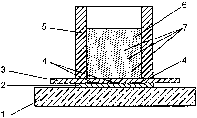
Fig. 1 illustrates a support plate 1 on which there is a
deposit of active substance 2. Above the deposit of active
substance 2 there is a membrane filter 3 which is joined to
the deposit of active substance 2 by means of a plurality of
adhesive bonds 4. Above the membrane filter 3 there is a
tubular vessel 5, into which a liquid 6 has been introduced
which contains cells 7 whose ability to migrate is to be
measured. The membrane filter 3 has an area which is equal to
at least 1.6 times the size of the base opening of the vessel
5.
The membrane filter 3 exerts a sucking action on the liquid 6
together with the cells 7 contained therein. As a result,
liquid also passes into the deposit of active substance 2
which is arranged beneath the membrane filter 3, during which
process fractions of the active substance are dissolved and
subsequently diffuse into the membrane filter 3 and the liquid
above it. As a result, the cells 7 are acted on in such a way
that their migration into the membrane filter 3 is influenced.
The fact that the area of the membrane filter 3 is at least
1.6 times as large as the base opening in the vessel 5 leads
to a significantly stronger sucking action being exerted on
the liquid 6, together with the cells 7 contained therein,
situated in the vessel 5 than would be the case if the
membrane filter is of approximately the same size as the base
opening of the vessel 5. As a result, the cells 7 are brought
into contact with the membrane filter 3 more quickly and can
penetrate into this filter more rapidly, so that the time
required for the measurement is reduced.
Since the ability of some types of cell to migrate may change
rapidly outside the organism, a reduced measurement time leads
to an improved determination of the diagnostically significant
CA 02336402 2001-O1-02
WO 00/17652 ~ ~ -5- PCT/AT99/00165
original readiness of the cells to migrate. Therefore, the
increased suction provided by the enlarged membrane filter
results in significantly more accurate measurement results
compared to the known prior art.
The fact that the deposit of active substance 2 is joined to
the membrane filter 3 by adhesive bonding so as to bear
tightly against it ensures that the active substance is
dissolved and then diffuses into the membrane filter 3 and
onward into the liquid 6 inside the vessel 5 in a controlled,
uniform manner, which is one of the preconditions for the
reproducibility and standardization of migration measurements.
However, the total area of the bonded surfaces 4 should cover
no more than 30% of the surfaces bearing against one another,
since otherwise the diffusion of the liquid into the deposit
of active substance 2 and, in addition, the diffusion of the
dissolved active substance out of the deposit of active
substance 2 into the membrane filter 3 would be considerably
restricted. The individual bonded areas 4 may be arranged in a
grid-like pattern.
After the migration process has ended, the cells which have
migrated into the membrane filter 3 are made visible, for
example by dyeing, and then their number, distribution and
shape are determined by means of a microscopic assessment
method. If the components of the apparatus are transparent or
can be made transparent, a transillumination method can be
employed for this purpose.
The basic structure and fundamental function of an apparatus
of this type have been described with reference to Fig. 1. By
contrast, Figs. 2 and 2a illustrate an apparatus of this type
which can be used to facilitate series of measurements. In
this apparatus, a plurality of the components illustrated in
CA 02336402 2001-O1-02
w0 00/17652 ~ -6- PCT/AT99/00165
Fig. 1 are combined to form one measurement unit, allowing a
simple, rapid and clear migration measurement to be carried
out.
This apparatus comprises a rectangular support plate 11 which
is preferably made from a transparent or translucent material.
A mat-like membrane filter 13, which covers a plurality of
deposits of active substance 12 which are spaced apart from
one another, is situated on top of this support plate 11. The
deposits of active substance 12 are joined to the membrane
filter 13 by means of a plurality of adhesive bonds. The
deposits of active substance 12 and those parts of the
membrane filter 13 which do not cover the deposits of active
substance 12 are also joined to the support plate 11 by
adhesive bonds.
In this exemplary embodiment, two rows of in each case three
vessels 15 and 15a, into which the liquid 16 containing the
cells 17 to be analyzed has been introduced, are situated
above the membrane filter 13. The individual vessels 15 and
15a in the two rows are connected to one another by means of
webs 18 and 18a to form units. As a result, during production
of the apparatus they can be placed onto the membrane filter
13 together and then adhesively bonded to the filter. In
addition, they can be detached from the membrane filter 13
together after the migration process and the preparation of
the cells which have migrated into the membrane filter 13. The
deposits of active substance 12 situated beneath the three
vessels 13 are laden with an active substance, whereas the
deposits of active substance situated beneath the vessels 15a
arranged parallel to the vessels 15 do not contain any active
substance, since they are used to measure the unstimulated,
spontaneous cell migration. The measurement which is carried
CA 02336402 2001-O1-02
WO 00/17652 ~ - -7- PCT/AT99/00165
out in triplicate simultaneously in the exemplary embodiment
is used to increase the measurement accuracy.
According to another exemplary embodiment, the deposits of
active substance 12 beneath the vessels 15 and 15a are each
laden with different active substances, so that it is possible
to compare the different actions of these substances on the
migration of the cells which are to be analyzed.
According to another method, after the migration process has
concluded, the cells which have migrated into the membrane
filter 13 are fixed and dyed as a result of suitable
substances being added to the vessels 15 and 15a. After
preparation has finished, the vessels 15 and 15a are detached
from the membrane filter 13. The cells which have migrated
into the membrane filter 13 are then accessible for
microscopic analysis.
In one exemplary embodiment, the vessels 15 and 15a have an
internal diameter of approximately 7 mm and a height of
approximately 9 mm. The membrane filter 13 and the deposits of
active substance 12 are each 140 ~m thick. The vessels 15 and
15a and the support plate 11 may be made from plastic or from
glass. The only required criterion is that they be neutral
with respect to the media used and the cells to be analyzed.