Note : Les descriptions sont présentées dans la langue officielle dans laquelle elles ont été soumises.
CA 02338984 2001-01-30
WO 00/76403 PCTIUSOO/12556
RETINAL TISSUE I14PLANTATION INSTRUMENT
BACKGROUND OF THE INVENTION
The present invention relates to an instrument for
implanting delicate tissue and/or materials in the human body,
more particularly to an instrument for surgically restoring
eyesight by implanting fetal retinal tissue into the
subretinal space in the back of the eye.
Most common eye problems, for example, myopia
(nearsightedness), hyperopia (farsightedness), astigmatism
(asymmetrical cornea, and presbyopia (the inability to focus
on an object at close range) are due to errors in the
refraction of light by the lens and cornea in the anterior
part of the eye. Generally, these problems can be corrected
by glasses, contact lenses, or corrective surgery.
However, blindness is most commonly due to damage of the
retina in the back of the eye and, more specifically, is
caused by abnormalities in the subretinal space under the
retina.
The transparent, layered retina processes light images
projected by the cornea and lens. The photoreceptor layer in
the back of the retina transforms the light into electrical
impulses. Other retinal layers transfer these impulses
through the optic nerve to the brain which interprets the
impulses into what we perceive as sight.
The subretinal space is the area between the retinal
pigment epithelium (RPE) and the photoreceptors of the retina.
Normally, the photoreceptors are in close contact with the
RPE. The RPE has many functions. It provides nutrition for
the photoreceptors, and also removes waste products from the
photoreceptors. In a normal eye, there are no blood vessels
in the subretinal space. However, in some retinal diseases,
blood vessels and connective tissue can grow in this space and
cause blindness. Under certain disease conditions, the
photoreceptors can be detached very easily from the RPE. The
photoreceptors will then degenerate, resulting in vision loss
or blindness, while the other layers of the retina may remain
-1-
CA 02338984 2001-01-30
WO 00/76403 PCT/US00/12556
functional. By replacing the diseased RPE and/or
photoreceptors that can hook up to the functional part of the
retina, vision may be restored.
The most frequent cause of legal blindness is macular
degeneration and retinitis pigmentosa. The macula is located
in the back of the eye in the central portion of the retina
and is responsible for central vision. In patients with
macular degeneration, there is initially a dysfunction of the
RPE in the macular region, which later leads to ingrowth of
blood vessels and destruction of the light-sensitive
photoreceptors in the overlying retina. This results in
impairment of central vision. Age related macular
degeneration is an example of an eye disease that can be
delayed by using the herein disclosed method and instrument.
Retinitis pigmentosa is a term for genetically caused
photoreceptor degeneration. In these patients, the
photoreceptors must be replaced. Again, the method and
instrument of the present invention can be utilized.
It is to be noted that surgical correction of diseases in
the subretinal space between the retina and the RPE is
rendered extremely difficult by the environment in which the
surgery must take place. Moreover, the surgical procedure
disclosed herein to implant fetal retinal tissue into the
subretinal space of the eye is complicated by the fact that
fetal retinal tissue is in the nature of a transparent
gelatinous mass and therefore extremely fragile.
SUMMARY OF THE INVENTION
In accordance with the present invention, I have
developed an improved implantation instrument capable of
handling fetal retinal tissue and placing this tissue into the
subretinal space between the retinal pigment epithelium and
the retina of the human eye.
I have discovered that intact sheets of fetal retinal
tissue can be transplanted into the subretinal space by
flattening and protecting it by a gel that disintegrates and
-2-
CA 02338984 2001-01-30
WO 00/76403 PCT/US00/12556
is subsequently absorbed by the recipient eye so as to leave
the transplant free. The transplant develops organized
parallel layers resembling normal retina, with fully developed
photoreceptors. The transplant can replace diseased
photoreceptors and/or RPE. Moreover, the fetal retinal tissue
is immunologically tolerated in the subretinal space and is
not subject to rejection provided there is little surgical
trauma.
The instrument of the instant invention comprises a
handpiece for the support of a mandrel, a sleeve support
telescoped over the mandrel, a tubular sleeve slidably
journaled on the sleeve support, a nozzle having an aperture
for the acceptance of retinal tissue mounted on the sleeve and
extending over the mandrel, and a toggle mechanism that
controls the position of the nozzle relative to the mandrel.
The handpiece, mandrel, sleeve support, sleeve and toggle
mechanism are preferably made of stainless steel to facilitate
autoclaving. The nozzle is molded from elastic plastic.
However, the instrument can be manufactured primarily from
plastic if desired, so as to be disposable.
Advancement and retraction of the sleeve and nozzle
relative to the handpiece and mandrel is controlled by the
toggle mechanism on the handpiece, one element of which is a
spring. When the spring element of the toggle mechanism is
pressed toward the handpiece, the sleeve and the nozzle
thereon move outwardly on the mandrel to a point where the
aperture in the nozzle is disposed outwardly of the tip of the
mandrel creating a space in the nozzle that accepts retinal
tissue therein.
When the tissue is in place inside the nozzle, the nozzle is
partially retracted to bias the retinal tissue to a position
adjacent the tip of the nozzle whereupon a toggle lock engages
a peg on the spring element so as to lock the spring element
and, therefore, the sleeve relative to the mandrel. The
surgeon inserts the instrument on the target and holds his
hand absolutely still. With a slight pressure on the spring
-3-
CA 02338984 2001-01-30
WO 00/76403 PCT/US00/12556
elemerit, without movement of the handpiece, the toggle lock is
released, and the sleeve and nozzle retract over the mandrel
under the bias of the toggle spring, placing the tissue at the
desired location.
'rhe position of the toggle lock can be regulated so as to
determine the space between the mandrel tip and the nozzle tip
thereby adapting the instrument to the size of the transplant.
Mandrels and nozzles can be customized in different sizes
and shapes for implantation of different kinds of fragile
tissue; gels containing different trophic factors or drugs; or
electronic microchips into the subretinal space. Mandrels and
nozzles can be produced in sterile packages for one-time use.
The details of the instrument of the invention are more
fully described in the following specification and drawings.
BRIEF DESCRIPTION OF THE DRAWINGS
FIG. 1 is a frontal view of the human eye;
FIG. 2 is a cross-sectional view of the human eye showing
the implantation instrument inserted through the pars plana
into the subretinal space;
FIG. 3 is an enlarged view taken within the circle "3"
of Fig. 2;
FIG. 4 is an enlarged view of the area within the circle
"4" prior to insertion of the instrument nozzle into the
subretinal space;
FIG. 5 is a view, similar to Fig. 4, with the instrument
nozzle in the subretinal space;
FIG. 6 is a view, similar to Fig. 5, with the instrument
nozzle retracted and the retinal tissue in the target area of
the subretinal space;
FIG. 7 is an elevational view of the instrument of the
invention in the loaded condition;
FIG. 8 is a cross sectional view of the instrument of
Fig. 7 after implantation of retinal tissue;
FIG. 9 is a view of a tissue prior to transfer through
the receiving aperture I the instrument nozzle;
-4-
CA 02338984 2001-01-30
WO 00/76403 PCT/US00/12556
FIG. 10 is a view of the retinal tissue after the first
increment of movement toward the tip of the instrument nozzle;
FIG. 11 is view of the retinal tissue in position in the
instrument nozzle for implantation; and
FIG. 12 in a view of the retinal tissue after discharge
from the nozzle of the instrument.
DETAILED DESCRIPTION OF THE PREFERRED EMBODIMENT(S)
The environment in which the present invention has
particular utility is illustrated in Fig. 1 and 2 of the
drawings. The front of the eye 50 is covered by a transparent
tissue, the cornea 52, surrounded by white conjunctive
tissue 54. The sclera 56 is hard fibrous tissue that covers
the exterior of the eyeball. The pupil 58 is the opening
through which light passes to the back of the eye. The
iris 60 changes the size of the pupil 58 to adjust to the
amount of light. The transparent lens 62 is located behind
the iris 60 and is suspended by a net of fibers 64. The
fibers 64 are attached to the ciliary body 66 that extends to
where the retina 68 begins. The part of the ciliary body 66
adjacent to the retina 68 is called pars plana 70. The
lens 62 focuses light rays onto the retina 68. The bulk of
the eyeball 50 behind the lens 62 is formed by the vitreous
chamber 72, which is filled with a colorless, gelatin like
substance.
The retina 68 covers most of the wall of the vitreous
chamber 72 and comprises transparent layers that extend
forwardly to the pars plana 70 and which processes light
images projected from the cornea 52 and the lens 62.
The rear of the retina 68 contains photoreceptors 74,
which transform light into electrical impulses. The electrical
impulses are carried by nerves in the retina 68 to the optic
nerve 76, which, in turn leads to the brain. A monolayer of
cells termed the retinal pigment epithelium (RPE) 77 resides
behind the retina 68. The choroid 78 is a layer of blood
-5-
__
CA 02338984 2001-01-30
WO 00/76403 PCT/USOO/12556
vessels behind the RPE 77, that supplies oxygen and nutrients
essential to the function of the eye 50. The RPE 77
transports these nutrients to the retina 68 and maintains a
barrier between choroid 78 and retina 68.
The region between the retina 68 and the RPE 77 is called
the subretinal space 80 (Fig 4-6). Normally, there is no
"space". However, the retina 68 detaches very easily from the
RPE 77 and it is in this "space" that the surgeon transplants
the new piece of retinal tissue to replace damaged
photoreceptors 74 and/or RPE 77.
The fovea 82 is a small depression in the center of the
retina 68 that is essential for sharp (focussed) vision as
well as color vision. The small area surrounding the fovea 82
is known as the macula 84 and is responsible for central
vision. The point at which the optic nerve 76 leaves the
retir-a 68 on its way to the brain is called optic disc 86.
In accordance with the present invention, surgical
correction of retinal diseases in the subretinal "space" 80
between the retina 68 and the RPE 77 is facilitated by a novel
implantation instrument 100.
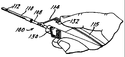
As seen in Fig. 7 through 11, the instrument 100
comprises a handpiece 102, a tubular sleeve support 104, a
mandrel 106 disposed internally of the sleeve support 104, a
sleeve 108 slidably journaled on the sleeve support 104, and a
nozzle 110 mounted on the sleeve 108 having an aperture 112
therein for the acceptance of retinal tissue 120. A toggle
mechanism comprising a rigid link 114 and a spring link 115
controls advancement and retraction of the sleeve 108 and
nozzle 110 relative to the mandrel 106. The handpiece 102,
sleeve support 104, mandrel 106, sleeve 108 and toggle
links 114 and 115 can be made of stainless steel to facilitate
autoclaving. The instrument can also be made disposable by
using plastic with some metal parts.
The mandrel 106 comprises an elongated flat and narrow
strip of steel that is fixed in the handpiece but is
longitudinally adjustable relative thereto. Extension or
-6-
CA 02338984 2001-01-30
WO 00/76403 PCT/USOO/12556
retraction of the mandrel 106 relative to the housing 102
regulates its longitudinal position relative to the length of
the plastic nozzle 110 in the retracted position.
The fit between the plastic nozzle 110 and the
mandrel 106 must permit relative movement therebetween,
whereby the aperture 112 in the nozzle 110 can be positioned
for the acceptance of the retinal tissue 120 and subsequent
discharge of the tissue 120.
The nozzle 110 is molded of elastic plastic, for example
fluorinated ethylene propylene, so as to have a curvature at
the tip 118 thereof in order to slide under the retina 68 into
the subretinal "space" 80. Because of its elasticity, the
curved nozzle tip 118, when retracted, will straighten out
over the mandrel 106 so as to deposit implant tissue at the
target area behind the retina 68.
Advancement and retraction of the sleeve 108 and
nozzle 110 is controlled by the rigid toggle link 114 and
spring element 115 on the handpiece 102. When the spring
element 115 of the toggle mechanism is pressed toward the
handpiece 102, the sleeve 108 and the nozzle 110 are driven
forwardly, relative to the mandrel 106, by the rigid toggle
link 114. When pressure on the spring element 115 of the
toggle mechanism is released, the sleeve 108 and the
nozzle 110 thereon is retracted over the mandrel 106, biasing
the retinal tissue 120 to the desired location.
In preparation for the use of tool of the invention, the
surgeon first places an incision in the pars plana 70 of the
eye 50. A small incision is then made in, for example, the
macular region 84 of the retina 68. If necessary, abnormal
tissue is removed from the subretinal "space" 80 between the
retina 68 and the RPE 77. The nozzle 110 of the
instrument 100, with the retinal tissue 120 enclosed in the
curved nozzle tip 118 thereof, is inserted through the
incision in the pars plana 70 and through the incision in the
retina 68 until the tip of the nozzle 118 is orientated
adjacent the target area in the subretinal "space" 80.
-7-
__
CA 02338984 2001-01-30
WO 00/76403 PCT/US00/12556
Slight pressure on the spring element 115 of the
instrument 100 then releases the toggle lock 130 from the
pin 132 allowing the spring element 115 and rigid link 114 of
the toggle mechanism to effect retraction of the nozzle 110
and deposition of the retinal tissue 120. It is to be noted
that the handpiece 102 and mandrel 106 of the instrument 100
are not required to move incident to deposition of the retinal
tissue 120 allowing the surgeon to precisely position the
tissue.
From the forgoing, it should be apparent that the
instrument of the present invention accepts retinal tissue in
an ef:ficient manner and thereafter precisely implants the
tissue into the eye. The surgeon has only to keep his hand
still and exert a slight pressure on the spring element 115 of
the instrument 100 to release the toggle lock 130 conditioning
the instrument 100 itself to effect retraction of the
nozzle 110 and placement of the implant on the target.
While the preferred embodiment of the invention has been
disclosed, it should be appreciated that the invention is
susceptible of modification without departing from the scope
of the following claims:
-8-
.._
