Sélection de la langue

Search

Sommaire du brevet 2345921 

Énoncé de désistement de responsabilité concernant l'information provenant de tiers

Une partie des informations de ce site Web a été fournie par des sources externes. Le gouvernement du Canada n'assume aucune responsabilité concernant la précision, l'actualité ou la fiabilité des informations fournies par les sources externes. Les utilisateurs qui désirent employer cette information devraient consulter directement la source des informations. Le contenu fourni par les sources externes n'est pas assujetti aux exigences sur les langues officielles, la protection des renseignements personnels et l'accessibilité.

Disponibilité de l'Abrégé et des Revendications

L'apparition de différences dans le texte et l'image des Revendications et de l'Abrégé dépend du moment auquel le document est publié. Les textes des Revendications et de l'Abrégé sont affichés :

  • lorsque la demande peut être examinée par le public;
  • lorsque le brevet est émis (délivrance).
(12) Brevet: (11) CA 2345921
(54) Titre français: FIL-GUIDE MODIFIE POUR DERIVATION D'ACCES AU VENTRICULE GAUCHE
(54) Titre anglais: MODIFIED GUIDEWIRE FOR LEFT VENTRICULAR ACCESS LEAD
Statut: Périmé et au-delà du délai pour l’annulation
Données bibliographiques
(51) Classification internationale des brevets (CIB):
  • A61M 25/01 (2006.01)
  • A61N 1/05 (2006.01)
(72) Inventeurs :
  • WESTLUND, RANDY W. (Etats-Unis d'Amérique)
  • TOCKMAN, BRUCE A. (Etats-Unis d'Amérique)
  • PETERFESO, RANDALL M. (Etats-Unis d'Amérique)
  • GREENLAND, JOHN S. (Etats-Unis d'Amérique)
  • HINDERS, MARY N. (Etats-Unis d'Amérique)
(73) Titulaires :
  • CARDIAC PACEMAKERS, INC.
(71) Demandeurs :
  • CARDIAC PACEMAKERS, INC. (Etats-Unis d'Amérique)
(74) Agent: SMART & BIGGAR LP
(74) Co-agent:
(45) Délivré: 2005-01-25
(86) Date de dépôt PCT: 1999-04-06
(87) Mise à la disponibilité du public: 1999-12-16
Requête d'examen: 2001-03-08
Licence disponible: S.O.
Cédé au domaine public: S.O.
(25) Langue des documents déposés: Anglais

Traité de coopération en matière de brevets (PCT): Oui
(86) Numéro de la demande PCT: PCT/US1999/007515
(87) Numéro de publication internationale PCT: WO 1999064100
(85) Entrée nationale: 2001-03-08

(30) Données de priorité de la demande:
Numéro de la demande Pays / territoire Date
09/097,101 (Etats-Unis d'Amérique) 1998-06-12
09/164,891 (Etats-Unis d'Amérique) 1998-10-01

Abrégés

Abrégé français

Cette invention se rapporte à un fil-guide amélioré (20) destiné à faciliter l'implantation d'une dérivation cardiaque et comprenant à cet effet trois sections (22, 24, 26). La zone la plus distale (22) est suffisamment souple pour empêcher tout traumatisme aux parois du vaisseau dans lequel sont insérés le fil-guide (20) et la dérivation. Une zone intermédiaire (24) est, en général, plus rigide et possède une section transversale inférieure ou égale à celle de la zone distale (22). La troisième zone (26) est encore plus rigide et elle est jointe à la zone intermédiaire par un épaulement (34) servant à transférer les forces à la dérivation.


Abrégé anglais


An improved guide wire (20) for assisting in implantation of a cardiac lead
includes three sections (22, 24, 26). The most distal zone
(22) is sufficiently floppy to prevent trauma to the vessel walls through
which the guide wire (20), and lead are inserted. An intermediate
zone (24) is generally stiffer, and has a cross section less than or equal to
the cross section of the distal zone (22). The third zone (26) is
stiffer yet, and is joined to the intermediate zone by a shoulder (34) to
transfer force to the lead.

Revendications

Note : Les revendications sont présentées dans la langue officielle dans laquelle elles ont été soumises.


-12-
CLAIMS:
1. An intravenous lead insertion assembly, comprising
a catheter having a proximal end and a distal end;
an intravenous lead at least partially located within the catheter and having
at
least one electrode and a lumen extending from a proximal end of the lead to
a distal end of the lead, the lumen having a first opening through the
proximal
end of the lead and a second opening through the distal end of the lead;
a guidewire for facilitating advancement of the intravenous lead into the
coronary sinus, said guidewire sized to extend through the first and second
openings in said lumen and past the distal end of the lead during implantation
of the lead, and being removable from the lead after placement of the lead in
the coronary sinus; and
a removal wire sized to extend through the first opening and into the lumen of
the intravenous lead, the removal wire having a distal end portion including a
lead contacting portion for contacting the lead to facilitate securing the
lead
during a removal of the catheter from around the lead.
2. The intravenous lead insertion assembly according to claim 1, wherein the
lumen of the intravenous lead is of a size to separately receive the guidewire
and the removal wire, but is not of a size to receive the guidewire and the
removal wire simultaneously.
3. The intravenous lead insertion assembly according to claim 1, wherein the
distal end of the intravenous lead extends distal of the distal end of the
catheter prior to removal of the catheter.
4. The intravenous lead insertion assembly according to claim 1, wherein the
contacting portion of the removal wire abuts a portion of the intravenous
lead.

-13-
5. The intravenous lead insertion assembly according to claim 1, wherein the
contacting portion of the removal wire contacts the lumen of intravenous lead.
6. The intravenous lead insertion assembly according to claim 1, wherein the
contacting portion of the removal wire includes a transition portion located
between a larger diameter section of the removal wire and a smaller diameter
section of the removal wire.
7. The intravenous lead insertion assembly according to claim 6, wherein the
lumen of the intravenous lead includes a first diameter portion and a second
diameter portion, the second diameter portion being smaller than the first
diameter and located distal of the first diameter.
8. The intravenous lead insertion assembly according to claim 7, wherein the
lumen of the intravenous lead includes a tapered portion between the first
and second diameter portions.
9. The intravenous lead insertion assembly according to claim 1, wherein the
contacting portion of the removal wire provides a frictional engagement with
the lumen of the intravenous lead.
10. The intravenous lead insertion assembly according to claim 1, wherein the
removal wire includes a coiled member forming a squared distal tip.
11. The intravenous lead insertion assembly according to claim 1, wherein the
lumen of the intravenous lead is formed by a helical coil and the contacting
portion engages the helical coil of the lead.
12. The intravenous lead insertion assembly according to claim 1, wherein the
lumen of the intravenous lead is formed by a helical coil.
13. An intravenous lead insertion assembly, comprising
a catheter having a proximal end and a distal end;

-14-
an intravenous cardiac pacing lead at least partially located within the
catheter and having at least one pacing electrode and a lumen extending
from a proximal end of the lead to a distal end of the lead, the lumen having
a
first opening through the proximal end of the lead and a second opening
through the distal end of the lead;
a guidewire for facilitating advancement of the intravenous lead into the
coronary sinus, said guidewire sized to extend through the first and second
openings in said lumen and past the distal end of the lead during implantation
of the lead, and being removable from the lead after placement of the lead in
the coronary sinus; and
a removal wire sized to extend through the first opening and into the lumen of
the intravenous lead, the removal wire having a distal end portion including a
lead contacting portion for contacting the lead to facilitate securing the
lead
during a removal of the catheter from around the lead.
14. The intravenous lead insertion assembly according to claim 13, wherein the
lumen of the intravenous lead is of a size to separately receive the guidewire
and the removal wire, but is not of a size to receive the guidewire and the
removal wire simultaneously.
15. The intravenous lead insertion assembly according to claim 13, wherein the
contacting portion of the removal wire abuts the intravenous lead.
16. The intravenous lead insertion assembly according to claim 13, wherein the
contacting portion of the removal wire includes a transition portion located
between a larger diameter section of the removal wire and a smaller diameter
section of the removal wire.
17. The intravenous lead insertion assembly according to claim 13, wherein the
lumen of the intravenous lead is formed by a helical coil.
18. An intravenous lead insertion assembly, comprising
a catheter having a proximal end and a distal end;

-15-
an intravenous cardiac lead at least partially located within the catheter and
having at least one defibrillation electrode and a lumen extending from a
proximal end of the lead to a distal end of the lead, the lumen having a first
opening through the proximal end of the lead and a second opening through
the distal end of the lead;
a guidewire for facilitating advancement of the intravenous lead into the
coronary sinus, said guidewire sized to extend through the first and second
openings in said lumen and past the distal end of the lead during implantation
of the lead, and being removable from the lead after placement of the lead in
the coronary sinus; and
a removal wire sized to extend through the first opening and into the lumen of
the intravenous lead, the removal wire having a distal end portion including a
lead contacting portion for contacting the lead to facilitate securing the
lead
during a removal of the catheter from around the lead.
19. The intravenous lead insertion assembly according to claim 18, wherein the
lumen of the intravenous lead is of a size to separately receive the guidewire
and the removal wire, but is not of a size to receive the guidewire and the
removal wire simultaneously.
20. The intravenous lead insertion assembly according to claim 18, wherein the
contacting portion of the removal wire abuts a portion of the intravenous
lead.
21. The intravenous lead insertion assembly according to claim 18, wherein the
contacting portion of the removal wire includes a transition portion located
between a larger diameter section of the removal wire and a smaller diameter
section of the removal wire.
22. The intravenous lead insertion assembly according to claim 18, wherein the
lumen of the lead is formed by a helical coil.

-16-
23. The intravenous lead insertion assembly of claim 1, wherein the removal
wire
includes a proximal portion and a distal portion, the distal portion being
tapered.
24. The intravenous lead insertion assembly of claim 13, wherein the removal
wire includes a proximal portion and a distal portion, the distal portion
being tapered.
25. The intravenous lead insertion assembly of claim 18, wherein the removal
wire includes a proximal portion and a distal portion, the distal portion
being tapered.

Description

Note : Les descriptions sont présentées dans la langue officielle dans laquelle elles ont été soumises.

CA 02345921 2003-12-22
WO 99/64100 ~ PCT/US99/075I5
MODIFIED GUIDEWZRE FOR LEFT VENTRICULAR ACCESB LEAD
BACKGROUND OF THE INVENTION
I. Field of the Invention
The invention relates to the implantation and
placement of cardiac leads used in combination with
cardiac rhythm management devices, e.g., heart pacemakers
or defibrillators, to monitor and control the rhythm of
the heart. This invention is more particularly directed
toward a guidewire configuration adapted to assist in the
implantation and placement of cardiac leads having
electrodes that are to reside in the distal branches of
the coronary venous system, the great cardiac vein, or
coronary sinus.
II. Discussion of the Prior Art
Placement of cardiac leads in the distal branches of
the coronary venous system, the great cardiac vein, or
the coronary sinus is a difficult task. Often when
deploying the lead there comes a point at which the lead
cannot be advanced further into the vascular system using
standard techniques and equipment. All too often this
point is not the optimal position for the lead's
electrode either sensing cardiac activity or delivering
therapy to the heart.
There are several reasons which make proper
placement of the lead difficult. These include (1)
friction between the vasculature and the lead; (2)
partial obstruction of the vasculature; (3) unusually
shaped bifurcations in the vasculature; and (4)
accumulative friction between lead, guide catheter and
guidewire. Prior efforts to resolve such problems
included the use of a stiffer guidewire. While stiffer
guidewires offer additional support, they may, impede
advancement due to their relative size with respect to

CA 02345921 2001-03-08
WO 99/64100 PCT/US99/07515
-2-
the lumen of the lead.
Additionally, when proper placement of the lead is
achieved, problems arise during guide catheter
extraction. All too often, the act of extracting the
guide catheter causes the lead to be dislodged from the
implanted position. Standard guidewires and stylets are
not suitable for maintaining position while the guide
catheter is removed due to insufficient stiffness, lack
of appropriate force transmission features, and friction
between the guidewire and lumen wall of the coronary vein
lead.
The present invention is deemed to be an improvement
over such prior art guidewires. It is more effective in
properly placing the lead. It is also less likely to
cause a properly placed lead to become dislodged during
extraction of the guide catheter and the guidewire
itself .
SUl~IARY OF THE INVENTION
In accordance with the present invention, there is
provided a guidewire comprised of at least three zones.
Each zone differs from the other two in stiffness and
flexibility. Each zone also has geometric
characteristics which assist in proper placement of the
lead and further assist in preventing dislodgement of the
lead as the guide catheter is extracted and as the
guidewire itself is extracted.
Specifically, the first and most distal zone is
intended to be sufficiently floppy to prevent trauma to
the surrounding vessel walls when the guidewire is used
for deploying a coronary vein lead. This zone includes a
spiral wound portion surrounding a thin, solid ribbon
core and a spherical tip. The second intermediate zone,
is generally stiffer and comprises a solid wire or spiral
wound wire having a cross-sectional diameter that does
not exceed the cross-sectional diameter of the first
zone. The third most proximal zone is of a larger
diameter and is stiffer than the first and second zones.

CA 02345921 2001-03-08
WO 99!64100 PCT/US99/07515
-3-
The third zone comprises a wire or tube which is
manipulated to apply advancement forces during deployment
of the lead and counter forces.during extraction of the
guide catheter. The diametric transition between the
second and third zones is abrupt and ideally corresponds
to a matching feature in the lead so that this transition
is the point where most of the advancement forces and
counter forces are transmitted to the lead.
Other improvements also exist. For example, the
improved guidewire (or portions thereof) can be provided
with a hydrophilic coating to produce a highly lubricious
surface. The presence of such a surface reduces friction
between the lumen wall of the lead and the guidewire
thereby reducing the risk that the lead will be dislodged
during extraction of the guidewire. Likewise, a locking
mechanism can be provided between the lumen of the lead
and the guidewire so that forces applied to the guidewire
are transmitted to the lead through the locking
mechanism.
DESCRIPTION OF THE DRAWINGS
The foregoing features, objects and advantages of
the present invention will become more clear to those
skilled in the art from the following detailed
description of a preferred embodiment, particularly when
considered in conjunction with the accompanying drawings
in which like numerals in the several views refer to
corresponding parts.
Figure 1 is a view showing an intravenous cardiac
lead having an electrode positioned in a coronary vein.
Figure 2 is a perspective view of a preferred
embodiment of the guidewire of the present invention.
Figure 3 is a cross-sectional view of a cardiac lead
with a guidewire positioned within the lumen of the lead.
Figure 4 is a plan view of an alternative embodiment
of a guidewire made in accordance with the present
invention.
Figure 5 is a cross-sectional view of a cardiac lead

CA 02345921 2001-03-08
WO 99/64100 PCT/US99/07515
-4-
with a guidewire of the type shown in Figure 4 positioned
within the lumen of the lead.
DETAILED DEBCRIPTION OF THE INVENTION
Figure 1 shows a human heart 1 with a coronary lead
10 passing through the superior vena cava 2, the right
atrium 3, and the coronary sinus 4 into the great vein of
the heart 5 so that an electrode 12 on the lead 10 is
properly positioned in a branch of the coronary vein.
When positioned as shown, the electrode 12 can be used to
sense the electrical activity of the heart or apply
stimulating pulses to the left ventricle 7 without the
electrode being positioned within the left ventricular
chamber. The present invention is directed toward
elongated wires useful in placing the electrode in the
vasculature. Such wires are generally referred to as
"guidewires". As used herein, the term "guidewire"
includes both wires used to position the electrode and
wires used to retain the electrode in the proper position
as a guide catheter is removed.
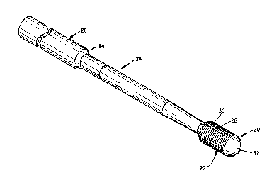
Figure 2 shows a first preferred embodiment of a
guidewire 20 advantageously used to position the coronary
lead 10 as shown in Figure 1 and retain the coronary lead
in that position as a guide catheter is removed.
Guidewire 20 has three zones, a distal zone 22,
intermediate zone 24, and a proximal zone 26.
The distal zone 22 preferably is 1-1.5 inches long.
The distal zone 22 is circular in cross-section and has a
cross-sectional diameter of approximately 0.014 inches.
The distal zone 22 comprises an internal shapeable ribbon
core member 28, a spiral winding 30 and a spherical tip
32. The internal ribbon core member 28, as it extends
distally, tapers from about 0.005 inches to about 0.001
inches. This construction is sufficiently floppy such
that there is no trauma induced by the guidewire to a
surrounding vessel wall. This construction also allows
it to be capable of being biased so as to aid in steering

CA 02345921 2001-03-08
WO 99/64100 PCT/US99/07515
-5-
through the vasculature.
The intermediate zone 24 is generally stiffer than
the distal zone 22. The intermediate zone comprises a
solid wire having a circular cross-section. The cross-
sectional diameter of the wire can vary depending upon
the performance needs, but should not exceed the cross-
sectional diameter of distal zone 22. The length of
intermediate zone 24 can also vary, but preferably will
be one to four inches long.
The proximal zone 26 is made of a wire or tubing and
is the stiffest and longest section of the guidewire 20.
The proximal zone 26, being the stiffest and most
proximal, is the portion handled and used to apply forces
during deployment and guide catheter extraction.
Preferably the overall length of the guidewire 20 will be
in the range of five feet. The cross-sectional diameter
of the proximal zone is larger than the cross-sectional
diameter of the distal zone 22 and the intermediate zone
24. For example, if the distal and intermediate zones
have a diameter of approximately 0.014 inches, the
proximal zone will have a diameter of approximately 0.022
inches. The diametrical transition between the proximal
and intermediate zones taper, though abruptly, from 0.022
inches to 0.014 inches. As discussed below, this
diametrical transition constitutes a shoulder 34 through
which most of the advancement and counter forces are
transmitted between the lead 10 and guidewire 20.
Figure 3 shows the guidewire 20 of Figure 2
positioned within a lumen 14 of the coronary lead 10.
The lumen 14 has a projection 16 which corresponds to the
shoulder 34 of the guidewire 20. When the shoulder 34
engages the projection 16, advancement forces applied to
the guidewire 20 are transferred to the lead 10 through
the shoulder 34 and projection 16 or reduction in lumen
diameter.
Figures 4 and 5 show a second preferred embodiment
of a guidewire 20, specifically referred to as a removal

CA 02345921 2001-03-08
WO 99/64100 PCT/US99/07515
-6-
wire. This embodiment is ideally suited for use during
removal of a guide catheter. As such, it may be used to
replace a standard guidewire after insertion of the lead
but before removal of the guide catheter. Again, the
guidewire 20 of this embodiment has three zones -- a
distal zone 22, an intermediate zone 24, and a proximal
zone 26. The guidewire 20 shown in Figure 4 is
dimensioned in a fashion similar to the guidewire shown
in the other three figures. The primary difference
between the removal wire shown in Figure 4 and the
guidewire shown in the other figures is that the tip 32
is not attached to the core 28 by a solder joint. Also,
the tip 32 of this embodiment is not intended to exit the
distal end of the lead 10, thus it is not shapeable or
steerable in the vasculature. It may be used to lock into
the lead 10 and transmit force to the lead tip, but only
as used in conjunction with the lead. The tip 32 of this
embodiment is not considered atraumatic whereas the tip
in the embodiment shown in Figure 2 is designed to be
extremely atraumatic. This spiral wound wire is secured
to the proximal zone 26 by a solder joint. The direction
of the winding will preferably be opposite that of any
winding 11 of the lead 10 itself. This allows for better
tracking through the central lumen of the lead. The
distal zone 22 is a continuation of the spiral winding of
the intermediate zone. However, the diameter of the
winding is increased to form the distal zone. The distal
tip 32 of the coil which forms the intermediate end
distal zones is cut square and not attached to a core
wire or the like. Thus, as the guide catheter is
withdrawn, the square cut of tip 32 seats in the taper of
the coil il of the lead preventing the lead from
withdrawing as the guide catheter is pulled free.
Furthermore, as the removal wire is pulled free from the
lead, the square, unattached coil tip 32 slightly
distends and easily frees itself from the tapered coil
section of the lead. This feature allows for easy,

CA 02345921 2001-03-08
WO 99/64100 PCTNS99107515
predictable removal of the removal wire from the lead,
thus preventing loss of lead position upon its
withdrawal.
In either embodiment, a hydrophilic coating can be
applied to create a surface 21 on the guidewire 20 that
is highly lubricious. Alternatively, all surfaces of
guidewire 20 can be coated with a hydrophilic coating or
polytetrafluoroethylene (PTFE), or some other dry
lubricious material, i.e. silicone film. This serves to
reduce friction between the guidewire 20 and lead lumen
14 thereby reducing the risk that the lead 10 will be
dislodged from its proper position as the guidewire 20 is
extracted.
What is claimed is:

Dessin représentatif
Une figure unique qui représente un dessin illustrant l'invention.
États administratifs

2024-08-01 : Dans le cadre de la transition vers les Brevets de nouvelle génération (BNG), la base de données sur les brevets canadiens (BDBC) contient désormais un Historique d'événement plus détaillé, qui reproduit le Journal des événements de notre nouvelle solution interne.

Veuillez noter que les événements débutant par « Inactive : » se réfèrent à des événements qui ne sont plus utilisés dans notre nouvelle solution interne.

Pour une meilleure compréhension de l'état de la demande ou brevet qui figure sur cette page, la rubrique Mise en garde , et les descriptions de Brevet , Historique d'événement , Taxes périodiques et Historique des paiements devraient être consultées.

Historique d'événement

Description Date
Le délai pour l'annulation est expiré 2011-04-06
Lettre envoyée 2010-04-06
Inactive : TME en retard traitée 2008-04-10
Lettre envoyée 2008-04-07
Inactive : CIB de MCD 2006-03-12
Accordé par délivrance 2005-01-25
Inactive : Page couverture publiée 2005-01-24
Préoctroi 2004-11-12
Inactive : Taxe finale reçue 2004-11-12
Lettre envoyée 2004-05-14
Un avis d'acceptation est envoyé 2004-05-14
Inactive : Approuvée aux fins d'acceptation (AFA) 2004-04-01
Modification reçue - modification volontaire 2003-12-22
Inactive : Dem. de l'examinateur par.30(2) Règles 2003-06-27
Lettre envoyée 2003-05-27
Avancement de l'examen jugé conforme - alinéa 84(1)a) des Règles sur les brevets 2003-05-27
Inactive : Taxe de devanc. d'examen (OS) traitée 2003-04-25
Inactive : Avancement d'examen (OS) 2003-04-25
Modification reçue - modification volontaire 2003-04-25
Lettre envoyée 2002-03-21
Inactive : Transfert individuel 2002-02-12
Inactive : Page couverture publiée 2001-06-20
Inactive : Lettre de courtoisie - Preuve 2001-06-19
Inactive : CIB en 1re position 2001-06-17
Inactive : Lettre de courtoisie - Preuve 2001-06-12
Inactive : Acc. récept. de l'entrée phase nat. - RE 2001-06-06
Demande reçue - PCT 2001-06-04
Toutes les exigences pour l'examen - jugée conforme 2001-03-08
Exigences pour une requête d'examen - jugée conforme 2001-03-08
Demande publiée (accessible au public) 1999-12-16

Historique d'abandonnement

Il n'y a pas d'historique d'abandonnement

Taxes périodiques

Le dernier paiement a été reçu le 2004-04-01

Avis : Si le paiement en totalité n'a pas été reçu au plus tard à la date indiquée, une taxe supplémentaire peut être imposée, soit une des taxes suivantes :

  • taxe de rétablissement ;
  • taxe pour paiement en souffrance ; ou
  • taxe additionnelle pour le renversement d'une péremption réputée.

Veuillez vous référer à la page web des taxes sur les brevets de l'OPIC pour voir tous les montants actuels des taxes.

Titulaires au dossier

Les titulaires actuels et antérieures au dossier sont affichés en ordre alphabétique.

Titulaires actuels au dossier
CARDIAC PACEMAKERS, INC.
Titulaires antérieures au dossier
BRUCE A. TOCKMAN
JOHN S. GREENLAND
MARY N. HINDERS
RANDALL M. PETERFESO
RANDY W. WESTLUND
Les propriétaires antérieurs qui ne figurent pas dans la liste des « Propriétaires au dossier » apparaîtront dans d'autres documents au dossier.
Documents

Pour visionner les fichiers sélectionnés, entrer le code reCAPTCHA :


Pour visualiser une image, cliquer sur un lien dans la colonne description du document. Pour télécharger l'image (les images), cliquer l'une ou plusieurs cases à cocher dans la première colonne et ensuite cliquer sur le bouton "Télécharger sélection en format PDF (archive Zip)" ou le bouton "Télécharger sélection (en un fichier PDF fusionné)".

Liste des documents de brevet publiés et non publiés sur la BDBC .

Si vous avez des difficultés à accéder au contenu, veuillez communiquer avec le Centre de services à la clientèle au 1-866-997-1936, ou envoyer un courriel au Centre de service à la clientèle de l'OPIC.


Description du
Document 
Date
(aaaa-mm-jj) 
Nombre de pages   Taille de l'image (Ko) 
Dessin représentatif 2001-06-20 1 8
Revendications 2003-04-25 4 158
Page couverture 2001-06-20 1 34
Abrégé 2001-03-08 1 49
Description 2001-03-08 7 327
Revendications 2001-03-08 4 146
Dessins 2001-03-08 5 70
Description 2003-12-22 7 323
Revendications 2003-12-22 5 171
Page couverture 2004-12-23 1 40
Avis d'entree dans la phase nationale 2001-06-06 1 203
Demande de preuve ou de transfert manquant 2002-03-11 1 108
Courtoisie - Certificat d'enregistrement (document(s) connexe(s)) 2002-03-21 1 113
Avis du commissaire - Demande jugée acceptable 2004-05-14 1 161
Avis concernant la taxe de maintien 2008-05-05 1 172
Quittance d'un paiement en retard 2008-05-05 1 165
Avis concernant la taxe de maintien 2010-05-18 1 171
Correspondance 2001-06-12 1 24
PCT 2001-03-08 12 481
Taxes 2003-04-07 1 30
Taxes 2002-04-02 1 32
Taxes 2004-04-01 1 34
Correspondance 2004-11-12 1 23
Taxes 2008-04-10 1 34
Taxes 2009-03-19 1 35