Sélection de la langue

Search

Sommaire du brevet 2351514 

Énoncé de désistement de responsabilité concernant l'information provenant de tiers

Une partie des informations de ce site Web a été fournie par des sources externes. Le gouvernement du Canada n'assume aucune responsabilité concernant la précision, l'actualité ou la fiabilité des informations fournies par les sources externes. Les utilisateurs qui désirent employer cette information devraient consulter directement la source des informations. Le contenu fourni par les sources externes n'est pas assujetti aux exigences sur les langues officielles, la protection des renseignements personnels et l'accessibilité.

Disponibilité de l'Abrégé et des Revendications

L'apparition de différences dans le texte et l'image des Revendications et de l'Abrégé dépend du moment auquel le document est publié. Les textes des Revendications et de l'Abrégé sont affichés :

  • lorsque la demande peut être examinée par le public;
  • lorsque le brevet est émis (délivrance).
(12) Demande de brevet: (11) CA 2351514
(54) Titre français: BOBINE DE RECEPTION EXTENSIBLE POUR IMAGERIE PAR RESONANCE MAGNETIQUE
(54) Titre anglais: EXPANDABLE MRI RECEIVING COIL
Statut: Réputée abandonnée et au-delà du délai pour le rétablissement - en attente de la réponse à l’avis de communication rejetée
Données bibliographiques
(51) Classification internationale des brevets (CIB):
  • A61B 05/055 (2006.01)
  • G01R 33/34 (2006.01)
(72) Inventeurs :
  • MINKOFF, LAWRENCE A. (Etats-Unis d'Amérique)
(73) Titulaires :
  • CARDIAC M.R.I. INC.
(71) Demandeurs :
  • CARDIAC M.R.I. INC. (Etats-Unis d'Amérique)
(74) Agent: BARRIGAR INTELLECTUAL PROPERTY LAW
(74) Co-agent:
(45) Délivré:
(86) Date de dépôt PCT: 1999-11-18
(87) Mise à la disponibilité du public: 2000-05-25
Requête d'examen: 2004-11-18
Licence disponible: S.O.
Cédé au domaine public: S.O.
(25) Langue des documents déposés: Anglais

Traité de coopération en matière de brevets (PCT): Oui
(86) Numéro de la demande PCT: PCT/US1999/027581
(87) Numéro de publication internationale PCT: US1999027581
(85) Entrée nationale: 2001-05-17

(30) Données de priorité de la demande:
Numéro de la demande Pays / territoire Date
60/108,968 (Etats-Unis d'Amérique) 1998-11-18

Abrégés

Abrégé français

L'invention concerne une bobine de réception (10) pour imagerie par résonance magnétique, comprenant un premier ballonnet (12) présentant un axe longitudinal. Une surface intérieure du premier ballonnet (12) définit une chambre intérieure gonflable. Un second ballonnet (14) présente un axe longitudinal. Ce second ballonnet (14) est placé autour du premier ballonnet (12). Une pluralité de rainures (16, 18, 20, 22) s'étendant longitudinalement sont disposées sur la surface extérieure du premier ballonnet (12), et sur la surface intérieure du second ballonnet (14). Un premier fil (28) est placé dans au moins l'une des rainures. Un second fil (30) est placé dans au moins une seconde rainure (16, 20, 22). Chacun des premier (28), et second (30) fils sont conçus pour être électriquement reliés à un appareil d'imagerie par résonance magnétique.


Abrégé anglais


A magnetic resonance image receiving coil (10) includes a first balloon (12)
having a longitudinal axis. An internal surface of the first ballon (12)
defines an internal inflatable chamber. A second balloon (14) has a
longitudinal axis. The second balloon (14) is disposed about the first balloon
(12). A plurality of longitudinally extending grooves (16, 18, 20, 22) are
disposed in one of an external surface of the first balloon (12), and the
internal surface of the second balloon (14). A first wire (28) is disposed in
at least one of the grooves. A second wire (30) is disposed in at least a
second one of the grooves (16, 20, 22). Each of the first wire (28), and the
second wire (30) is adapted to be electrically connected to an MRI apparatus.

Revendications

Note : Les revendications sont présentées dans la langue officielle dans laquelle elles ont été soumises.


9
WHAT IS CLAIMED IS:
1. A magnetic resonance image receiving coil comprising:
a first balloon having a longitudinal axis, an internal surface of said first
balloon defining an internal inflatable chamber;
a second balloon having a longitudinal axis, said second balloon being
disposed about said first balloon;
a plurality of longitudinally extending grooves, said grooves being disposed
in one of an external surface of said first balloon and an internal surface of
said second
balloon;
a first wire disposed in at least one of said grooves; and
a second wire disposed in at least a second one of said grooves, each of said
first wire and said second wire having means for being electrically connected
to an MRI
apparatus.
2. The magnetic resonance image receiving coil in accordance with claim
1, wherein said plurality of grooves include four grooves that are disposed at
approximately
90° intervals so that a first pair of said grooves are disposed
generally diametrically opposite
one another and a second pair of said grooves are disposed generally
diametrically opposite
one another.
3. The magnetic resonance image receiving coil in accordance with claim
2, wherein said first wire is disposed in said first pair of said grooves.

10
4. The magnetic resonance image receiving coil in accordance with claim
3, wherein said second wire is disposed in said second pair of said grooves.
5. The magnetic resonance image receiving coil in accordance with claim
4, wherein said wires are fixedly connected to said grooves.
6. The magnetic resonance image receiving coil in accordance with claim
5, wherein said wires are glued within said grooves.
7. The magnetic resonance image receiving coil in accordance with claim
5, wherein said grooves are disposed in said external surface of said first
balloon.
8. The magnetic resonance image receiving coil in accordance with claim
5, wherein said grooves are disposed in said internal surface of said second
balloon.
9. The magnetic resonance image receiving coil in accordance with claim
7, wherein an elastic shaft is disposed within said first balloon.
10. The magnetic resonance image receiving coil in accordance with claim
8, wherein an elastic shaft is disposed within said first balloon.
11. The magnetic resonance image receiving coil in accordance with claim

11
1, wherein said first and second wires are coaxial cable.
12. The magnetic resonance image receiving coil in accordance with claim
11, wherein each of said first and second wires are connected to a tuning
capacitor in a
parallel circuit.
13. The magnetic resonance image receiving coil in accordance with claim
11, wherein each of said first and second wires are connected to a tuning
capacitor in a
parallel circuit.
14. A magnetic resonance image receiving coil comprising:
a first balloon having a longitudinal axis, an internal surface of said first
balloon defining an internal inflatable chamber;
a second balloon having a longitudinal axis, said second balloon being
disposed about said first balloon;
a sheath being disposed between said first balloon and said second balloon,
said sheath having an internal surface and an external surface;
a plurality of longitudinally extending grooves, said grooves being disposed
in one of said internal surface and said external surface of said sheath;
a first wire disposed in at least one of said grooves; and
a second wire disposed in at least a second one of said grooves, each of said
first wire and said second wire having means for being electrically connected
to an MRI
apparatus.

12
15. The magnetic resonance image receiving coil in accordance with claim
14, wherein said plurality of grooves include four grooves that are disposed
at approximately
90° intervals so that a first pair of said grooves are disposed
generally diametrically opposite
one another and a second pair of said grooves are disposed generally
diametrically opposite
one another.
16. The magnetic resonance image receiving coil in accordance with claim
15, wherein said first wire is disposed in said first pair of said grooves.
17. The magnetic resonance image receiving coil in accordance with claim
16, wherein said second wire is disposed in said second pair of said grooves.
18. The magnetic resonance image receiving coil in accordance with claim
19, wherein said wires are fixedly connected to said grooves.
19. The magnetic resonance image receiving coil in accordance with claim
18, wherein said wires are glued within said grooves.
20. The magnetic resonance image receiving coil in accordance with claim
18, wherein said grooves are disposed in said external surface of said sheath.
21. The magnetic resonance image receiving coil in accordance with claim
18, wherein said grooves are disposed in said internal surface of said sheath.

13~
22. The magnetic resonance image receiving coil in accordance with claim
20, wherein an elastic shaft is disposed within said sheath.
23. The magnetic resonance image receiving coil in accordance with claim
21, wherein an elastic shaft is disposed within said first balloon.
24. The magnetic resonance image receiving coil in accordance with claim
14, wherein said first and second wires are coaxial cable.
25. The magnetic resonance image receiving coil in accordance with claim
24, wherein each of said first and second wires are connected to a tuning
capacitor in a
parallel circuit.
26. The magnetic resonance image receiving coil in accordance with claim
24, wherein each of said first and second wires are connected to a tuning
capacitor in a
parallel circuit.
27. A magnetic resonance image receiving coil comprising:
a first balloon having an internal surface, an external surface and a
longitudinal axis, said internal surface of said first balloon defining an
internal inflatable
chamber;
a plurality of guide tubes being connected to said external surface of said
first
balloon;
a first wire disposed in at least one of said guide tubes; and

14
a second wire disposed in at least a second one of said guide tubes, each of
said first wire and said second wire having means for being electrically
connected to an MRI
apparatus.
28. The magnetic resonance image receiving coil in accordance with claim
27, wherein said plurality of guide tubes include two guide tubes that are
disposed at
approximately 90° intervals with respect to each other.
29. The magnetic resonance image receiving coil in accordance with claim
27, wherein an elastic shaft is disposed within said first balloon.
30. The magnetic resonance image receiving coil in accordance with claim
28, wherein an elastic shaft is disposed within said first balloon.
31. The magnetic resonance image receiving coil in accordance with claim
27, wherein said first and second wires are coaxial cable.
32. The magnetic resonance image receiving coil in accordance with claim
31, wherein each of said first and second wires are connected to a tuning
capacitor in a
parallel circuit.
33. The magnetic resonance image receiving coil in accordance with claim
31, wherein each of said first and second wires are connected to a tuning
capacitor in a
parallel circuit.

Description

Note : Les descriptions sont présentées dans la langue officielle dans laquelle elles ont été soumises.


CA 02351514 2001-05-17
WO 00/28896 PCT/US99/27581
10 EXPANDABLE MRI RECEIVING COIL
This application claims priority pursuant to 35 U.S.C. ~ 119, based upon U.S.
Provisional Application Serial No. 60/108,968, filed November 18,1998, the
entire disclosure
of which is hereby incorporated by reference.
BACKGROUND OF THE IlV'VENTION
1. Field of the Invention
The present invention relates to an expandable MRI receiving coil. More
specifically, the present invention relates to an expandable internal MRI
receiving coil that
has a first wire loop and a second wire loop, such that the plane of the first
wire loop is
positioned 90° from the plane of the second wire loop to produce a
signal that is 90° out
of phase with respect to the signal produced by the second wire loop.
2. Discussion of the Related Art
Currently there are over 1.2 million angiography procedures performed
annually in the United States. These procedures are performed to provide
images of the
cardiac system to physicians. But traditional X-ray angiography will only
provide a
physician with information regarding blood flow, and the amount of an
occlusion in the

CA 02351514 2001-05-17
WO 00/Z8896 PCT/US99/27581
2
vessel. Moreover, the reasons for an occlusion may not be apparent because no
information
regarding the underlying biochemistry of the occlusion is provided by these
conventional
techniques.
Magnetic resonance imaging is based on the chemistry of the observed tissue.
Therefore, MRI provides not only more detailed information of the structures
being imaged,
but also provides information on the chemistry of the imaged structures. For
example, most
heart attacks occur in vessels that are less than 50% occluded with plaque.
But there are
different types of plaque. One type of plaque is very stable and is not likely
to cause
problems. However, another type of plaque is unstable, if it becomes pitted or
rough it is
possible for blood to clot and occlude the vessel. These different types of
plaque that are
contained within the blood vessels can be identified by MRI as has been
described, for
example, by J.F. Toussaint et al., Circulation, Vol. 94, pp. 932-938 (1996).
Conventionally, MR imaging of the heart has been achieved with the use of a
body coil
(i.e., a receiving coil that completely surrounds the torso) and specialized
surface coils
designed for cardiac use. However, an external body coil provides a relatively
low signal
to noise (SNR) when the object to be imaged is small and distant from the coil
as is the
heart (especially the rear portion thereof) and the aorta. Surface coils do
increase the SNR
in those regions close to the coil, but not to those at any distance from the
coil.
Thus, in producing an MR image, it is desirable to increase the SNR as much
as possible. As a general rule, the closer the receiving coil is to the object
to be imaged,
the better the SNR will be. Thus, to produce an image of the heart and/or the
aorta, it is
preferable to place a receiving coil within the body (i.e., an internal
receiving coil).
Additionally, for internal receiving coils, the larger the diameter of the
receiving coil, the
larger its area will be thereby improving its SNR.

CA 02351514 2001-05-17
WO OO/Z8896 PCTlUS99/27581
3
SUMMARY OF THE INVENTION
It is an object of the present invention to obtain an MR image of an object
deep within the body having a relatively high SNR. This is accomplished by
using a
receiving coil that can be passed through the esophagus into a position
adjacent to the heart
and its surrounding vessels so that an MR image of the heart, the aortic arch
and the other
major vessels of the heart can be made. The receiving coil has a pair of loops
that are
oriented 90° relative to each other so that their respective signals
are 90° out of phase and
the resultant combined image from these signals will be more symmetrical.
BRIEF DESCRIPTION OF THE DRAWING FIGURES
The above and still further objects, features and advantages of the present
invention will become apparent upon consideration of the following detailed
description of
a specific embodiment thereof, especially when taken in conjunction with the
accompanying
drawings wherein like reference numerals in the various figures are utilized
to designate like
components, and wherein:
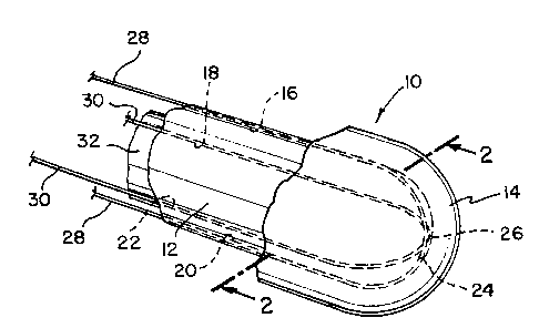
Figure 1 is a partial perspective view of an expandable MRI balloon
receiving coil in accordance with the present invention;
Figure 2A is a cross-sectional view of one embodiment of the present
invention, taken along line 2-2 of Figure 1 and looking in the direction of
the arrows;
Figure 2B is a cross-sectional view of another embodiment of the present
invention, taken along line 2-2 of Figure 1 and looking in the direction of
the arrows;
Figure 2C is a cross-sectional view of yet another embodiment of the present
invention, taken along line 2-2 of Figure 1 and looking in the direction of
the arrows;
Figure 2D is a cross-sectional view of another embodiment of the present

CA 02351514 2001-05-17
WO 00/28896 PCT/US99/27581
4
invention, taken along line 2-2 of Figure 1 and looking in the direction of
the arrows;
Figure 3 is a schematic illustration of the wires in the form of two loops of
coaxial cable connected in series with a tuning capacitor;
Figure 4 is a schematic illustration of the wires in the form of two loops of
coaxial cable connected in parallel with a tuning capacitor;
Figure 5 is a cross-sectional view of the MRI probe showing only one coil,
its tuning capacitor, central shaft, and the internal and external balloons;
Figure 6 is a perspective view of the internal balloon and the wire loops in
quadrature; and
Figure 7 is a view of two wire loops shown in quadrature, without the central
shaft, internal and external balloon.
DETAILED DESCRIPTION OF THE PREFERRED EMBODIMENT
Referring now to Figure 1, a partial perspective view of an MRI probe 10
is illustrated. Probe 10 includes an inner balloon 12 and an outer balloon 14.
In a first embodiment, which is illustrated in Figure 2A, balloon 12 has four
axially extending grooves 16, 18, 20 and 22 in its outer surface. Groove 16 is
disposed
generally diametrically opposite from groove 20. Likewise, groove 18 is
disposed generally
diametrically opposite from groove 22. Thus, adjacent grooves are disposed at
90°
intervals. At the distal end 24 of inner balloon 12, grooves 16, 18, 20, 22
curve radially
inwardly and intersect at the distal tip or apex 26 of inner balloon 12. Thus,
as viewed
from the front, grooves 16, 18, 20 and 22 appear to intersect at 90°
angles, thereby
resembling cross hairs. A first wire 28 is placed within grooves 16, 20. A
second wire 30
is placed within grooves 18, 22. Wires 28, 30 are insulated from one another
at least at

CA 02351514 2001-05-17
WO 00/28896 PCT/US99/27581
their point of intersection at distal tip 26. Wires 28, 30 are fixedly held
within grooves 16,
20, 18 and 22. In a currently preferred embodiment, wires 28, 30 are glued
within their
respective grooves 16, 20 and 18, 22, respectively.
A shaft 32 is disposed within inner balloon I2. If shaft 32 is used, it is
5 preferably a plastic tube of appropriate size and is formed from an elastic
material that has
sufficient flexibility to allow probe 10 to enter the human body through
either the mouth or
nose and, thereafter, be placed within the esophagus. Shaft 32 preferably has
an outer
diameter of less than 3/ 16" if it is to enter into the mouth and less than '/
" if is to be inserted
into the nose. An annular space 34 is disposed between shaft 32 and inner
balloon 12.
Annular space 34 is, at its proximal end, fluidly connected to a conduit (not
shown), which
is connected to a source of fluid pressure to selectively inflate and deflate
the inner balloon
as desired. Additionally, as those skilled in the art will readily recognize,
wires 28, 30 form
two loops that are electrically connected at their proximal end via interface
circuits for
impendence matching (not shown). The interface circuits are then electrically
connected to
a conventional MRI apparatus (e.g., an MRI spectrometer) to produce an image
based upon
a signal received by wires 28, 30.
Wires loops 28, 30 are preferably each formed from coaxial cable that may
be connected to a tuning capacitor 60 either as shown in Fig. 3 in a series
circuit or as shown
in Fig. 4 in a parallel circuit. In the currently preferred embodiment, the
parallel circuit is
used because it provides at least twice the SNR of a series circuit. In any of
the below
embodiments, wire loops 28, 30 are each preferably formed from coaxial cable,
which has
an outer conductor 70 and an inner conductor 71. For both wire loops 28, 30,
the
approximate midpoints of the outer conductor 70 has a gap 75. While gap 75 is
provided
at or near the point of intersection of wires 28,30, the wires are still
insulated from one

CA 02351514 2001-05-17
WO 00/28896 PCT/US99/27581
6
another. Wires 28,30 are disposed at approximately 90° intervals. Thus,
the signal
produced by wire 28 and 30 are said to be in quadrature. Therefore, the
resulting image
produced from the signals received from wires 28, 30 is more symmetrical than
a
conventional receiving coil. The MRI apparatus can be, for example, a GE
Signa, 1.5 Tesla,
which is commercially available from General Electric Company.
In operation, the probe 10 is initially in a deflated state and the outer
surface
of outer balloon 14 is preferably well lubricated with a conventional,
sterile, water-soluable
lubricant. The distal end 24 of the probe is then inserted into the body
through either the
mouth or the nose. Distal end 24 is further inserted into the body until it
passes into the
esophagus. The receiving coil is placed in the desired position within the
esophagus, as
close to the object to be imaged as possible. For example, for the closest
approach to the
heart and the aortic arch, the receiver coil should be placed within the
esophagus behind and
under the heart and the aortic arch. The balloon assembly is inflated to
maintain the position
of the receiver coil within the esophagus and so that the receiver coil will
be as large in
diameter as possible without causing harm to the esophagus. Of course, the
amount that the
balloon is inflated will vary from patient to patient, but will typically will
be on the order
of about 'h inch in diameter by 5 inches in length when inflated.
The receiving coil alone may be sufficient to obtain an adequate image of the
aortic arch. Alternatively, an external surface MRI receiving coil may be
placed on the
patient to produce a combined image from the internal probe 10 and the
external receiving
coil (not shown). A method of generating a combined image of the heart and the
vessels
emanating from the heart, from the combination of a first image from a coil
placed within
the body and a second image from a coil placed externally to the body is
disclosed in
Applicants' copending application serial number 09/081,908, entitled "Cardiac
MRI With

CA 02351514 2001-05-17
WO 00128896 PC'f/US99/27581
7
An Internal Receiving Coil and An External Receiving Coil", filed on May 20,
1998, the
disclosure of which is hereby fully incorporated by reference.
Referring now to Figure 2B, an alternate embodiment of probe 10' is
illustrated. In this embodiment, an intermediate tubular sheath 36 is disposed
between inner
balloon 12 and outer balloon 14. Sheath 36 is formed with grooves 38, 40, 42,
44 to receive
wires 28, 30. Sheath 36 is made from an elastic material, such as, for
example, latex, to
permit tubular sheath 36 to expand when inner balloon 14 is inflated once the
probe has
been placed in the esophagus.
Referring now to Figure 2C, a further alternate embodiment of probe 10" is
illustrated. In this embodiment, a plurality of guide tubes 46, 48 are placed
on the exterior
surface of balloon 12. Each guide tube extends about the closed distal end 24
of balloon 12.
Thus, each guide tube has a first portion that is disposed on one external
side of balloon 12
and a second portion that is disposed on a generally diametrically opposite
external side of
balloon 12. Wire 28 is inserted into guide tube 46. Similarly, wire 30 is
placed within guide
tube 48. Thus, when probe 10" is placed within the esophagus, balloon 12 may
be inflated
to maintain the position of wires 28, 30, which together form the receiving
coil within the
esophagus so that the receiver coil will have as large a diameter as possible
without causing
harm to the esophagus.
Referring now to Figure 2D, a further alternative embodiment of probe 10"'
is illustrated. Grooves 50, 52, 54 and 56 are disposed within the inner
cylindrical surface
of outer balloon 14. Wire 28 is placed within grooves 50, 54. Similarly, wire
30 is placed
within grooves 52, 56. In use, probe 10"' operates in a manner similar to the
embodiments
illustrated in Figures 2A, 2B and 2C. In other words, once the probe has been
placed within
the esophagus, the annular space between shaft 32 and inner balloon 12 is
inflated thereby

CA 02351514 2001-05-17
WO 00/28896 PCT/US99/27581
8
causing the entire probe to stably maintain the position of the receiving coil
within the
esophagus so that the receiving coil has as large a diameter as possible
without causing harm
to the esophagus. The receiving coil may then be used to obtain an image of,
for example,
the heart and/or the aortic arch.
Referring now to Figure 5, a cross-sectional view of the MRI probe I00 is
illustrated. Here a single wire loop 128 or 130 (referred to as 128, 130 in
Figure 5) is
illustrated inflated on inner balloon 112. Both the wire loop and the inner
balloon are
covered by the outer balloon 114. Both the inner and outer balloons are
subsequently
attached at both ends to the central tubular shaft 132. Wire loop 128 or 130
also penetrates
into the central tube 132 at both ends. At the proximal end, where the loop
128 or 130
penetrates into the central tube 132 , the wire 128 or 130 continues down
through central
shaft 132 and out of its proximal end to the MRI spectrometer.
Referring now to Figure 6, a perspective view of the wire loops 28, 30 and
inflated inner balloon 12 is illustrated. Wire loops 28, 30 are shown in
quadrature, with
outer balloon 14 being removed for the sake of clarity on the drawings.
Referring now to
Figure 7, only the wire loops 28, 30 are shown for the sake of clarity. Wire
loops 28, 30
are shown in quadrature.
Having described the presently preferred exemplary embodiment of an
expandable MRI receiving coil in accordance with the present invention, it is
believed that
other modifications, variations and changes will be suggested to those skilled
in the art in
view of the teachings set forth herein. It is, therefore, to be understood
that all such
modifications, variations, and changes are believed to fall within the scope
of the present
invention as defined by the appended claims.
Dessin représentatif
Une figure unique qui représente un dessin illustrant l'invention.
États administratifs

2024-08-01 : Dans le cadre de la transition vers les Brevets de nouvelle génération (BNG), la base de données sur les brevets canadiens (BDBC) contient désormais un Historique d'événement plus détaillé, qui reproduit le Journal des événements de notre nouvelle solution interne.

Veuillez noter que les événements débutant par « Inactive : » se réfèrent à des événements qui ne sont plus utilisés dans notre nouvelle solution interne.

Pour une meilleure compréhension de l'état de la demande ou brevet qui figure sur cette page, la rubrique Mise en garde , et les descriptions de Brevet , Historique d'événement , Taxes périodiques et Historique des paiements devraient être consultées.

Historique d'événement

Description Date
Demande non rétablie avant l'échéance 2006-11-20
Le délai pour l'annulation est expiré 2006-11-20
Inactive : CIB de MCD 2006-03-12
Réputée abandonnée - omission de répondre à un avis sur les taxes pour le maintien en état 2005-11-18
Lettre envoyée 2004-12-03
Requête d'examen reçue 2004-11-18
Exigences pour une requête d'examen - jugée conforme 2004-11-18
Toutes les exigences pour l'examen - jugée conforme 2004-11-18
Inactive : IPRP reçu 2004-02-25
Lettre envoyée 2003-12-11
Exigences de rétablissement - réputé conforme pour tous les motifs d'abandon 2003-11-27
Réputée abandonnée - omission de répondre à un avis sur les taxes pour le maintien en état 2003-11-18
Lettre envoyée 2003-01-17
Inactive : Grandeur de l'entité changée 2003-01-16
Exigences de rétablissement - réputé conforme pour tous les motifs d'abandon 2002-12-12
Réputée abandonnée - omission de répondre à un avis sur les taxes pour le maintien en état 2002-11-18
Inactive : Page couverture publiée 2001-09-24
Lettre envoyée 2001-09-21
Inactive : CIB en 1re position 2001-08-19
Inactive : Transfert individuel 2001-08-07
Inactive : Lettre de courtoisie - Preuve 2001-07-31
Inactive : Notice - Entrée phase nat. - Pas de RE 2001-07-26
Demande reçue - PCT 2001-07-23
Demande publiée (accessible au public) 2000-05-25

Historique d'abandonnement

Date d'abandonnement Raison Date de rétablissement
2005-11-18
2003-11-18
2002-11-18

Taxes périodiques

Le dernier paiement a été reçu le 2004-11-05

Avis : Si le paiement en totalité n'a pas été reçu au plus tard à la date indiquée, une taxe supplémentaire peut être imposée, soit une des taxes suivantes :

  • taxe de rétablissement ;
  • taxe pour paiement en souffrance ; ou
  • taxe additionnelle pour le renversement d'une péremption réputée.

Les taxes sur les brevets sont ajustées au 1er janvier de chaque année. Les montants ci-dessus sont les montants actuels s'ils sont reçus au plus tard le 31 décembre de l'année en cours.
Veuillez vous référer à la page web des taxes sur les brevets de l'OPIC pour voir tous les montants actuels des taxes.

Historique des taxes

Type de taxes Anniversaire Échéance Date payée
Taxe nationale de base - petite 2001-05-17
Enregistrement d'un document 2001-08-07
TM (demande, 2e anniv.) - petite 02 2001-11-19 2001-11-16
TM (demande, 3e anniv.) - générale 03 2002-11-18 2002-11-15
Rétablissement 2002-12-12
Rétablissement 2003-11-27
TM (demande, 4e anniv.) - générale 04 2003-11-18 2003-11-27
TM (demande, 5e anniv.) - générale 05 2004-11-18 2004-11-05
Requête d'examen - générale 2004-11-18
Titulaires au dossier

Les titulaires actuels et antérieures au dossier sont affichés en ordre alphabétique.

Titulaires actuels au dossier
CARDIAC M.R.I. INC.
Titulaires antérieures au dossier
LAWRENCE A. MINKOFF
Les propriétaires antérieurs qui ne figurent pas dans la liste des « Propriétaires au dossier » apparaîtront dans d'autres documents au dossier.
Documents

Pour visionner les fichiers sélectionnés, entrer le code reCAPTCHA :


Pour visualiser une image, cliquer sur un lien dans la colonne description du document. Pour télécharger l'image (les images), cliquer l'une ou plusieurs cases à cocher dans la première colonne et ensuite cliquer sur le bouton "Télécharger sélection en format PDF (archive Zip)" ou le bouton "Télécharger sélection (en un fichier PDF fusionné)".

Liste des documents de brevet publiés et non publiés sur la BDBC .

Si vous avez des difficultés à accéder au contenu, veuillez communiquer avec le Centre de services à la clientèle au 1-866-997-1936, ou envoyer un courriel au Centre de service à la clientèle de l'OPIC.


Description du
Document 
Date
(aaaa-mm-jj) 
Nombre de pages   Taille de l'image (Ko) 
Dessin représentatif 2001-08-29 1 15
Description 2001-05-16 8 355
Abrégé 2001-05-16 1 61
Dessins 2001-05-16 4 107
Revendications 2001-05-16 6 184
Rappel de taxe de maintien due 2001-07-25 1 112
Avis d'entree dans la phase nationale 2001-07-25 1 194
Courtoisie - Certificat d'enregistrement (document(s) connexe(s)) 2001-09-20 1 137
Courtoisie - Lettre d'abandon (taxe de maintien en état) 2003-01-15 1 176
Avis de retablissement 2003-01-16 1 167
Courtoisie - Lettre d'abandon (taxe de maintien en état) 2003-12-10 1 177
Avis de retablissement 2003-12-10 1 167
Rappel - requête d'examen 2004-07-19 1 117
Accusé de réception de la requête d'examen 2004-12-02 1 177
Courtoisie - Lettre d'abandon (taxe de maintien en état) 2006-01-15 1 174
Correspondance 2001-07-25 1 25
PCT 2001-05-16 7 323
Taxes 2002-11-14 1 36
Taxes 2002-12-11 1 46
Taxes 2003-11-26 1 34
Taxes 2001-11-15 1 35
PCT 2001-05-17 3 148
Taxes 2004-11-04 1 33